


Brain connectivity loss due to traumatic brain injury, stroke or multiple sclerosis can have serious consequences on life quality and a measurable impact upon neural and cognitive function. Though brain network properties are known to be affected disproportionately by injuries to certain gray matter regions, the manner in which white matter insults affect such properties remains poorly understood. Here, network-theoretic analysis allows us to identify the existence of a macroscopic neural connectivity core in the adult human brain which is particularly sensitive to network lesioning. The systematic lesion analysis of brain connectivity matrices from diffusion neuroimaging over a large sample (N=110) reveals that the global vulnerability of brain networks can be predicated upon the extent to which injuries disrupt this connectivity core, which is found to be quite distinct from the set of connections between rich club nodes in the brain. Thus, in addition to connectivity within the rich club, the brain as a network also contains a distinct core scaffold of network edges consisting of WM connections whose damage dramatically lowers the integrative properties of brain networks. This pattern of core white matter fasciculi whose injury results in major alterations to overall network integrity presents new avenues for clinical outcome prediction following brain injury by relating lesion locations to connectivity core disruption and implications for recovery. The findings of this study contribute substantially to current understanding of the human white matter connectome, its sensitivity to injury, and clarify a long-standing debate regarding the relative prominence of gray vs. white matter regions in the context of brain structure and connectomic architecture.
The First Map Showing All the Connections in Your Brain's White Matter
Scientists have created the first map of the ways that the white matter in our brains connects with itself, and with our grey matter. The takeaway? It

The neuroimaging correlates of depression established across six large-scale population datasets
Depression has been linked to reduced size of subcortical regions and abnormal functional connectivity in frontal and default mode networks. However, recent meta-analyses have failed to identify significant converging correlates of depression across the literature such that a conclusive mapping of the neuroimaging correlates of depression remains elusive. We leveraged 23,417 participants across 6 population datasets to comprehensively establish the neuroimaging correlates of depression. We found reductions in gray matter volume/cortical surface area associated with depression in the frontal cortex, anterior cingulate and insula, confirming previous studies showing the importance of prefrontal and default mode regions in depression. Our findings demonstrate multiple surprising results, including a lack of depression correlates in subcortical brain regions and significant depression correlates in somatomotor and visual regions. Overall, these results shed new light on key brain regions involved in the pathophysiology of depression, updating our understanding of the neuroimaging correlates of depression symptoms.
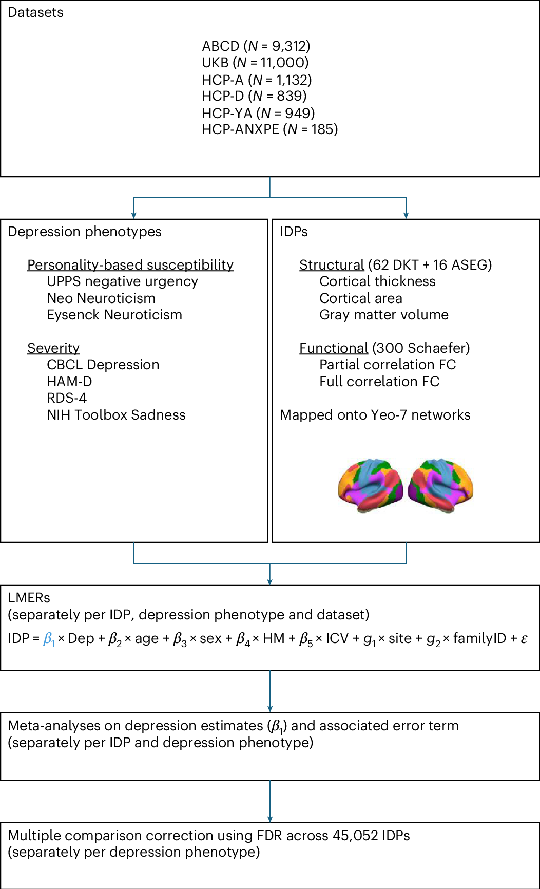
The neuroimaging correlates of depression established across six large-scale population datasets
Depression has been linked to reduced size of subcortical regions and abnormal functional connectivity in frontal and default mode networks. However, recent meta-analyses have failed to identify significant converging correlates of depression across the literature such that a conclusive mapping of the neuroimaging correlates of depression remains elusive. We leveraged 23,417 participants across 6 population datasets to comprehensively establish the neuroimaging correlates of depression. We found reductions in gray matter volume/cortical surface area associated with depression in the frontal cortex, anterior cingulate and insula, confirming previous studies showing the importance of prefrontal and default mode regions in depression. Our findings demonstrate multiple surprising results, including a lack of depression correlates in subcortical brain regions and significant depression correlates in somatomotor and visual regions. Overall, these results shed new light on key brain regions involved in the pathophysiology of depression, updating our understanding of the neuroimaging correlates of depression symptoms.
Brain Hyperconnectivity in Children with Autism and its Links to Social Deficits
In this study, Supekar, Menon, and colleagues shed light on a longstanding question about the nature of brain connectivity aberrations in autism. Using functional neuroimaging data, they demonstrate that the brains of children with autism are hyperconnected in ways that are related to the severity of social impairment exhibited by these children. They replicate these findings in two additional cohorts of children, challenging current theories of brain connectivity in autism and providing unique insights into the brain mechanisms underlying the disorder.

Embracing the suboptimal organization of the human brain
Human brain architecture is guiding brain-inspired artificial intelligence (AI) and has been treated as an optimal template, whose deviations could mark different psychiatric and neurological conditions. We argue this premise is wrong: under any single goal (e.g., minimal wiring cost or maximal communication efficiency), the human connectome is suboptimal. Instead, its organization reflects multi-objective trade-offs navigated over evolution and development under biological and environmental constraints. For psychopathology, atypical trajectories are not distances from an ideal brain but reweighted compromises in the same trade-off space. For neuro-AI, directly duplicating the brain’s connectivity risks copying its irrelevant compromises. Treating brains and models as products of multi-objective optimization and co-tuning relevant objectives offers a more powerful framework for interpreting clinical phenotypes and designing next-generation AI.

Innate connectivity patterns drive the development of the visual word form area
What determines the functional organization of cortex? One hypothesis is that innate connectivity patterns, either structural or functional connectivity, set up a scaffold upon which functional specialization can later take place. We tested this hypothesis by asking whether the visual word form area (VWFA), an experience-driven region, was already functionally connected to proto language networks in neonates scanned within one week of birth. Using the data from the Human Connectone Project (HCP) and the Developing Human Connectome Project (dHCP), we calculated intrinsic functional connectivity during resting-state functional magnetic resonance imaging (fMRI), and found that neonates showed similar functional connectivity patterns to adults. We observed that (1) language regions connected more strongly with the putative VWFA than other adjacent ventral visual regions that also show foveal bias, and (2) the VWFA connected more strongly with frontotemporal language regions than with regions adjacent to these language regions. These data suggest that the location of the VWFA is earmarked at birth due to its connectivity with the language network, providing evidence that innate connectivity instructs the later refinement of cortex.

GitHub - AIM-KannLab/BrainIAC | Giovanni Giulietti
One of the biggest hurdles in neuroimaging AI has always been the scarcity of labeled data, especially for rare conditions. But a recent study published in Nature Neuroscience introduces a potential solution: BrainIAC (Brain Imaging Adaptive Core). Unlike traditional models that are trained for one specific task, BrainIAC is a "foundation model", a large-scale AI model trained on vast amounts of data that can perform many different tasks and be adapted to specific applications. It was pretrained on a massive dataset of 48965 brain structural MRI sequences (T1w, T2w, FLAIR, T1CE) using self-supervised learning, allowing it to understand the "big picture" of brain anatomy across different ages and conditions. It has been tested across seven diverse and clinically meaningful applications: - brain age prediction - IDH mutation classification - Mild Cognitive Impairment (MCI) classification - diffuse glioma overall survival prediction - MR sequence classification - time-to-stroke prediction - tumor segmentation BrainIAC consistently outperformed traditional supervised learning approaches and other medical-specific models. The real "magic" happens in low-data scenarios. While traditional AI usually needs thousands of examples, BrainIAC can adapt to new tasks using only a few samples. BrainIAC can be used for structural brain MRI analysis through two main methods: - Web Usage (BrainIAC platform): the easiest way to use BrainIAC, providing a user-friendly interface to upload structural brain MRI data and run inference. - Local usage (GitHub repository): for more advanced and customized analysis. It allows complete control over the data preprocessing, model training, and inference pipelines. It requires Python 3.9+ and NVIDIA GPU with CUDA 11.0+. 🔗 BrainIAC paper: https://lnkd.in/dq6Gprgt 🔗 BrainIAC web platform https://lnkd.in/dpreadMp 🔗 BrainIAC GitHub repo: https://lnkd.in/duwSEg4V #MedicalAI #Neuroscience #HealthTech #BrainIAC #Radiology #Innovation #DeepLearning #PrecisionMedicine

The brain functional connectome is robustly altered by lack of sleep
Sleep is a universal phenomenon necessary for maintaining homeostasis and function across a range of organs. Lack of sleep has severe health-related consequences affecting whole-body functioning, yet no other organ is as severely affected as the brain. The neurophysiological mechanisms underlying these deficits are poorly understood. Here, we characterize the dynamic changes in brain connectivity profiles inflicted by sleep deprivation and how they deviate from regular daily variability. To this end, we obtained functional magnetic resonance imaging data from 60 young, adult male participants, scanned in the morning and evening of the same day and again the following morning. 41 participants underwent total sleep deprivation before the third scan, whereas the remainder had another night of regular sleep. Sleep deprivation strongly altered the connectivity of several resting-state networks, including dorsal attention, default mode, and hippocampal networks. Multivariate classification based on connectivity profiles predicted deprivation state with high accuracy, corroborating the robustness of the findings on an individual level. Finally, correlation analysis suggested that morning-to-evening connectivity changes were reverted by sleep (control group)-a pattern which did not occur after deprivation. We conclude that both, a day of waking and a night of sleep deprivation dynamically alter the brain functional connectome.
Neuroscientists Roll Out First Comprehensive Atlas of Brain Cells - Slashdot
A slew of 17 studies reveals the first comprehensive list of all cell types in the primary motor complex, offering "a starting point for tracing cellular networks to understand how they control our body and mind and how they are disrupted in mental and physical disorders," reports ScienceDaily. From...

Open-source software reveals complete 3D architecture of brain cells
The neurons in our brain that underlie thought connect to each other using tiny branch-like structures on their surfaces known as dendritic spines. Now scientists at Columbia's Zuckerman Institute and ...

Chemotherapy Disrupts Brain Connectivity - Neuroscience News
New research reveals that chemotherapy can lead to rapid and widespread changes in brain connectivity in breast cancer patients.
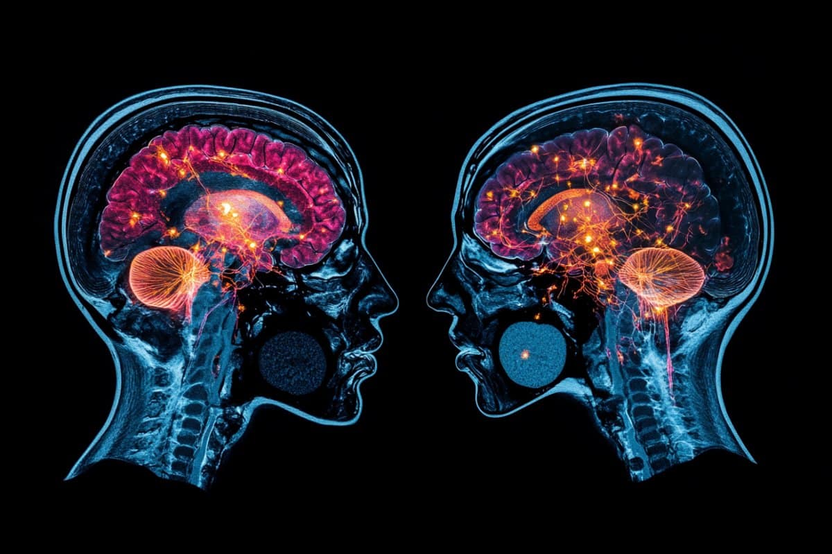
fMRI Data Reveals the Number of Parallel Processes Running in the Brain
The human brain carries out many tasks at the same time, but how many? Now fMRI data has revealed just how parallel gray matter is.
The Global Brain Algorithm
<p>This is a collection of explanations on the global brain algorithm. We explain core concepts in simple terms that help to understand the algorithms as a whole.</p>
Your Brain Is Shaped Like Nobody Else’s - Nautilus
Every brain’s white matter is different—and that might hold the key to better treatments.
Natural process activating brain's immune cells could point way to repairing damaged brain tissue
The brain's key "breeder" cells, it turns out, do more than that. They secrete substances that boost the numbers and strength of critical brain-based immune cells believed to play a vital role in brain ...
Intelligence Network in the Human Brain Discovered - Neuroscience News
Researchers identify two gene clusters in the brain linked to human intelligence.
