

Sleep is a universal phenomenon necessary for maintaining homeostasis and function across a range of organs. Lack of sleep has severe health-related consequences affecting whole-body functioning, yet no other organ is as severely affected as the brain. The neurophysiological mechanisms underlying these deficits are poorly understood. Here, we characterize the dynamic changes in brain connectivity profiles inflicted by sleep deprivation and how they deviate from regular daily variability. To this end, we obtained functional magnetic resonance imaging data from 60 young, adult male participants, scanned in the morning and evening of the same day and again the following morning. 41 participants underwent total sleep deprivation before the third scan, whereas the remainder had another night of regular sleep. Sleep deprivation strongly altered the connectivity of several resting-state networks, including dorsal attention, default mode, and hippocampal networks. Multivariate classification based on connectivity profiles predicted deprivation state with high accuracy, corroborating the robustness of the findings on an individual level. Finally, correlation analysis suggested that morning-to-evening connectivity changes were reverted by sleep (control group)-a pattern which did not occur after deprivation. We conclude that both, a day of waking and a night of sleep deprivation dynamically alter the brain functional connectome.
Why Sleep Deprivation Eases Depression
Glial activity reveals how sleep deprivation elevates mood

Bad sleep 'dramatically' alters body
A run of poor sleep can have a dramatic effect on the internal workings of the human body, say UK researchers.

Are two halves better than one whole? A comparison of the amount and quality of sleep obtained by healthy adult males living on split and consolidated sleep-wake schedules
The aim of this study was to compare the quantity/quality of sleep obtained by people living on split and consolidated sleep-wake schedules. The study had a between-groups design, with 13 participants in a consolidated condition (all males, mean age of 22.5yr) and 16 participants in a split condition (all males, mean age of 22.6yr). Both conditions employed forced desynchrony protocols with the activity:rest ratio set at 2:1, but the consolidated condition had one sleep-wake cycle every 28h (9.33+18.67), while the split condition had one sleep-wake cycle every 14h (4.67+9.33). Sleep was assessed using polysomnography. Participants in the split and consolidated conditions obtained 4.0h of sleep per 14h and 7.6h of sleep per 28h, respectively. Some differences between the groups indicated that sleep quality was lower in the split condition than the consolidated condition: the split sleeps had longer sleep onset latency (9.7 vs. 4.3min), more arousals (7.4 vs. 5.7 per hour in bed), and a greater percentage of stage 1 sleep (4.1% vs. 3.1%), than the consolidated sleeps. Other differences between the groups indicated that sleep quality was higher in the split condition than the consolidated condition: the split sleeps had a lower percentage of wake after sleep onset sleep (11.7% vs. 17.6%), and a greater percentage of slow wave sleep (30.2% vs. 23.8%), than the consolidated sleeps. These results indicate that the split schedule was not particularly harmful, and may have actually been beneficial, to sleep. Split work-rest schedules can be socially disruptive, but their use may be warranted in work settings where shiftworkers are separated from their normal family/social lives (e.g., fly-in fly-out mining) or where the need for family/social time is secondary to the task (e.g., emergency response to natural disasters).
The neuroimaging correlates of depression established across six large-scale population datasets
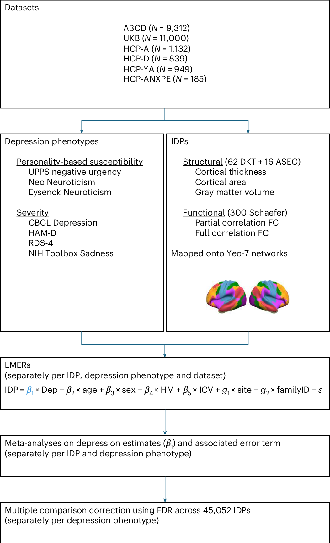
Depression has been linked to reduced size of subcortical regions and abnormal functional connectivity in frontal and default mode networks. However, recent meta-analyses have failed to identify significant converging correlates of depression across the literature such that a conclusive mapping of the neuroimaging correlates of depression remains elusive. We leveraged 23,417 participants across 6 population datasets to comprehensively establish the neuroimaging correlates of depression. We found reductions in gray matter volume/cortical surface area associated with depression in the frontal cortex, anterior cingulate and insula, confirming previous studies showing the importance of prefrontal and default mode regions in depression. Our findings demonstrate multiple surprising results, including a lack of depression correlates in subcortical brain regions and significant depression correlates in somatomotor and visual regions. Overall, these results shed new light on key brain regions involved in the pathophysiology of depression, updating our understanding of the neuroimaging correlates of depression symptoms.
The neuroimaging correlates of depression established across six large-scale population datasets
Depression has been linked to reduced size of subcortical regions and abnormal functional connectivity in frontal and default mode networks. However, recent meta-analyses have failed to identify significant converging correlates of depression across the literature such that a conclusive mapping of the neuroimaging correlates of depression remains elusive. We leveraged 23,417 participants across 6 population datasets to comprehensively establish the neuroimaging correlates of depression. We found reductions in gray matter volume/cortical surface area associated with depression in the frontal cortex, anterior cingulate and insula, confirming previous studies showing the importance of prefrontal and default mode regions in depression. Our findings demonstrate multiple surprising results, including a lack of depression correlates in subcortical brain regions and significant depression correlates in somatomotor and visual regions. Overall, these results shed new light on key brain regions involved in the pathophysiology of depression, updating our understanding of the neuroimaging correlates of depression symptoms.
Depression linked to internal jet lag study finds
Circadian rhythms controlling sleep were out of sync in a quarter of young people with depression in this Sydney-based study. Read more

Innate connectivity patterns drive the development of the visual word form area
What determines the functional organization of cortex? One hypothesis is that innate connectivity patterns, either structural or functional connectivity, set up a scaffold upon which functional specialization can later take place. We tested this hypothesis by asking whether the visual word form area (VWFA), an experience-driven region, was already functionally connected to proto language networks in neonates scanned within one week of birth. Using the data from the Human Connectone Project (HCP) and the Developing Human Connectome Project (dHCP), we calculated intrinsic functional connectivity during resting-state functional magnetic resonance imaging (fMRI), and found that neonates showed similar functional connectivity patterns to adults. We observed that (1) language regions connected more strongly with the putative VWFA than other adjacent ventral visual regions that also show foveal bias, and (2) the VWFA connected more strongly with frontotemporal language regions than with regions adjacent to these language regions. These data suggest that the location of the VWFA is earmarked at birth due to its connectivity with the language network, providing evidence that innate connectivity instructs the later refinement of cortex.


Why We Sleep, and Why We Often Can’t
Does our contemporary obsession with sleep obscure what makes it special in the first place?

Down From the Trees, Humans Finally Got a Decent Night’s Sleep (Published 2015)
Humans sleep more deeply but for shorter periods than other primates’ habits, a study finds. The pattern may have helped humans evolve more powerful brains.

'Sleep should be prescribed': what those late nights out could be costing you
Leading neuroscientist Matthew Walker on why sleep deprivation is increasing our risk of cancer, heart attack and Alzheimer’s – and what you can do about it

Scientists discover how deep sleep builds muscle, burns fat, and boosts the brain
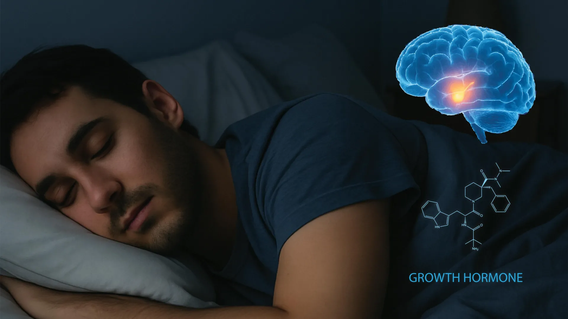
Scientists have discovered a surprising two-way relationship between sleep and growth hormone, revealing how the brain carefully balances physical recovery with wakefulness. The findings could help explain why poor sleep harms metabolism and brain health, potentially opening the door to new treatments for sleep and neurological disorders.
Sleep as a Competitive Advantage
Too many of us live by the myth that one less hour of sleep gives us one more hour of productivity, Tony Schwartz writes in the Life@Work column. In reality, lost sleep not only leaves us more fatigued, but also takes a toll on our cognitive capacity.

Sleep as a Competitive Advantage
Too many of us live by the myth that one less hour of sleep gives us one more hour of productivity, Tony Schwartz writes in the Life@Work column. In reality, lost sleep not only leaves us more fatigued, but also takes a toll on our cognitive capacity.
A Good Night’s Sleep Is Tied to Interruptions, Not Just Hours
New research shows that waking up several times in a night’s sleep can cause bad moods and limit one’s ability to focus and think.

Sleep's memory role discovered
Scientists in China and the US use advanced microscopy to discover exactly how a good night's sleep improves learning and memory.
