

The most detailed vascular image of a living human brain we've seen, captured with ultrasound through the skull — and the pipeline behind it.
Aleph
We're a research lab working on new interfaces for the brain. We think our telepathic future is both imminent and wonderful.
Foundation Model Predicts Brain Responses to Visual and Auditory Stimuli | Elisa Cascardi posted on the topic | LinkedIn
Thrilled to share this work with the world! Today, we're releasing a foundation model that predicts how the human brain responds to almost any sight or sound -- and replace the need for human scans to significantly fast-track neuroscience and clinical research. 🧠 With this model, we can simulate brain responses to advance our understanding of the brain -- without the need for costly human brain scans 🌐 By using improved understanding of how efficient our brains perceive the world around us, we can guide the development of more advanced AI systems 👩⚕️ With computer-simulated experimentation, we can now speedup clinical research to diagnose neurological diseases and find treatments faster We've open sourced the model and code for researchers to use and build on, and an interactive demo for you to learn more -- see below! 📄 Paper: https://lnkd.in/e7cbunJp 💻 Code: https://lnkd.in/ebwBVuJp ▶️ Demo: https://lnkd.in/eEUVxP4S 🤗 Model: https://lnkd.in/e2T8nPJP So thrilled to be a part of this team with Stéphane d'Ascoli Jean-Rémi King Jérémy RAPIN Yohann Benchetrit Teon Brooks Katelyn Begany Joséphine Raugel Hubert Banville and for the great teamwork with Diego Marcos Dominic Giardini bringing this research to life! #neuroscience #AI #aiforscience #opensource #neuroAI
This Is Your Brain on Podcasts (Published 2016)
Different people’s brains absorb a compelling story in the roughly the same complicated pattern, using many parts of the brain, researchers found.

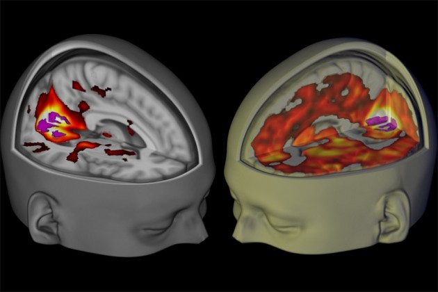
Brain scans reveal how LSD affects consciousness
Drug researcher David Nutt discusses brain-imaging studies with hallucinogens.
The Brainmade Mark
When you see this logo on any artwork, whether painting, poetry, or prose, you know that it was made by a human just like you.
Foundation Models in Medical Imaging: A Review and Outlook
Foundation models (FMs) are changing the way medical images are analyzed by learning from large collections of unlabeled data. Instead of relying on manually annotated examples, FMs are...

The brain's 7D sandcastles could be the key to consciousness | New Scientist
We’ve glimpsed mind-bending geometric structures that fleetingly encode our thoughts, memories and feelings – and could solve the greatest mystery of all

Data Hosting & Sharing: The Open MEG Archive (OMEGA)
Welcome to the Open MEG Archive (OMEGA) The Open MEG Archive (OMEGA) is the fruit of a collaboration between the McConnell Brain Imaging Centre (BIC) of the Montreal Neurological Institute-Hospital

Cog Neuro Hub OPMEG Workshop
The inner screen model of consciousness: applying the free energy principle directly to the study of conscious experience
This paper presents a model of consciousness that follows directly from the free- energy principle (FEP). We first rehearse the classical and quantum formulations of the FEP. In particular, we consider the “inner screen hypothesis” that follows from the quantum information theoretic version of the FEP. We then review applications of the FEP to the known sparse (nested and hierarchical) neuro-anatomy of the brain. We focus on the holographic structure of the brain, and how this structure supports (overt and covert) action.
The brain in the machine: How AI could help explain how we think | IBM
Scientists are using large AI models to predict patterns of brain activity at scale, a development that researchers say is pushing neuroscience toward a new kind of digital imaging.

NEJM retracts case study for AI-manipulated imagery
An “Images in Clinical Medicine” item in the New England Journal of Medicine has been retracted after the authors acknowledged using AI to alter the photo. Y. Wang, X. Mu/© The New Engl…
Virtual brain twins for stimulation in epilepsy
Estimating the epileptogenic zone network (EZN) is an important part of the diagnosis of drug-resistant focal epilepsy and has a pivotal role in treatment and intervention. Virtual brain twins provide a modeling method for personalized diagnosis and treatment. They integrate patient-specific brain topography with structural connectivity from anatomical neuroimaging such as magnetic resonance imaging, and dynamic activity from functional recordings such as electroencephalography (EEG) and stereo-EEG (SEEG). Seizures show rich spatial and temporal features in functional recordings, which can be exploited to estimate the EZN. Stimulation-induced seizures can provide important and complementary information. Here we consider invasive SEEG stimulation and non-invasive temporal interference stimulation as a complementary approach. This paper offers a high-resolution virtual brain twin framework for EZN diagnosis based on stimulation-induced seizures. It provides an important methodological and conceptual basis to make the transition from invasive to non-invasive diagnosis and treatment of drug-resistant focal epilepsy.

Reticulum Network
For the better part of a generation, we have been taught to visualize the digital world through the lens of hierarchy. The mental maps we carry are dominated by a single, misleading image: The Cloud.
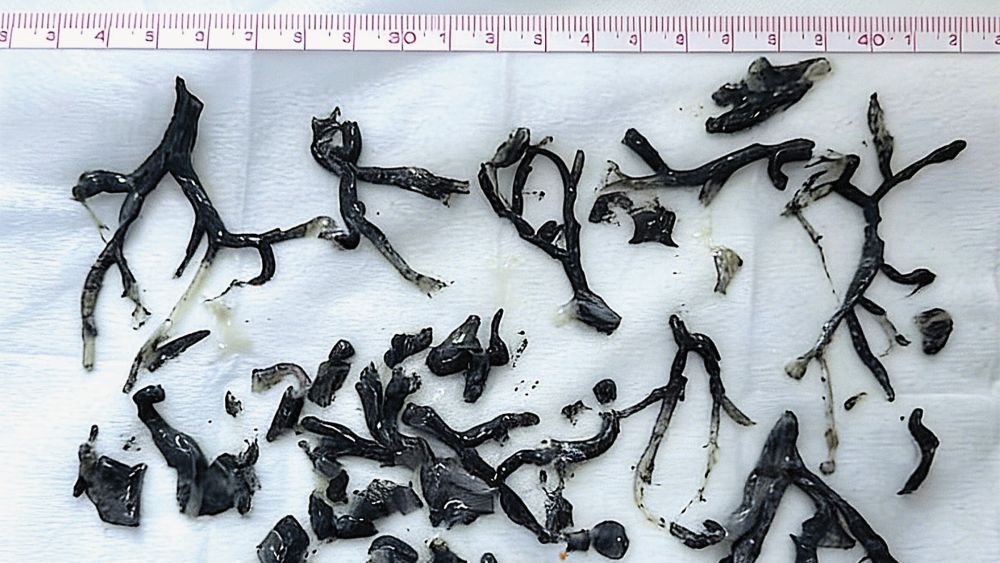
Any experts on retinal arteries in rats here, please? Some images in PMID: 30695730 look unexpectedly similar to me. The author claims these arteries are expected to look so similar. What do you think? pubpeer.com/publications/878202C68F72B479…
