

Any experts on retinal arteries in rats here, please? Some images in PMID: 30695730 look unexpectedly similar to me. The author claims these arteries are expected to look so similar. What do you think? pubpeer.com/publications/878202C68F72B479…
Jan 17, 2025 at 4:34 PM
Will Their Bionic Eyes Be Obsolete and Unsupported Too?
As another retinal implant company goes broke, the tech’s future dims

Ultrasound imaging of the brain — Aleph
The most detailed vascular image of a living human brain we've seen, captured with ultrasound through the skull — and the pipeline behind it.
Effects of carbon monoxid upon the eye
This paper gives a general survey of the subject and includes a graphic description of the subjective and general nutritional effects of carbon monoxid poisoning, by one of the victims; and incidentally throws light on the origin of a story of a haunted house. It gives an account of the ocular symptoms of such poisoning, revealing a distinct tendency of the poison to cause neuritis and especially to affect the optic nerve.
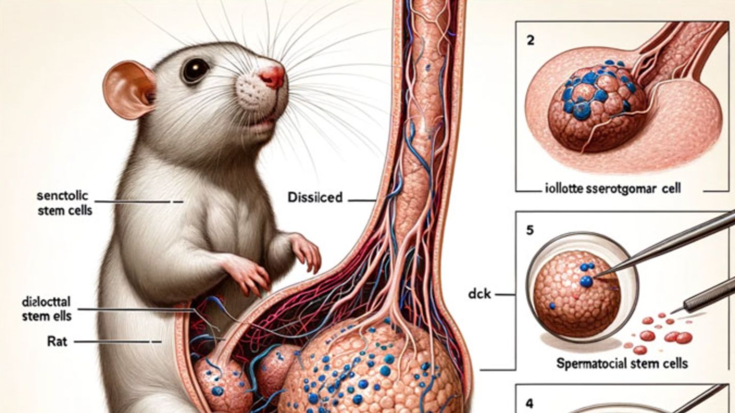
Study Featuring AI-Generated Giant Rat Penis Retracted, Journal Apologizes
A peer-reviewed study featured nonsensical AI images including a giant rat penis in the latest example of how generative AI has seeped into academia.
Computer Vision Syndrome
In no point in human history have people been staring at bright screens just a few feet from their face like they are today. Computer vision syndrome or CVS (no…

Designed to Deceive: Do These People Look Real to You? (Published 2020)
The people in this story may look familiar, like ones you’ve seen on Facebook or Twitter or Tinder. But they don’t exist. They were born from the mind of a computer, and the technology behind them is improving at a startling pace.

A running list of ATproto ideas - Tynan's Leaflets
Updated when the brain has more
Tipographia
Visual archive of graphic design, typography and things printed in Portugal. A collection by...

What's My JND?
Find your Just Noticeable Difference in colour perception. How small a colour difference can you actually see?

Special Dyslexia Fonts Are Based on Voodoo Pseudoscience
Link to: https://www.edutopia.org/article/do-dyslexia-fonts-actually-work

Open-source software reveals complete 3D architecture of brain cells
The neurons in our brain that underlie thought connect to each other using tiny branch-like structures on their surfaces known as dendritic spines. Now scientists at Columbia's Zuckerman Institute and ...

Substrates vision statement
Submitted to Substrates-25 Workshop Jonathan EdwardsMay 12, 2025

How Do Geckos Blink? A Comprehensive Guide to Gecko Eye Anatomy and Behavior. | ThePetFAQ
Geckos are unique animals that belong to the family Gekkonidae. They are known for their adhesive toe pads that allow them to climb walls and ceilings, but
Once you see it, you can't unsee it

W Social is TruthSocial with a European accent.
wecanjustdothings.leaflet.pubOnce you see it, you can't unsee it
W Social is TruthSocial with a European accent.
wecanjustdothings.leaflet.pub

