


Abstract There are many works dealing with the dynamics of bone remodeling, proposing increasingly complex and complete models. In the recent years, the efforts started to focus on developing models that not only reproduce the temporal evolution, but also include the spatial aspects of this phenomenon. In this work, we propose the spatial extension of an existing model that includes the dynamics of osteocytes. The spatial dependence is modeled in terms of a linear diffusion, as proposed in previous works dealing with related problems. The resulting model is then written in its variational form, and fully discretized using the well‐known finite element method and a combination of the implicit and explicit Euler schemes. The numerical algorithm is then analyzed, proving some a priori error estimates and its linear convergence. Finally, we extend the examples already published for the temporal model to one and two dimensions, showing the dynamics of the solution in the spatial domain.
Towards a new spatial representation of bone remodeling
Irregular bone remodeling is associated with a number of bone diseases such as osteoporosis and multiple myeloma. Computational and mathematical modeling can aid in therapy and treatment as well as understanding fundamental biology. Different approaches to modeling give insight into different aspects of a phenomena so it is useful to have an arsenal of various computational and mathematical models. Here we develop a mathematical representation of bone remodeling that can effectively describe many aspects of the complicated geometries and spatial behavior observed. There is a sharp interface between bone and marrow regions. Also the surface of bone moves in and out, i.e. in the normal direction, due to remodeling. Based on these observations we employ the use of a level-set function to represent the spatial behavior of remodeling. We elaborate on a temporal model for osteoclast and osteoblast population dynamics to determine the change in bone mass which influences how the interface between bone and marrow changes. We exhibit simulations based on our computational model that show the motion of the interface between bone and marrow as a consequence of bone remodeling. The simulations show that it is possible to capture spatial behavior of bone remodeling in complicated geometries as they occur in vitro and in vivo. By employing the level set approach it is possible to develop computational and mathematical representations of the spatialbehavior of bone remodeling. By including in this formalism further details, such as more complex cytokine interactions and accurate parameter values, it is possible to obtain simulations of phenomena related to bone remodeling with spatial behavior much as in vitro and in vivo. This makes it possible to perform in silica experiments more closely resembling experimental observations.
A review of recent developments in mathematical modeling of bone remodeling
In this article, we summarize the developments in the mathematical modeling of the mechanics of bone and related biological phenomena. We will devote special attention to the results of the last 10–15 years, although we will cover some relevant classical work to better frame the more recent researches. We will propose a division of the literature based on the main aim of the model (mechanical/biomathematical) and the type of biological phenomena considered (stimulus, growth, cell population dynamics). Finally, we will suggest some possible directions for future investigations.

The Cellular Dynamics of Bone Remodeling: A Mathematical Model
The mechanical properties of vertebrate bone are largely determined by a process which involves the complex interplay of three different cell types. This process is called bone remodeling and occurs asynchronously at multiple sites in the mature skeleton. The cells involved are bone resorbing osteoclasts, bone matrix producing osteoblasts, and mechanosensing osteocytes. These cells communicate with each other by means of autocrine and paracrine signaling factors and operate in complex entities, the so-called bone multicellular units (BMUs). To investigate the BMU dynamics in silico, we develop a novel mathematical model resulting in a system of nonlinear partial differential equations (PDEs) with time delays. The model describes the osteoblast and osteoclast populations together with the dynamics of the key messenger molecule RANKL and its decoy receptor OPG. Scaling theory is used to address parameter sensitivity and predict the emergence of pathological remodeling regimes. The model is studied numerically in one and two space dimensions using finite difference schemes in space and explicit delay equation solvers in time. The computational results are in agreement with in vivo observations and provide new insights into the role of the RANKL/OPG pathway in the spatial regulation of bone remodeling.

A new biological bone remodeling in silico model combined with advanced discretization methods
Abstract Bone remodeling remains a highly researched topic investigated by many strands of science. The main purpose of this work is formulating a new computational framework for biological simulation, extending the version of the bone remodeling model previously proposed by Komarova. Thus, considering only the biological aspect of the remodeling process, the action of osteoclasts and osteoblasts is taken into account as well as its impact on bone mass. It is conducted a spatiotemporal analysis of a remodeling cycle obtaining a dynamic behavior of bone cells very similar to the biological process already described in the literature. The numerical example used is based on bone images obtained with scanning electron microscopy. During simulation, it is possible to observe the variation of bone's architecture through isomaps. These maps are obtained through the combination of biological bone remodeling models with three distinct numerical techniques—finite element method (FEM), radial point interpolation method (RPIM), and natural neighbor radial point interpolation method (NNRPIM). A study combining these numerical techniques allows to compare their performance. Ultimately, this work supports the inclusion of meshless methods due to their smoother results and its easiness to be combined with medical images from CT scans and MRI.

<i>In silico</i> models of bone remodeling from macro to nano—from organ to cell
Abstract Computational modeling is a tool through which researchers can achieve a greater understanding of the mechanisms governing biological systems. In the field of bone biology, a plethora of models exist which attempt to replicate and investigate bone's dynamic behavior at different scales. At organ level, models are continuum based and describe the variation of bone's apparent density as a function of both biological and external mechanical stimuli. At tissue level, models include bone microarchitecture and more descriptive parameters such as trabecular thickness, osteoclast resorption depth, and activation frequency. Finally, at cell level, models employ partial differential equations to describe complex cellular interactions in the temporal domain. Although informative, these models exist in isolation. Consequently, their interpretation is limited. In this review, we present an overview of the organ‐, tissue‐, and cell‐level models and assess their ability to reflect bone's metabolic processes reliably. Existing interscale synergies are then presented along with a computational framework which could be exploited to achieve a fully integrated, multiscale modeling approach. WIREs Syst Biol Med 2011 3 241–251 DOI: 10.1002/wsbm.115 This article is categorized under: Analytical and Computational Methods > Computational Methods Models of Systems Properties and Processes > Organ, Tissue, and Physiological Models

A review of mathematical modeling of bone remodeling from a systems biology perspective
Bone remodeling is an essential, delicately balanced physiological process of coordinated activity of bone cells that remove and deposit new bone tissue in the adult skeleton. Due to the complex nature of this process, many mathematical models of bone remodeling have been developed. Each of these models has unique features, but they have underlying patterns. In this review, the authors highlight the important aspects frequently found in mathematical models for bone remodeling and discuss how and why these aspects are included when considering the physiology of the bone basic multicellular unit, which is the term used for the collection of cells responsible for bone remodeling. The review also emphasizes the view of bone remodeling from a systems biology perspective. Understanding the systemic mechanisms involved in remodeling will help provide information on bone pathology associated with aging, endocrine disorders, cancers, and inflammatory conditions and enhance systems pharmacology. Furthermore, some features of the bone remodeling cycle and interactions with other organ systems that have not yet been modeled mathematically are discussed as promising future directions in the field.

A review of mathematical modeling of bone remodeling from a systems biology perspective
Bone remodeling is an essential physiological process in the adult skeleton. Due to the complex nature of this process, many mathematical models of bone remodeling have been developed. Each of these models has unique features, but they have underlying patterns. In this review, the authors highlight the important aspects frequently found in mathematical models for bone remodeling and discuss how and why these aspects are included when considering the physiology of the bone basic multicellular unit, which is the term used for the collection of cells responsible for bone remodeling. The review also emphasizes the view of bone remodeling from a systems biology perspective. Understanding the systemic mechanisms involved in remodeling will help provide information on bone pathology associated with aging, endocrine disorders, cancers, and inflammatory conditions and enhance systems pharmacology. Furthermore, some features of the bone remodeling cycle and interactions with other organ systems that have not yet been modeled mathematically are discussed as promising future directions in the field.
Mathematical Modeling of Spatio-Temporal Dynamics of a Single Bone Multicellular Unit
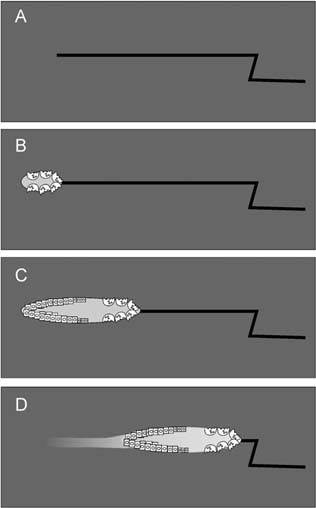
Abstract During bone remodeling, bone-resorbing osteoclasts and bone-forming osteoblasts are organized in bone multicellular units (BMUs), which travel at a rate of 20–40 μm/d for 6–12 mo, maintaining a cylindrical structure. However, the interplay of local BMU geometry with biochemical regulation is poorly understood. We developed a mathematical model of BMU describing changes in time and space of the concentrations of proresorptive cytokine RANKL and its inhibitor osteoprotegerin (OPG), in osteoclast and osteoblast numbers, and in bone mass. We assumed that osteocytes surrounding a microfracture produce RANKL, which attracted osteoclasts. OPG and RANKL were produced by osteoblasts and diffused through bone, RANKL was eliminated by binding to OPG and RANK. Osteoblasts were coupled to osteoclasts through paracrine factors. The evolution of the BMU arising from this model was studied using numerical simulations. Our model recapitulated the spatio-temporal dynamics observed in vivo in a cross-section of bone. In response to a RANKL field, osteoclasts moved as a well-confined cutting cone. The coupling of osteoclasts to osteoblasts allowed for sufficient recruitment of osteoblasts to the resorbed surfaces. The RANKL field was the highest at the microfracture in front of the BMU, whereas the OPG field peaked at the back of the BMU, resulting in the formation of a RANKL/OPG gradient, which strongly affected the rate of BMU progression and its size. Thus, the spatial organization of a BMU provides important constraints on the roles of RANKL and OPG as well as possibly other regulators in determining the outcome of remodeling in the BMU.
Optimizing the dynamics of bone turnover with genetic algorithm
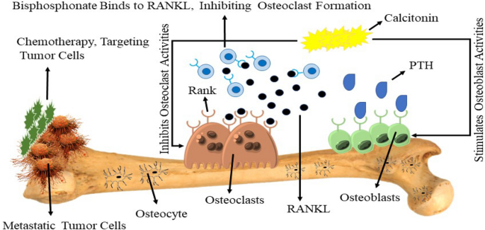
Control systems and the modeling strategies are not only limited to engineering problems. These approaches can be used in the field of bio-mathematics as well and modern studies have promoted this approach to a great extent. The computational modeling and simulation of bone metastasis is painful yet critical after cancer invades the body. This vicious cycle is complex, and several research centers worldwide are devoted to understanding the dynamics and setting up a treatment strategy for this life-threatening behavior of cancer. Cancerous cells activation and the corresponding process of metastasis is reported to boost during the periodic waves of COVID-19, due to the inflammatory nature of the infection associated with SARS-2 and its variants. The bone cells are comprised of two types of cells responsible for bone formation and resorption. The computational framework of such cells, in spatial form, can help the researchers forecast the bone dynamics in a robust manner where the impact of cancer is incorporated into the computational model as a source of perturbation. A series of computational models are presented to explore the complex behavior of bone metastasis with COVID-19 induced infection. The finite difference algorithm is used to simulate the nonlinear computational model. The results obtained are in close agreement with the experimental findings. The computational results can help explore the vicious cycle’s fate and help set up control strategies through drug therapies.
Spatio-temporal simulations of bone remodelling using a bone cell population model based on cell availability
Here we developed a spatio-temporal bone remodeling model to simulate the action of Basic Multicelluar Units (BMUs). This model is based on two major extensions of a temporal-only bone cell population model (BCPM). First, the differentiation into mature resorbing osteoclasts and mature forming osteoblasts from their respective precursor cells was modelled as an intermittent process based on precursor cells availability. Second, the interaction between neighbouring BMUs was considered based on a ``metabolic cost'' argument which warrants that no new BMU will be activated in the neighbourhood of an existing BMU. With the proposed model we have simulated the phases of the remodelling process obtaining average periods similar to those found in the literature: resorption ($\sim22$ days) - reversal ($\sim$8 days) - formation ($\sim$ 65 days) - quiescence (560 - 600 days) and an average BMU activation frequency of $\sim$1.6 BMUs/year/mm$^3$. We further show here that the resorption and formation phases of the BMU become coordinated only by the presence of TGF-$\upbeta$ (transforming growth factor $\upbeta$, i.e. a major coupling factor stored in the bone matrix. TGF-$\upbeta$ is released through resorption so upregulating osteoclast apoptosis and accumulation of osteoblast precursors, i.e. facilitating the transition from the resorption to the formation phase at a given remodelling site. Finally, we demonstrate that this model can explain targeted bone remodelling as the BMUs are steered towards damaged bone areas in order to commence bone matrix repair.

Mathematical modeling of postmenopausal osteoporosis and its treatment by the anti‐catabolic drug denosumab
SUMMARY Denosumab, a fully human monoclonal antibody, has been approved for the treatment of postmenopausal osteoporosis. The therapeutic effect of denosumab rests on its ability to inhibit osteoclast differentiation. Here, we present a computational approach on the basis of coupling a pharmacokinetics model of denosumab with a pharmacodynamics model for quantifying the effect of denosumab on bone remodeling. The pharmacodynamics model comprises an integrated systems biology‐continuum micromechanics approach, including a bone cell population model, considering the governing biochemical factors of bone remodeling (including the action of denosumab), and a multiscale micromechanics‐based bone mechanics model, for implementing the mechanobiology of bone remodeling in our model. Numerical studies of postmenopausal osteoporosis show that denosumab suppresses osteoclast differentiation, thus strongly curtailing bone resorption. Simulation results also suggest that denosumab may trigger a short‐term bone volume gain, which is, however, followed by constant or decreasing bone volume. This evolution is accompanied by a dramatic decrease of the bone turnover rate by more than one order of magnitude. The latter proposes dominant occurrence of secondary mineralization (which is not anymore impeded through cellular activity), leading to higher mineral concentration per bone volume. This explains the overall higher bone mineral density observed in denosumab‐related clinical studies. Copyright © 2013 John Wiley & Sons, Ltd.

A mechano-chemo-biological model for bone remodeling with a new mechano-chemo-transduction approach
Bone remodeling is a fundamental biological process that develops in bone tissue along its whole lifetime. It refers to a continuous bone transformation with new bone formation and old bone resorption that changes the internal microstructure and composition of the tissue. The main objectives of bone remodeling are: repair of the internal microcracks; adaptation of the macroscopic stiffness and strength to the actual changing mechanical demands; and control of the calcium homeostasis. Understanding this process and predicting its evolution is critical to reduce the effects of long-term disuse as happens during periods of reduced mobility. It is also important in the design of bone implants to avoid long-term stress shielding. Many mathematical models have been proposed from the earliest purely phenomenological to the latest that include biological knowledge. However, there still exists a lack of connection between the mechanical driving force and the biochemical and cell processes it triggers. Here, and following previous works that model independently the mechanobiological and biochemical processes in bone remodeling, we present a more complete model, useful for both cortical and trabecular bone, that uses a new mechanotransduction approach based on the effect of strains onto the bonding–unbonding rate of RANK/RANKL/OPG receptor–ligand reactions. We compare the results of this model with previous ones, showing a good agreement in similar conditions. We also apply it to realistic situations such as a femoral bone after implantation of a hip prosthesis, getting similar results to the clinical ones in the final bone density distribution. Finally, we extend this approach to the anisotropic case, getting not only the mean density, but also the directional homogenization of the microstructure. This biochemical approach permits, not only to predict the bone evolution under changes in the mechanical loads, but also, to consider anabolic and catabolic drugs to control bone density, such as those used in osteoporosis.

A mathematical model of bone remodeling dynamics for normal bone cell populations and myeloma bone disease
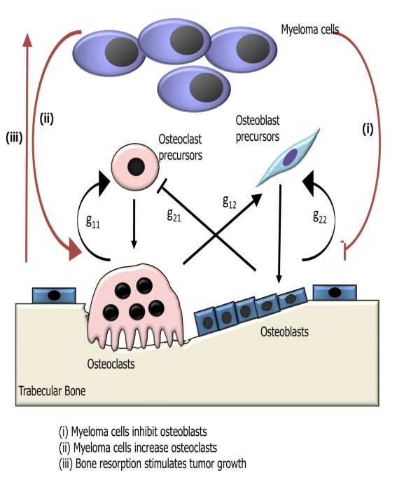
Multiple myeloma is a hematologic malignancy associated with the development of a destructive osteolytic bone disease.
Dynamic Biochemical and Cellular Models of Bone Physiology: Integrating Remodeling Processes, Tumor Growth, and Therapy
Bone is an activeCoelho, Rui M. Neto, Joana P. connective tissue composed of different types of cells. The dynamic behavior of bone remodeling processes is typically represented throughValério, Duarte differential equations, which represent the physiological phenomena occurring in this organ. These models takeVinga, Susana into account the tight biochemical regulation between osteoclasts and osteoblasts and have also been enriched with variables and parameters related to bone pathologies and treatment. This chapter reviews some of the more recent models describing bone physiology, focusing on those that include the main cellular processes, along the biochemical control, and also the pharmacokinetic/pharmacodynamic (PK/PD) of the most common treatments for diseases such as cancer. These models are then compared in terms of the simulations obtained and, finally, some highlights on integrating them with the biomechanical component of the system which will be given. These models are expected to provide a valuable insight into this complex system and to support the development of clinical decision systems for bone pathologies.

A novel mathematical model of bone remodelling cycles for trabecular bone at the cellular level
After an initial phase of growth and development, bone undergoes a continuous cycle of repair, renewal and optimisation by a process called remodelling. This paper describes a novel mathematical model of the trabecular bone remodelling cycle. It is essentially formulated to simulate a remodelling event at a fixed position in the bone, integrating bone removal by osteoclasts and formation by osteoblasts. The model is developed to construct the variation in bone thickness at a particular point during the remodelling event, derived from standard bone histomorphometric analyses. The novelties of the approach are the adoption of a predator–prey model to describe the dynamic interaction between osteoclasts and osteoblasts, using a genetic algorithm–based solution; quantitative reconstruction of the bone remodelling cycle; and the introduction of a feedback mechanism in the bone formation activity to co-regulate bone thickness. The application of the model is first demonstrated by using experimental data recorded for normal (healthy) bone remodelling to predict the temporal variation in the number of osteoblasts and osteoclasts. The simulated histomorphometric data and remodelling cycle characteristics compare well with the specified input data. Sensitivity studies then reveal how variations in the model’s parameters affect its output; it is hoped that these parameters can be linked to specific biochemical factors in the future. Two sample pathological conditions, hypothyroidism and primary hyperparathyroidism, are examined to demonstrate how the model could be applied more broadly, and, for the first time, the osteoblast and osteoclast populations are predicted for these conditions. Further data are required to fully validate the model’s predictive capacity, but this work shows it has potential, especially in the modelling of pathological conditions and the optimisation of the treatment of those conditions.

Modeling Osteocyte Network Formation: Healthy and Cancerous Environments
Advanced cancers, such as prostate and breast cancers, commonly metastasize to bone. In the bone matrix, dendritic osteocytes form a spatial network allowing communication between osteocytes and the osteoblasts located on the bone surface. This communication network facilitates coordinated bone remodelling. In the presence of a cancerous microenvironment, the morphology of this network changes. Commonly osteocytes appear to be either overdifferentiated (i.e., there are more dendrites than healthy bone) or underdeveloped(i.e., dendrites do not fully form). In addition to structural changes, histological sections from metastatic breast cancer xenografted mice show that number of osteocytes per unit area is different between healthy bone and cancerous bone. We present a stochastic agent-based model for bone formation incorporating osteoblasts and osteocytes that allows us to probe both network structure and density of osteocytes in bone. Our model both allows for the simulation of our spatial network model and analysis of mean-field equations in the form of integro-partial differential equations. We considered variations of our model to study specific physiological hypotheses related to osteoblast differentiation; for example predicting how changing biological parameters, such as rates of bone secretion, rates of cancer formation and rates of osteoblast differentiation can allow for qualitatively different network morphologies. We then used our model to explore how commonly applied therapies such as bisphosphonates (e.g. zoledronic acid) impact osteocyte network formation.
