

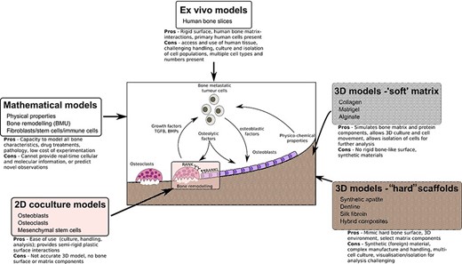
ABSTRACT Bone is the most common site for cancer metastasis. Understanding the interactions within the complex, heterogeneous bone–tumor microenvironment is essential for the development of new therapeutics. Various animal models of tumor‐induced bone disease are routinely used to provide valuable information on the relationship between cancer cells and the skeleton. However, new model systems exist that offer an alternative approach to the use of animals and might more accurately reveal the cellular interactions occurring within the human bone–tumor niche. This review highlights replacement models that mimic the bone microenvironment and where cancer metastases and tumor growth might be assessed alongside bone turnover. Such culture models include the use of calcified regions of animal tissue and scaffolds made from bone mineral hydroxyapatite, synthetic polymers that can be manipulated during manufacture to create structures resembling trabecular bone surfaces, gel composites that can be modified for stiffness and porosity to resemble conditions in the tumor–bone microenvironment. Possibly the most accurate model system involves the use of fresh human bone samples, which can be cultured ex vivo in the presence of human tumor cells and demonstrate similar cancer cell–bone cell interactions as described in vivo. In addition, the use of mathematical modeling and computational biology approaches provide an alternative to preliminary animal testing. The use of such models offers the capacity to mimic significant elements of the human bone–tumor environment, and complement, refine, or replace the use of preclinical models. © 2020 The Authors. JBMR Plus published by Wiley Periodicals, Inc. on behalf of American Society for Bone and Mineral Research.
Dynamic Biochemical and Cellular Models of Bone Physiology: Integrating Remodeling Processes, Tumor Growth, and Therapy
Bone is an activeCoelho, Rui M. Neto, Joana P. connective tissue composed of different types of cells. The dynamic behavior of bone remodeling processes is typically represented throughValério, Duarte differential equations, which represent the physiological phenomena occurring in this organ. These models takeVinga, Susana into account the tight biochemical regulation between osteoclasts and osteoblasts and have also been enriched with variables and parameters related to bone pathologies and treatment. This chapter reviews some of the more recent models describing bone physiology, focusing on those that include the main cellular processes, along the biochemical control, and also the pharmacokinetic/pharmacodynamic (PK/PD) of the most common treatments for diseases such as cancer. These models are then compared in terms of the simulations obtained and, finally, some highlights on integrating them with the biomechanical component of the system which will be given. These models are expected to provide a valuable insight into this complex system and to support the development of clinical decision systems for bone pathologies.

An Integrated Computational Model of the Bone Microenvironment in Bone-Metastatic Prostate Cancer
Abstract Bone metastasis will impact most men with advanced prostate cancer. The vicious cycle of bone degradation and formation driven by metastatic prostate cells in bone yields factors that drive cancer growth. Mechanistic insights into this vicious cycle have suggested new therapeutic opportunities, but complex temporal and cellular interactions in the bone microenvironment make drug development challenging. We have integrated biologic and computational approaches to generate a hybrid cellular automata model of normal bone matrix homeostasis and the prostate cancer-bone microenvironment. The model accurately reproduces the basic multicellular unit bone coupling process, such that introduction of a single prostate cancer cell yields a vicious cycle similar in cellular composition and pathophysiology to models of prostate-to-bone metastasis. Notably, the model revealed distinct phases of osteolytic and osteogenic activity, a critical role for mesenchymal stromal cells in osteogenesis, and temporal changes in cellular composition. To evaluate the robustness of the model, we assessed the effect of established bisphosphonate and anti-RANKL therapies on bone metastases. At approximately 100% efficacy, bisphosphonates inhibited cancer progression while, in contrast with clinical observations in humans, anti-RANKL therapy fully eradicated metastases. Reducing anti-RANKL yielded clinically similar results, suggesting that better targeting or dosing could improve patient survival. Our work establishes a computational model that can be tailored for rapid assessment of experimental therapies and delivery of precision medicine to patients with prostate cancer with bone metastases. Cancer Res; 74(9); 2391–401. ©2014 AACR.
Mathematical modelling of the pathogenesis of multiple myeloma‐induced bone disease
SUMMARY Multiple myeloma (MM) is the second most common haematological malignancy and results in destructive bone lesions. The interaction between MM cells and the bone microenvironment plays an important role in the development of the tumour cells and MM‐induced bone disease and forms a ‘vicious cycle’ of tumour development and bone destruction, intensified by suppression of osteoblast activity and promotion of osteoclast activity. In this paper, a mathematical model is proposed to simulate how the interaction between MM cells and the bone microenvironment facilitates the development of the tumour cells and the resultant bone destruction. It includes both the roles of inhibited osteoblast activity and stimulated osteoclast activity. The model is able to mimic the temporal variation of bone cell concentrations and resultant bone volume after the invasion and then removal of the tumour cells and explains why MM‐induced bone lesions rarely heal even after the complete removal of MM cells. The behaviour of the model compares well with published experimental data. The model serves as a first step to understand the development of MM‐induced bone disease and could be applied further to evaluate the current therapies against MM‐induced bone disease and even suggests new potential therapeutic targets. © 2014 The Authors. International Journal for Numerical Methods in Biomedical Engineering published by John Wiley & Sons Ltd

Modeling Osteocyte Network Formation: Healthy and Cancerous Environments
Advanced cancers, such as prostate and breast cancers, commonly metastasize to bone. In the bone matrix, dendritic osteocytes form a spatial network allowing communication between osteocytes and the osteoblasts located on the bone surface. This communication network facilitates coordinated bone remodelling. In the presence of a cancerous microenvironment, the morphology of this network changes. Commonly osteocytes appear to be either overdifferentiated (i.e., there are more dendrites than healthy bone) or underdeveloped(i.e., dendrites do not fully form). In addition to structural changes, histological sections from metastatic breast cancer xenografted mice show that number of osteocytes per unit area is different between healthy bone and cancerous bone. We present a stochastic agent-based model for bone formation incorporating osteoblasts and osteocytes that allows us to probe both network structure and density of osteocytes in bone. Our model both allows for the simulation of our spatial network model and analysis of mean-field equations in the form of integro-partial differential equations. We considered variations of our model to study specific physiological hypotheses related to osteoblast differentiation; for example predicting how changing biological parameters, such as rates of bone secretion, rates of cancer formation and rates of osteoblast differentiation can allow for qualitatively different network morphologies. We then used our model to explore how commonly applied therapies such as bisphosphonates (e.g. zoledronic acid) impact osteocyte network formation.

A mathematical model for describing the metastasis of cancer in bone tissue
Metastasis is the rapid proliferation of cancer cells (secondary tumour) at a specific place, generally leading to death. This occurs at anatomical parts providing the necessary environment for vas...

<i>In silico</i> models of bone remodeling from macro to nano—from organ to cell
Abstract Computational modeling is a tool through which researchers can achieve a greater understanding of the mechanisms governing biological systems. In the field of bone biology, a plethora of models exist which attempt to replicate and investigate bone's dynamic behavior at different scales. At organ level, models are continuum based and describe the variation of bone's apparent density as a function of both biological and external mechanical stimuli. At tissue level, models include bone microarchitecture and more descriptive parameters such as trabecular thickness, osteoclast resorption depth, and activation frequency. Finally, at cell level, models employ partial differential equations to describe complex cellular interactions in the temporal domain. Although informative, these models exist in isolation. Consequently, their interpretation is limited. In this review, we present an overview of the organ‐, tissue‐, and cell‐level models and assess their ability to reflect bone's metabolic processes reliably. Existing interscale synergies are then presented along with a computational framework which could be exploited to achieve a fully integrated, multiscale modeling approach. WIREs Syst Biol Med 2011 3 241–251 DOI: 10.1002/wsbm.115 This article is categorized under: Analytical and Computational Methods > Computational Methods Models of Systems Properties and Processes > Organ, Tissue, and Physiological Models

Bone metastases
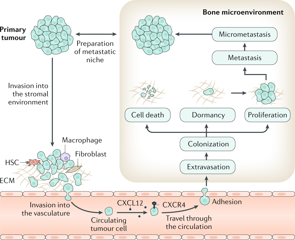
Bone is the most frequent site for metastasis for many cancers, notably for tumours originating in the breast and the prostate. Tumour cells can escape from the primary tumour site and colonize the bone microenvironment. Within the bone, these disseminated tumour cells, as well as those arising in the context of multiple myeloma, may assume a state of dormancy, remaining quiescent for years before resuming proliferation and causing overt metastasis, which causes bone destruction via activation of osteoclast-mediated osteolysis. This structural damage can lead to considerable morbidity, including pain, fractures and impaired quality of life. Although treatment of bone metastases and myeloma bone disease is rarely curative, disease control is often possible for many years through the use of systemic anticancer treatments on a background of multidisciplinary supportive care. This care should include bone-targeted agents to inhibit tumour-associated osteolysis and prevent skeletal morbidity as well as use of appropriate local treatments such as radiation therapy, orthopaedic surgery and specialist palliative care to minimize the impact of metastatic bone disease on physical functioning. In this Primer, we provide an overview of the clinical features, the pathophysiology and the specific treatment approaches to prevent and treat bone metastases from solid tumours as well as myeloma bone disease.
Osteoprotegerin in Bone Metastases: Mathematical Solution to the Puzzle
Bone is a common site for cancer metastasis. To create space for their growth, cancer cells stimulate bone resorbing osteoclasts. Cytokine RANKL is a key osteoclast activator, while osteoprotegerin (OPG) is a RANKL decoy receptor and an inhibitor of osteoclastogenesis. Consistently, systemic application of OPG decreases metastatic tumor burden in bone. However, OPG produced locally by cancer cells was shown to enhance osteolysis and tumor growth. We propose that OPG produced by cancer cells causes a local reduction in RANKL levels, inducing a steeper RANKL gradient away from the tumor and towards the bone tissue, resulting in faster resorption and tumor expansion. We tested this hypothesis using a mathematical model of nonlinear partial differential equations describing the spatial dynamics of OPG, RANKL, PTHrP, osteoclasts, tumor and bone mass. We demonstrate that at lower expression rates, tumor-derived OPG enhances the chemotactic RANKL gradient and osteolysis, whereas at higher expression rates OPG broadly inhibits RANKL and decreases osteolysis and tumor burden. Moreover, tumor expression of a soluble mediator inducing RANKL in the host tissue, such as PTHrP, is important for correct orientation of the RANKL gradient. A meta-analysis of OPG, RANKL and PTHrP expression in normal prostate, carcinoma and metastatic tissues demonstrated an increase in expression of OPG, but not RANKL, in metastatic prostate cancer, and positive correlation between OPG and PTHrP in metastatic prostate cancer. The proposed mechanism highlights the importance of the spatial distribution of receptors, decoys and ligands, and can be applied to other systems involving regulation of spatially anisotropic processes.
A review of mathematical modeling of bone remodeling from a systems biology perspective
Bone remodeling is an essential, delicately balanced physiological process of coordinated activity of bone cells that remove and deposit new bone tissue in the adult skeleton. Due to the complex nature of this process, many mathematical models of bone remodeling have been developed. Each of these models has unique features, but they have underlying patterns. In this review, the authors highlight the important aspects frequently found in mathematical models for bone remodeling and discuss how and why these aspects are included when considering the physiology of the bone basic multicellular unit, which is the term used for the collection of cells responsible for bone remodeling. The review also emphasizes the view of bone remodeling from a systems biology perspective. Understanding the systemic mechanisms involved in remodeling will help provide information on bone pathology associated with aging, endocrine disorders, cancers, and inflammatory conditions and enhance systems pharmacology. Furthermore, some features of the bone remodeling cycle and interactions with other organ systems that have not yet been modeled mathematically are discussed as promising future directions in the field.

A mechano-chemo-biological model for bone remodeling with a new mechano-chemo-transduction approach
Bone remodeling is a fundamental biological process that develops in bone tissue along its whole lifetime. It refers to a continuous bone transformation with new bone formation and old bone resorption that changes the internal microstructure and composition of the tissue. The main objectives of bone remodeling are: repair of the internal microcracks; adaptation of the macroscopic stiffness and strength to the actual changing mechanical demands; and control of the calcium homeostasis. Understanding this process and predicting its evolution is critical to reduce the effects of long-term disuse as happens during periods of reduced mobility. It is also important in the design of bone implants to avoid long-term stress shielding. Many mathematical models have been proposed from the earliest purely phenomenological to the latest that include biological knowledge. However, there still exists a lack of connection between the mechanical driving force and the biochemical and cell processes it triggers. Here, and following previous works that model independently the mechanobiological and biochemical processes in bone remodeling, we present a more complete model, useful for both cortical and trabecular bone, that uses a new mechanotransduction approach based on the effect of strains onto the bonding–unbonding rate of RANK/RANKL/OPG receptor–ligand reactions. We compare the results of this model with previous ones, showing a good agreement in similar conditions. We also apply it to realistic situations such as a femoral bone after implantation of a hip prosthesis, getting similar results to the clinical ones in the final bone density distribution. Finally, we extend this approach to the anisotropic case, getting not only the mean density, but also the directional homogenization of the microstructure. This biochemical approach permits, not only to predict the bone evolution under changes in the mechanical loads, but also, to consider anabolic and catabolic drugs to control bone density, such as those used in osteoporosis.

Control of Bone Matrix Properties by Osteocytes
Osteocytes make up 90-95% of the cellular content of bone and form a rich dendritic network with a vastly greater surface area than either osteoblasts or osteoclasts. Osteocytes are well positioned to play a role in bone homeostasis by interacting directly with the matrix; however, the ability for these cells to modify bone matrix remains incompletely understood. With techniques for examining the nano- and microstructure of bone matrix components including hydroxyapatite and type I collagen becoming more widespread, there is great potential to uncover novel roles for the osteocyte in maintaining bone quality. In this review, we begin with an overview of osteocyte biology and the lacunar-canalicular system. Next, we describe recent findings from in vitro models of osteocytes, focusing on the transitions in cellular phenotype as they mature. Finally, we describe historical and current research on matrix alteration by osteocytes in vivo, focusing on the exciting potential for osteocytes to directly form, degrade, and modify the mineral and collagen in their surrounding matrix.

A review of mathematical modeling of bone remodeling from a systems biology perspective
Bone remodeling is an essential physiological process in the adult skeleton. Due to the complex nature of this process, many mathematical models of bone remodeling have been developed. Each of these models has unique features, but they have underlying patterns. In this review, the authors highlight the important aspects frequently found in mathematical models for bone remodeling and discuss how and why these aspects are included when considering the physiology of the bone basic multicellular unit, which is the term used for the collection of cells responsible for bone remodeling. The review also emphasizes the view of bone remodeling from a systems biology perspective. Understanding the systemic mechanisms involved in remodeling will help provide information on bone pathology associated with aging, endocrine disorders, cancers, and inflammatory conditions and enhance systems pharmacology. Furthermore, some features of the bone remodeling cycle and interactions with other organ systems that have not yet been modeled mathematically are discussed as promising future directions in the field.
Computational Modeling of Interactions between Multiple Myeloma and the Bone Microenvironment
Multiple Myeloma (MM) is a B-cell malignancy that is characterized by osteolytic bone lesions. It has been postulated that positive feedback loops in the interactions between MM cells and the bone microenvironment form reinforcing ‘vicious cycles’, resulting in more bone resorption and MM cell population growth in the bone microenvironment. Despite many identified MM-bone interactions, the combined effect of these interactions and their relative importance are unknown. In this paper, we develop a computational model of MM-bone interactions and clarify whether the intercellular signaling mechanisms implemented in this model appropriately drive MM disease progression. This new computational model is based on the previous bone remodeling model of Pivonka et al. [1], and explicitly considers IL-6 and MM-BMSC (bone marrow stromal cell) adhesion related pathways, leading to formation of two positive feedback cycles in this model. The progression of MM disease is simulated numerically, from normal bone physiology to a well established MM disease state. Our simulations are consistent with known behaviors and data reported for both normal bone physiology and for MM disease. The model results suggest that the two positive feedback cycles identified for this model are sufficient to jointly drive the MM disease progression. Furthermore, quantitative analysis performed on the two positive feedback cycles clarifies the relative importance of the two positive feedback cycles, and identifies the dominant processes that govern the behavior of the two positive feedback cycles. Using our proposed quantitative criteria, we identify which of the positive feedback cycles in this model may be considered to be ‘vicious cycles’. Finally, key points at which to block the positive feedback cycles in MM-bone interactions are identified, suggesting potential drug targets.
Optimizing the dynamics of bone turnover with genetic algorithm
Control systems and the modeling strategies are not only limited to engineering problems. These approaches can be used in the field of bio-mathematics as well and modern studies have promoted this approach to a great extent. The computational modeling and simulation of bone metastasis is painful yet critical after cancer invades the body. This vicious cycle is complex, and several research centers worldwide are devoted to understanding the dynamics and setting up a treatment strategy for this life-threatening behavior of cancer. Cancerous cells activation and the corresponding process of metastasis is reported to boost during the periodic waves of COVID-19, due to the inflammatory nature of the infection associated with SARS-2 and its variants. The bone cells are comprised of two types of cells responsible for bone formation and resorption. The computational framework of such cells, in spatial form, can help the researchers forecast the bone dynamics in a robust manner where the impact of cancer is incorporated into the computational model as a source of perturbation. A series of computational models are presented to explore the complex behavior of bone metastasis with COVID-19 induced infection. The finite difference algorithm is used to simulate the nonlinear computational model. The results obtained are in close agreement with the experimental findings. The computational results can help explore the vicious cycle’s fate and help set up control strategies through drug therapies.
Dynamics of Bone Cell Interactions and Differential Responses to PTH and Antibody-Based Therapies
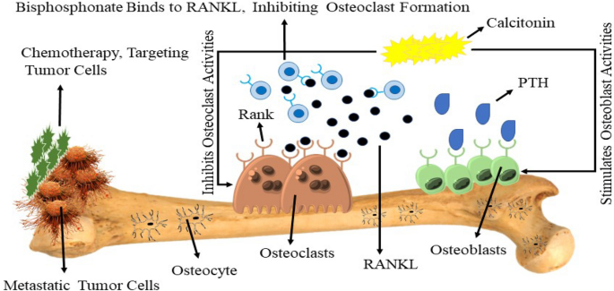
We propose a mathematical model describing the dynamics of osteoblasts and osteoclasts in bone remodeling. The goal of this work is to develop an integrated modeling framework for bone remodeling and bone cell signaling dynamics that could be used to explore qualitatively combination treatments for osteoporosis in humans. The model has been calibrated using 57 checks from the literature. Specific global optimization methods based on qualitative objectives have been developed to perform the model calibration. We also added pharmacokinetics representations of three drugs to the model, which are teriparatide (PTH(1–34)), denosumab (a RANKL antibody) and romosozumab (a sclerostin antibody), achieving excellent goodness-of-fit of human clinical data. The model reproduces the paradoxical effects of PTH on the bone mass, where continuous administration of PTH results in bone loss but intermittent administration of PTH leads to bone gain, thus proposing an explanation of this phenomenon. We used the model to simulate different categories of osteoporosis. The main attributes of each disease are qualitatively well captured by the model, for example changes in bone turnover in the disease states. We explored dosing regimens for each disease based on the combination of denosumab and romosozumab, identifying adequate ratios and doses of both drugs for subpopulations of patients in function of categories of osteoporosis and the degree of severity of the disease.

Mathematical modeling of postmenopausal osteoporosis and its treatment by the anti‐catabolic drug denosumab
SUMMARY Denosumab, a fully human monoclonal antibody, has been approved for the treatment of postmenopausal osteoporosis. The therapeutic effect of denosumab rests on its ability to inhibit osteoclast differentiation. Here, we present a computational approach on the basis of coupling a pharmacokinetics model of denosumab with a pharmacodynamics model for quantifying the effect of denosumab on bone remodeling. The pharmacodynamics model comprises an integrated systems biology‐continuum micromechanics approach, including a bone cell population model, considering the governing biochemical factors of bone remodeling (including the action of denosumab), and a multiscale micromechanics‐based bone mechanics model, for implementing the mechanobiology of bone remodeling in our model. Numerical studies of postmenopausal osteoporosis show that denosumab suppresses osteoclast differentiation, thus strongly curtailing bone resorption. Simulation results also suggest that denosumab may trigger a short‐term bone volume gain, which is, however, followed by constant or decreasing bone volume. This evolution is accompanied by a dramatic decrease of the bone turnover rate by more than one order of magnitude. The latter proposes dominant occurrence of secondary mineralization (which is not anymore impeded through cellular activity), leading to higher mineral concentration per bone volume. This explains the overall higher bone mineral density observed in denosumab‐related clinical studies. Copyright © 2013 John Wiley & Sons, Ltd.
