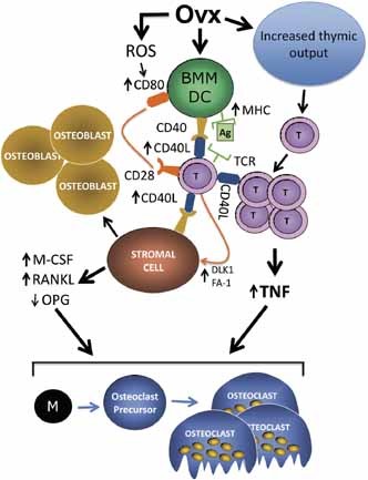
More than a decade has passed since the publication of the first report implicating T cells in the bone loss induced by ovariectomy (ovx).1 Since then ther
Bone versus immune system
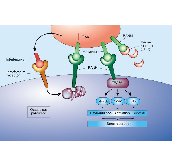
A molecule on activated T cells triggers bone loss by switching on bone-resorbing cells. Fortunately, it seems that this mechanism is kept in check by another molecule, secreted by the T cells.
Mice lacking DKK1 in T cells exhibit high bone mass and are protected from estrogen-deficiency-induced bone loss
Molecular Physiology; Immunology; Cell Biology;

Estrogen and the skeleton
Estrogen is the major hormonal regulator of bone metabolism in women and men. Therefore, there is considerable interest in unraveling the pathways by which estrogen exerts its protective effects on bone. Although the major consequence of the loss of estrogen is an increase in bone resorption, estrogen deficiency is associated with a gap between bone resorption and formation, indicating that estrogen is also important for maintaining bone formation at the cellular level. Direct estrogen effects on osteocytes, osteoclasts, and osteoblasts lead to inhibition of bone remodeling, decreased bone resorption, and maintenance of bone formation, respectively.

Immunoporosis: Immunology of Osteoporosis—Role of T Cells
The role of immune system in various bone pathologies such as osteoporosis, osteoarthritis and rheumatoid arthritis is now well established. This had led to the emergence of a modern field of systems biology called as osteoimmunology, an integrated research between fields of immunology and bone biology under one umbrella. Osteoporosis is one of the most common inflammatory bone-loss conditions with more than 200 million individuals affected worldwide. T helper cells along with various other immune cells are major players involved in bone homeostasis. In the present review, we specifically discuss the role of various defined T lymphocyte subsets (Th cells comprising Th1, Th2, Th9, Th17, Th22, regulatory T cells, follicular helper T cells, natural killer T cells, γδ T cells and CD8+ T cells) in the pathophysiology of osteoporosis. The study of the specific role of immune system in osteoporosis has now been proposed by our group as “Immunoporosis: The immunology of osteoporosis” with special emphasis on the role of various subsets of T lymphocytes. The establishment of this new field had been the need of the hour due to the emergence of novel roles of various T cell lymphocytes in accelerated bone loss observed during osteoporosis. Activated T cells either directly or indirectly through the secretion of various cytokines and factors modulate bone health and thereby regulate bone remodelling. Various studies have summarized the role of inflammation in pathogenesis of osteoporosis, but very few reports had delineated the precise role of various T cell subsets in the pathobiology of osteoporosis. The present review thus for the first time clearly highlights and summarizes the role of various T lymphocytes in the development and pathophysiology of osteoporosis, giving birth to a new field of biology termed as “Immunoporosis”. This novel field will thus provide an overview of the nexus between the cellular components of both bone and immune systems, responsible for the observed bone loss in osteoporosis. A molecular insight into the upcoming and novel field of immunoporosis would thus lead to development of innovative approaches for the prevention and treatment of osteoporosis.

Lactulose Suppresses Osteoclastogenesis and Ameliorates Estrogen Deficiency-Induced Bone Loss in Mice
<p>Postmenopausal osteoporosis is characterized by excess osteoclastogenesis which leads to net bone loss and brittle fractures. Studies have demonstrated that estrogen deficiency-associated bone loss is microbiota-dependent and could be prevented by probiotics and prebiotics. In this study, we report that orally administered lactulose (20 g/kg, 6 weeks) orally administered significantly inhibited osteoclastogenesis, bone resorption, and prevented ovariectomy (OVX)-induced bone loss in mice. Lactulose increased intestinal <i>Claudin 2, 3 and 15,</i> compared to the OVX group, and lowered pro-osteoclastogenic cytokines levels including tumor necrosis factor-α, interleukin(IL)-6, receptor activator of nuclear factor kappa-Β ligand (RANKL), and IL-17 as well as increased the anti-inflammatory cytokine IL-10 in the intestine, peripheral blood, and bone marrow. Lactulose significantly preserved the number of Foxp3<sup>+</sup> Treg cells in the intestines compared with that in OVX mice. Lactulose altered the composition of intestinal microbiota measured by 16s rDNA sequencing and increased intestinal and serum short-chain fatty acids (SCFAs) levels including acetate, propionate and butyrate which were decreased in OVX mice as measured by gas chromatography. Oral administration of lactulose for 2 weeks significantly lowered the level of bone resorption marker C-telopeptide of type 1 collagen-1 in healthy male young volunteers (aging 20-25 years). In conclusion, lactulose inhibited osteoclastogenesis and bone resorption by altering the intestinal microbiota and increasing SCFAs. Lactulose could serve as an ideal therapeutic agent for postmenopausal osteoporosis.</p>
The Role of Estrogen in the Control of Rat Osteocyte Apoptosis
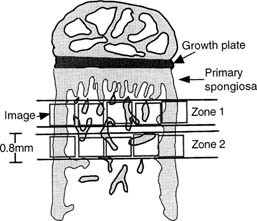
Abstract We have previously shown that estrogen withdrawal by gonadotrophin-releasing hormone analogs (GnRHa) induces osteocyte death via apoptosis in human bone. Although it is likely that the increase in osteocyte death via apoptosis was related to the loss of estrogen, these experiments could not rule out a direct role for the GnRHa. Therefore, in this study, we have used a rat model of ovariectomy (OVX) to determine whether the effect of estrogen withdrawal extends to other species and to clarify the role of estrogen in the maintenance of osteocyte viability. Twelve 9-week-old rats were divided into three treatment groups: sham operated (SHAM) (n = 4), OVX (n = 4), and OVX + estrogen (E2) (25 μg/day) (n = 4). At 3 weeks following the start of treatment, tibial bones were removed. The percentage of osteocytes displaying DNA breaks, using an in situ nick-translation method, was significantly higher in the OVX group compared with the SHAM control in both cortical bone (10.04% vs. 2.31%, respectively; p < 0.0001) and trabecular bone (6.44% vs. 1.58%, respectively; p = 0.003). Addition of estrogen in the OVX animals completely abrogated the increase in osteocyte apoptosis in cortical bone (0.78%) and trabecular bone (1.17%). The percentage of apoptotic osteocytes decreased with increasing distance from the primary/secondary spongiosa interface below the growth plate in the OVX model and the OVX + E2 model. Nuclear morphology and electrophoresis of DNA confirmed the presence of apoptotic cells in the samples. In conclusion, OVX in the rat results in an increase in osteocyte apoptosis as a direct or indirect result of E2 loss. Addition of estrogen in the OVX animals prevents this increase in osteocyte apoptosis. These data confirm an important role for estrogen in the control of osteocyte apoptosis and the maintenance of osteocyte viability. Estrogen deficiency might, through compromising the viability of osteocyte networks, reduce the ability of bone to respond appropriately to loading.
Osteoimmunology: shared mechanisms and crosstalk between the immune and bone systems
Osteoimmunology is an interdisciplinary field, that covers the shared mechanisms and interactions between bone cells and immune cellsReceptor activator of nuclear factor-κB (NF-κB) ligand (RANKL) is an osteoclast-differentiation factor that links the activated immune system and bone loss. In addition, abnormal bone homeostasis has been observed in various mice deficient in immunomodulatory molecules.Osteoclast differentiation is dependent on the transcription factor nuclear factor of activated T cells, cytoplasmic 1 (NFATc1), which is induced and activated by RANKL and its co-stimulatory (immunoglobulin-like) receptors.Interleukin-17 (IL-17)-producing T helper cells (TH17 cells) are the key T-cell subset that links T-cell activation and bone destruction in autoimmune arthritis.Bone cells are involved in the maintenance and mobilization of haematopoietic stem cells.Osteoimmunology is becoming increasingly important for understanding the pathogenesis of, and developing new therapeutic strategies for, diseases that affect both systems.
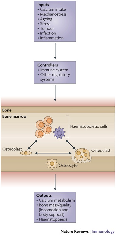
Physiological and pathophysiological bone turnover — role of the immune system
Receptor activator of NF-κB ligand (RANKL) and osteoprotegerin (OPG) are key downstream effectors of bone resorption; tumour necrosis factor (TNF) might synergize with RANKL to superinduce osteoclastic bone resorptionB cells, regulated by T cells, are a key source of basal OPG whereas activated T cells and B cells are key sources of TNF and RANKL in inflammatory conditionsShort-term antiresorptive therapies might safely prevent bone loss associated with combination antiretroviral therapyAnergic and parathyroid-hormone-treated T cells secrete the protein Wnt-10b, which promotes bone formationNovel therapeutic strategies targeting the immune system might promote bone formation and decrease bone resorption to manage osteoporotic bone loss and prevent fracture
Critical Role of Estrogens on Bone Homeostasis in Both Male and Female: From Physiology to Medical Implications
Bone is a multi-skilled tissue, protecting major organs, regulating calcium phosphate balance and producing hormones. Its development during childhood determines height and stature as well as resistance against fracture in advanced age. Estrogens are key regulators of bone turnover in both females and males. These hormones play a major role in longitudinal and width growth throughout puberty as well as in the regulation of bone turnover. In women, estrogen deficiency is one of the major causes of postmenopausal osteoporosis. In this review, we will summarize the main clinical and experimental studies reporting the effects of estrogens not only in females but also in males, during different life stages. Effects of estrogens on bone involve either Estrogen Receptor (ER)α or ERβ depending on the type of bone (femur, vertebrae, tibia, mandible), the compartment (trabecular or cortical), cell types involved (osteoclasts, osteoblasts and osteocytes) and sex. Finally, we will discuss new ongoing strategies to increase the benefit/risk ratio of the hormonal treatment of menopause.
Osteoimmunology: The Nexus between bone and immune system
Osteoimmunology is an interdisciplinary research field which combines the existing fields of osteology (bone biology) and immunology under one umbrella. The observation that contributed enormously to the emergence of osteoimmunology as an independent field of investigation was the enhanced bone loss in various inflammatory bone diseases such as rheumatoid arthritis, osteoporosis and periodontitis. T helper cells (Th1, Th2, Treg and Th17) along with various other immune cells (B cells, DC, macrophages etc.) are actively involved in bone homeostasis. The present review thus provides an overview of the nexus between these two prominent systems (Bone and Immune system) of an organism, which reside in a common niche (bone marrow) and thus cross-communicate to modulate their respective development. Investigations in the field of osteoimmunology thus promise the advent of new era in the field with novel therapeutics for bone loss in various inflammatory conditions. A molecular insight into the field of osteoimmunology can lead to novel approaches for the prevention and treatment of diverse inflammatory conditions such as osteoporosis, rheumatoid arthritis and osteoarthritis.
Associations Between Inflammatory Mediators and Bone Outcomes in Postmenopausal Women: A Cross-Sectional Analysis of Baseline Data from the Prune Study
These findings suggest that inflammatory markers may be an important mediator for postmenopausal bone loss.

Bone Remodeling in Post-menopausal Osteoporosis
Bone mass in the skeleton is dependent on the coordinated activities of bone-forming osteoblasts and bone-resorbing osteoclasts in discrete bone multi-cellular units. Remodeling of bone in these units is important not only for maintaining bone mass, but also to repair microdamage, to prevent accumulation of too much old bone, and for mineral homeostasis. The activities of osteoblasts and osteoclasts are controlled by a variety of hormones and cytokines, as well as by mechanical loading. Most importantly, sex hormones are very crucial for keeping bone mass in balance, and the lack of either estrogen or testosterone leads to decreased bone mass and increased risk for osteoporosis. The prevalence of osteoporotic fractures is increasing dramatically in the Western part of the world and is a major health problem in many countries. In the present review, the cellular and molecular mechanisms controlling bone remodeling and the influence of sex hormones on these processes are summarized. In a separate paper in this issue, the pathogenesis of post-menopausal osteoporosis will be compared with that of inflammation-induced bone remodeling, including the evidence for and against the hypothesis that concomitant post-menopausal osteoporotic disease influences the progression of periodontal disease.

Updates on Osteoimmunology: What's New on the Cross-Talk Between Bone and Immune System
The term osteoimmunology was coined many years ago to describe the research field that deals with the cross-regulation between bone cells and the immune system. As a matter of fact, many factors that are classically considered immune-related, such as interleukins (i.e. IL-6, -11,-17 and -23), tumor necrosis factor (TNF)-α, receptor-activator of nuclear factor kappa B (RANK) and its ligand (RANKL), nuclear factor of activated t-cell, cytoplasmatic-1 (NFATc1) and others have all been found to be crucial in osteoclast and osteoblast biology. Conversely, bone cells, which we used to think would only regulate each other and take care of remodeling bone, actually regulate immune cells, by creating the so-called “endosteal niche”. Both osteoblasts and osteoclasts participate to this niche, either by favoring engraftment, or mobilization of hematopoietic stem cells (HSCs). In this review, we will describe the main milestones at the base of the osteoimmunology and present the key cellular players of the bone-immune system cross-talk, including HSCs, osteoblasts, osteoclasts, bone marrow macrophages, osteomacs, T- and B-lymphocytes, dendritic cells and neutrophils. We will also briefly describe some pathological conditions in which the bone-immune system cross-talk plays a crucial role, with the final aim to portray the state of the art in the mechanisms regulating the bone-immune system interplay, and some of the latest molecular players in the field. This is important to encourage investigation in this field, to identify new targets in the treatment of bone and immune diseases.

OSTEOIMMUNOLOGY: Interplay Between the Immune System and Bone Metabolism
Studies of bone and the immune system have converged in recent years under the banner of osteoimmunology. The immune system is spawned in the bone marrow reservoir, and investigators now recognize that important niches also exist there for memory lymphocytes. At the same time, various factors produced during immune responses are capable of profoundly affecting regulation of bone. Mechanisms have evolved to prevent excessive interference by the immune system with bone homeostasis, yet pathologic bone loss is a common sequela associated with autoimmunity and cancer. There are also developmental links, or parallels, between bone and the immune system. Cells that regulate bone turnover share a common precursor with inflammatory immune cells and may restrict themselves anatomically, in part by utilizing a signaling network analogous to lymphocyte costimulation. Efforts are currently under way to further characterize how these two organ systems overlap and to develop therapeutic strategies that benefit from this understanding.

Osteoclast differentiation and activation
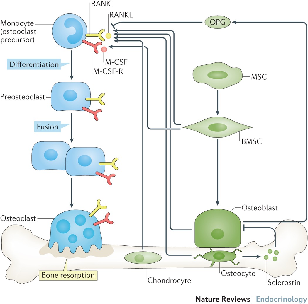
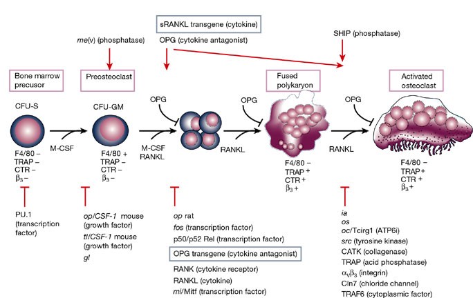
Osteoclasts are specialized cells derived from the monocyte/macrophage haematopoietic lineage that develop and adhere to bone matrix, then secrete acid and lytic enzymes that degrade it in a specialized, extracellular compartment. Discovery of the RANK signalling pathway in the osteoclast has provided insight into the mechanisms of osteoclastogenesis and activation of bone resorption, and how hormonal signals impact bone structure and mass. Further study of this pathway is providing the molecular basis for developing therapeutics to treat osteoporosis and other diseases of bone loss.
Transforming Growth Factor-β1 to the Bone
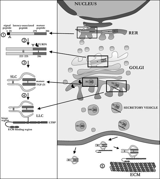
TGF-β1 is a ubiquitous growth factor that is implicated in the control of proliferation, migration, differentiation, and survival of many different cell types. It influences such diverse processes as embryogenesis, angiogenesis, inflammation, and wound healing. In skeletal tissue, TGF-β1 plays a major role in development and maintenance, affecting both cartilage and bone metabolism, the latter being the subject of this review. Because it affects both cells of the osteoblast and osteoclast lineage, TGF-β1 is one of the most important factors in the bone environment, helping to retain the balance between the dynamic processes of bone resorption and bone formation. Many seemingly contradictory reports have been published on the exact functioning of TGF-β1 in the bone milieu. This review provides an overall picture of the bone-specific actions of TGF-β1 and reconciles experimental discrepancies that have been reported for this multifunctional cytokine.