

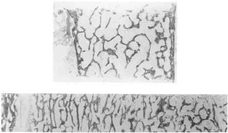
Before publication of the original version of this report in 1987, practitioners of bone histomorphometry communicated with each other in a variety of arca
Guidelines for assessment of bone microstructure in rodents using micro–computed tomography
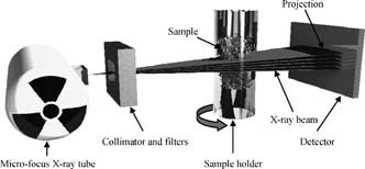
Abstract Use of high-resolution micro–computed tomography (µCT) imaging to assess trabecular and cortical bone morphology has grown immensely. There are several commercially available µCT systems, each with different approaches to image acquisition, evaluation, and reporting of outcomes. This lack of consistency makes it difficult to interpret reported results and to compare findings across different studies. This article addresses this critical need for standardized terminology and consistent reporting of parameters related to image acquisition and analysis, and key outcome assessments, particularly with respect to ex vivo analysis of rodent specimens. Thus the guidelines herein provide recommendations regarding (1) standardized terminology and units, (2) information to be included in describing the methods for a given experiment, and (3) a minimal set of outcome variables that should be reported. Whereas the specific research objective will determine the experimental design, these guidelines are intended to ensure accurate and consistent reporting of µCT-derived bone morphometry and density measurements. In particular, the methods section for papers that present µCT-based outcomes must include details of the following scan aspects: (1) image acquisition, including the scanning medium, X-ray tube potential, and voxel size, as well as clear descriptions of the size and location of the volume of interest and the method used to delineate trabecular and cortical bone regions, and (2) image processing, including the algorithms used for image filtration and the approach used for image segmentation. Morphometric analyses should be based on 3D algorithms that do not rely on assumptions about the underlying structure whenever possible. When reporting µCT results, the minimal set of variables that should be used to describe trabecular bone morphometry includes bone volume fraction and trabecular number, thickness, and separation. The minimal set of variables that should be used to describe cortical bone morphometry includes total cross-sectional area, cortical bone area, cortical bone area fraction, and cortical thickness. Other variables also may be appropriate depending on the research question and technical quality of the scan. Standard nomenclature, outlined in this article, should be followed for reporting of results. © 2010 American Society for Bone and Mineral Research
Ten‐Year Simulation of the Effects of Denosumab on Bone Remodeling in Human Biopsies
ABSTRACT Postmenopausal osteoporosis is a disease manifesting in degradation of bone mass and microarchitecture, leading to weakening and increased risk of fracture. Clinical trials are an essential tool for evaluating new treatments and may provide further mechanistic understanding of their effects in vivo. However, the histomorphometry from clinical trials is limited to 2D images and reflects single time points. Biochemical markers of bone turnover give global insight into a drug's action, but not the local dynamics of the bone remodeling process and the cells involved. Additionally, comparative trials necessitate separate treatment groups, meaning only aggregated measures can be compared. In this study, in silico modeling based on histomorphometry and pharmacokinetic data was used to assess the effects of treatment versus control on μCT scans of the same biopsy samples over time, matching the changes in bone volume fraction observed in biopsies from denosumab and placebo groups through year 10 of the FREEDOM Extension trial. In the simulation, treatment decreased osteoclast number, which led to a modest increase in trabecular thickness and osteocyte stress shielding. Long‐term bone turnover suppression led to increased RANKL production, followed by a small increase in osteoclast number at the end of the 6‐month–dosing interval, especially at the end of the Extension study. Lack of treatment led to a significant loss of bone mass and structure. The study's results show how in silico models can generate predictions of denosumab cellular action over a 10‐year period, matching static and dynamic morphometric measures assessed in clinical biopsies. The use of in silico models with clinical trial data can be a method to gain further insight into fundamental bone biology and how treatments can perturb this. With rigorous validation, such models could be used for informing the design of clinical trials, such that the number of participants could be reduced to a minimum to show efficacy. © 2021 The Authors. JBMR Plus published by Wiley Periodicals LLC on behalf of American Society for Bone and Mineral Research.
Integrated Bone Tissue Physiology: Anatomy and Physiology
New perspectives on body size and shape evolution in dinosaurs
ABSTRACT Diversity in the body shapes and sizes of dinosaurs was foundational to their widespread success during the Mesozoic era. The ability to quantify body size and form reliably is therefore critical to the study of dinosaur biology and evolution. Body mass estimates for any given fossil animal are, in theory, most informative when derived from volumetric models that account for the three‐dimensional shapes of the entire body. In addition to providing estimates of total body mass, volumetric approaches can be used to determine the inertial properties of specific body segments and the overall distribution of mass throughout the body, each of which are essential for the modelling and interpretation of form–function relationships and their associations with ecology. However, the determination of body volumes in fossil taxa is often subjective, and may be sensitive to varied artistic inference. This highlights the need for an approach to body mass estimation in which body segment volumes are systematically constrained by quantitative scaling relationships between the hard tissues that fossilise and the soft tissues only observable in extant taxa. To this end, we used recently published skeletal to soft tissue volumetric scaling factors derived from CT data of extant sauropsids to estimate body segment mass properties from skeletal models of 52 non‐avian dinosaurs representing the majority of major clades and body plans. The body masses estimated by this study range from less than 200 g in the tiny avialan Yixianornis to over 60 tonnes in the giant sauropod Patagotitan , which is currently the largest dinosaur known from mostly complete skeletal remains. From our models, we infer that many previous reconstructions of soft tissue envelopes may be too small, and that many dinosaurs were therefore heavier than previous estimates. Our models generally overlap with the range of body mass estimates derived from limb bone shaft dimensions, but with considerable quantitative variability among major clades. This suggests that different taxa either differed in skeletal to soft tissue volume ratios, or that their limb bone dimensions varied relative to body mass, perhaps related to differences in locomotor dynamics and postural evolution. Our models also allowed us to investigate variation in mass distribution and body proportions across different dinosaurs from a perspective grounded in extant anatomical data, framing long‐standing hypotheses about their form, function, and behaviour in a quantitative context. For example, reconstructed disparity in whole‐body centres of mass reflects a broad array of postures in different dinosaur clades, while the lack of strong positive allometry in the dimensions of the weight‐bearing limb segments relative to total body mass corroborates previous studies suggesting an overall decrease in dinosaur locomotor performance as body size increased.

Predicting Nonlinear Changes in Bone Mineral Density Over Time Using a Multiscale Systems Pharmacology Model
A mathematical model component that extends an existing physiologically based multiscale systems pharmacology model (MSPM) of calcium and bone homeostasis was developed, enabling prediction of nonlinear changes in lumbar spine bone mineral density (LSBMD). Data for denosumab, a monoclonal antibody osteoporosis treatment, dosed at several levels and regimens, was used for fitting the BMD component. Bone marker and LSBMD data extracted from the literature described on/off‐treatment effects of denosumab over 48 months [Miller, P.D. et al . Effect of denosumab on bone density and turnover in postmenopausal women with low bone mass after long‐term continued, discontinued, and restarting of therapy: a randomized blinded phase 2 clinical trial. Bone 43, 222–229 (2008)]. An indirect model linking bone markers to LSBMD was embedded in the existing MSPM, reasonably predicting nonlinear increases in LSBMD during treatment (24 months); LSBMD declines following discontinuation and increases upon treatment reinstitution. This study demonstrates the utility of MSPM extension to describe a phenomena of interest not originally in a model, and the ability of this updated MSPM to predict nonlinear longitudinal changes in the clinically relevant endpoint, LSBMD, with denosumab treatment. CPT: Pharmacometrics & Systems Pharmacology (2012) 1, e14; doi: 10.1038/psp.2012.15 ; advance online publication 14 November 2012

Bone mineral density and frequency of osteoporosis in female patients with rheumatoid arthritis: Results from 394 patients in the Oslo County rheumatoid arthritis register
Objective To examine the bone mineral density (BMD), frequency of osteoporosis, and risk factors for BMD reduction in a representative population of female rheumatoid arthritis (RA) patients ages 20...

Prevalence and clinical prediction of osteoporosis in a contemporary cohort of patients with rheumatoid arthritis
Abstract. Objective. Osteoporosis has previously been reported to be twice as common in patients with RA as in controls, but these studies predate the intr
Physiological Bone Remodeling: Systemic Regulation and Growth Factor Involvement
Bone remodeling is essential for adult bone homeostasis. It comprises two phases: bone formation and resorption. The balance between the two phases is crucial for sustaining bone mass and systemic mineral homeostasis. This review highlights recent work on physiological bone remodeling and discusses our knowledge of how systemic and growth factors regulate this process.

Osteonal and hemi‐osteonal remodeling: The spatial and temporal framework for signal traffic in adult human bone
Abstract The bone replacement process in the adult skeleton is known as remodeling. When bone is removed by osteoclasts, new bone is laid down by osteoblasts in the same place, because the load bearing requirement is unchanged. Bone is usually replaced because it is too old to carry out its function, which is mainly mechanical in cortical bone and mainly support for homeostasis and hematopoiesis in cancellous bone. Remodeling always begins on a quiescent bone surface, separated from the marrow by flat lining cells that are one of the two modes of terminal differentiation of osteoblasts. Lining cells are gatekeepers, able to be informed of the need for remodeling, and to either execute or mediate all four components of its activation‐selection and preparation of the site, recruitment of mononuclear preosteoclasts, budding of new capillaries, and attraction of preosteoclasts to the chosen site where they fuse into multinucleated osteoclasts. In cortical bone, osteonal remodeling is carried out by a complex and unique structure, the basic multicellular unit (BMU) that comprises a cutting cone of osteoclasts in front, a closing cone lined by osteoblasts following behind, and connective tissue, blood vessels and nerves filling the cavity. The BMU maintains its size, shape and internal organization for many months as it travels through bone in a controlled direction. Individual osteoclast nuclei are short‐lived, turning over about 8% per d, replaced by new preosteoclasts that originated in the bone marrow and travel in the circulation to the site of resorption. Refilling of bone at each successive cross‐sectional location is accomplished by a team of osteoblasts, probably originating from precursors within the local connective tissue, all assembled within a narrow window of time, at the right location, and in the right orientation to the surface. Each osteoblast team forms bone most rapidly at its onset and slows down progressively. Some of the osteoblasts are buried as osteocytes, some die, and the remainder gradually assume the shape of lining cells. Cancellous bone is more accessible to study than cortical bone, but is geometrically complex. Although remodeling conforms to the same sequence of surface activation, resorption and formation, its three‐dimensional organization is difficult to visualize from two‐dimensional histologic sections. But the average sizes of resorption sites, formation sites, and completed structural units increase progressively, as they do in cortical bone, indicating that the cancellous BMU travels across the surface digging a trench rather than a tunnel, but maintaining its size, shape and individual identity by the continuous recruitment of new cells, just as in cortical bone, a process that can be visualized as hemiosteonal remodeling. The conclusion that all remodeling is carried out by individual BMUs has important implications for bone biology, since many questions about how BMUs operate cannot be answered by studying either intact organisms or isolated cell systems. Many different steps in remodeling and many factors that influence each step have been identified, but very little is known about how the process is regulated in vivo to achieve its biologic purposes; most factors studied to date are likely permissive rather than regulatory in nature. Based on the proposed conceptual model of the BMU, much in vitro experimentation is relevant to the growth, modeling and repair of bone, but not to its remodeling in the adult skeleton. Further progress in the understanding of in vivo physiology will require the characterization of gene expression in individual cells to be related to the spatial and temporal organization of the BMU. This is likely to be possible only for osteonal remodeling in cortical bone in which, because of its geometric simplicity, individual BMUs can consistently be observed in two‐dimensional, longitudinal sections. © 1994 Wiley‐Liss, Inc.

Critical Role of Estrogens on Bone Homeostasis in Both Male and Female: From Physiology to Medical Implications
Bone is a multi-skilled tissue, protecting major organs, regulating calcium phosphate balance and producing hormones. Its development during childhood determines height and stature as well as resistance against fracture in advanced age. Estrogens are key regulators of bone turnover in both females and males. These hormones play a major role in longitudinal and width growth throughout puberty as well as in the regulation of bone turnover. In women, estrogen deficiency is one of the major causes of postmenopausal osteoporosis. In this review, we will summarize the main clinical and experimental studies reporting the effects of estrogens not only in females but also in males, during different life stages. Effects of estrogens on bone involve either Estrogen Receptor (ER)α or ERβ depending on the type of bone (femur, vertebrae, tibia, mandible), the compartment (trabecular or cortical), cell types involved (osteoclasts, osteoblasts and osteocytes) and sex. Finally, we will discuss new ongoing strategies to increase the benefit/risk ratio of the hormonal treatment of menopause.

A multiscale mechanobiological model of bone remodelling predicts site-specific bone loss in the femur during osteoporosis and mechanical disuse
We propose a multiscale mechanobiological model of bone remodelling to investigate the site-specific evolution of bone volume fraction across the midshaft of a femur. The model includes hormonal regulation and biochemical coupling of bone cell populations, the influence of the microstructure on bone turnover rate, and mechanical adaptation of the tissue. Both microscopic and tissue-scale stress/strain states of the tissue are calculated from macroscopic loads by a combination of beam theory and micromechanical homogenisation. This model is applied to simulate the spatio-temporal evolution of a human midshaft femur scan subjected to two deregulating circumstances: (i) osteoporosis and (ii) mechanical disuse. Both simulated deregulations led to endocortical bone loss, cortical wall thinning and expansion of the medullary cavity, in accordance with experimental findings. Our model suggests that these observations are attributable to a large extent to the influence of the microstructure on bone turnover rate. Mechanical adaptation is found to help preserve intracortical bone matrix near the periosteum. Moreover, it leads to non-uniform cortical wall thickness due to the asymmetry of macroscopic loads introduced by the bending moment. The effect of mechanical adaptation near the endosteum can be greatly affected by whether the mechanical stimulus includes stress concentration effects or not.

Toward a Mathematical Modeling of Diseases’ Impact on Bone Remodeling: Technical Review
A wide variety of bone diseases have hitherto been discovered, such as osteoporosis, Patget’s disease, osteopetrosis, and metastatic bone disease, which is not well defined. Some of these diseases are secondary to other pathologies, including cancer, or to some clinical treatments. To better understand bone behavior and prevent its deterioration, bone biomechanics have been the subject of mathematical modeling that exponentially increased over the last years. These models are more and more involving the impact of bone diseases on its quality. The current paper provides a meaningful and critical analysis of previously developed bone remodeling mathematical models, particularly those addressing bone diseases. Besides, mechanistic pharmacokinetic/pharmacodynamic (PK/PD) models, which englobe either bone disease and its treatments’ effect on bone health. Therefore, the review starts by presenting bone remodeling cycle and mathematical models describing this process, followed by introducing some bone diseases and discussing models of pathological mechanisms affecting bone, and ends by exhibiting the available bone treatment procedures and the PK/PD models considering these lasts.

A novel mathematical model of bone remodelling cycles for trabecular bone at the cellular level
After an initial phase of growth and development, bone undergoes a continuous cycle of repair, renewal and optimisation by a process called remodelling. This paper describes a novel mathematical model of the trabecular bone remodelling cycle. It is essentially formulated to simulate a remodelling event at a fixed position in the bone, integrating bone removal by osteoclasts and formation by osteoblasts. The model is developed to construct the variation in bone thickness at a particular point during the remodelling event, derived from standard bone histomorphometric analyses. The novelties of the approach are the adoption of a predator–prey model to describe the dynamic interaction between osteoclasts and osteoblasts, using a genetic algorithm–based solution; quantitative reconstruction of the bone remodelling cycle; and the introduction of a feedback mechanism in the bone formation activity to co-regulate bone thickness. The application of the model is first demonstrated by using experimental data recorded for normal (healthy) bone remodelling to predict the temporal variation in the number of osteoblasts and osteoclasts. The simulated histomorphometric data and remodelling cycle characteristics compare well with the specified input data. Sensitivity studies then reveal how variations in the model’s parameters affect its output; it is hoped that these parameters can be linked to specific biochemical factors in the future. Two sample pathological conditions, hypothyroidism and primary hyperparathyroidism, are examined to demonstrate how the model could be applied more broadly, and, for the first time, the osteoblast and osteoclast populations are predicted for these conditions. Further data are required to fully validate the model’s predictive capacity, but this work shows it has potential, especially in the modelling of pathological conditions and the optimisation of the treatment of those conditions.
Bone Dynamics in Metabolic Bone Disease:
Postmenopausal osteoporosis
Osteoporosis is a metabolic bone disorder that is characterized by low bone mass and micro-architectural deterioration of bone tissue. Fractures of the proximal femur, the vertebrae and the distal radius are the most frequent osteoporotic fractures, although most fractures in the elderly are probably at least partly related to bone fragility. The incidence of fractures varies greatly by country, but on average up to 50% of women >50 years of age are at risk of fractures. Fractures severely affect the quality of life of an individual and are becoming a major public health problem owing to the ageing population. Postmenopausal osteoporosis, resulting from oestrogen deficiency, is the most common type of osteoporosis. Oestrogen deficiency results in an increase in bone turnover owing to effects on all types of bone cells. The imbalance in bone formation and resorption has effects on trabecular bone (loss of connectivity) and cortical bone (cortical thinning and porosity). Osteoporosis is diagnosed using bone density measurements of the lumbar spine and proximal femur. Preventive strategies to improve bone health include diet, exercise and abstaining from smoking. Fractures may be prevented by reducing falls in high-risk populations. Several drugs are licensed to reduce fracture risk by slowing down bone resorption (such as bisphosphonates and denosumab) or by stimulating bone formation (such as teriparatide). Improved understanding of the cellular basis for osteoporosis has resulted in new drugs targeted to key pathways, which are under development.

Bone Remodeling in Post-menopausal Osteoporosis
Bone mass in the skeleton is dependent on the coordinated activities of bone-forming osteoblasts and bone-resorbing osteoclasts in discrete bone multi-cellular units. Remodeling of bone in these units is important not only for maintaining bone mass, but also to repair microdamage, to prevent accumulation of too much old bone, and for mineral homeostasis. The activities of osteoblasts and osteoclasts are controlled by a variety of hormones and cytokines, as well as by mechanical loading. Most importantly, sex hormones are very crucial for keeping bone mass in balance, and the lack of either estrogen or testosterone leads to decreased bone mass and increased risk for osteoporosis. The prevalence of osteoporotic fractures is increasing dramatically in the Western part of the world and is a major health problem in many countries. In the present review, the cellular and molecular mechanisms controlling bone remodeling and the influence of sex hormones on these processes are summarized. In a separate paper in this issue, the pathogenesis of post-menopausal osteoporosis will be compared with that of inflammation-induced bone remodeling, including the evidence for and against the hypothesis that concomitant post-menopausal osteoporotic disease influences the progression of periodontal disease.
