


Bone is an activeCoelho, Rui M. Neto, Joana P. connective tissue composed of different types of cells. The dynamic behavior of bone remodeling processes is typically represented throughValério, Duarte differential equations, which represent the physiological phenomena occurring in this organ. These models takeVinga, Susana into account the tight biochemical regulation between osteoclasts and osteoblasts and have also been enriched with variables and parameters related to bone pathologies and treatment. This chapter reviews some of the more recent models describing bone physiology, focusing on those that include the main cellular processes, along the biochemical control, and also the pharmacokinetic/pharmacodynamic (PK/PD) of the most common treatments for diseases such as cancer. These models are then compared in terms of the simulations obtained and, finally, some highlights on integrating them with the biomechanical component of the system which will be given. These models are expected to provide a valuable insight into this complex system and to support the development of clinical decision systems for bone pathologies.
Toward a Mathematical Modeling of Diseases’ Impact on Bone Remodeling: Technical Review
A wide variety of bone diseases have hitherto been discovered, such as osteoporosis, Patget’s disease, osteopetrosis, and metastatic bone disease, which is not well defined. Some of these diseases are secondary to other pathologies, including cancer, or to some clinical treatments. To better understand bone behavior and prevent its deterioration, bone biomechanics have been the subject of mathematical modeling that exponentially increased over the last years. These models are more and more involving the impact of bone diseases on its quality. The current paper provides a meaningful and critical analysis of previously developed bone remodeling mathematical models, particularly those addressing bone diseases. Besides, mechanistic pharmacokinetic/pharmacodynamic (PK/PD) models, which englobe either bone disease and its treatments’ effect on bone health. Therefore, the review starts by presenting bone remodeling cycle and mathematical models describing this process, followed by introducing some bone diseases and discussing models of pathological mechanisms affecting bone, and ends by exhibiting the available bone treatment procedures and the PK/PD models considering these lasts.

A review of mathematical modeling of bone remodeling from a systems biology perspective
Bone remodeling is an essential, delicately balanced physiological process of coordinated activity of bone cells that remove and deposit new bone tissue in the adult skeleton. Due to the complex nature of this process, many mathematical models of bone remodeling have been developed. Each of these models has unique features, but they have underlying patterns. In this review, the authors highlight the important aspects frequently found in mathematical models for bone remodeling and discuss how and why these aspects are included when considering the physiology of the bone basic multicellular unit, which is the term used for the collection of cells responsible for bone remodeling. The review also emphasizes the view of bone remodeling from a systems biology perspective. Understanding the systemic mechanisms involved in remodeling will help provide information on bone pathology associated with aging, endocrine disorders, cancers, and inflammatory conditions and enhance systems pharmacology. Furthermore, some features of the bone remodeling cycle and interactions with other organ systems that have not yet been modeled mathematically are discussed as promising future directions in the field.

Mathematical modelling of the pathogenesis of multiple myeloma‐induced bone disease
SUMMARY Multiple myeloma (MM) is the second most common haematological malignancy and results in destructive bone lesions. The interaction between MM cells and the bone microenvironment plays an important role in the development of the tumour cells and MM‐induced bone disease and forms a ‘vicious cycle’ of tumour development and bone destruction, intensified by suppression of osteoblast activity and promotion of osteoclast activity. In this paper, a mathematical model is proposed to simulate how the interaction between MM cells and the bone microenvironment facilitates the development of the tumour cells and the resultant bone destruction. It includes both the roles of inhibited osteoblast activity and stimulated osteoclast activity. The model is able to mimic the temporal variation of bone cell concentrations and resultant bone volume after the invasion and then removal of the tumour cells and explains why MM‐induced bone lesions rarely heal even after the complete removal of MM cells. The behaviour of the model compares well with published experimental data. The model serves as a first step to understand the development of MM‐induced bone disease and could be applied further to evaluate the current therapies against MM‐induced bone disease and even suggests new potential therapeutic targets. © 2014 The Authors. International Journal for Numerical Methods in Biomedical Engineering published by John Wiley & Sons Ltd

A review of mathematical modeling of bone remodeling from a systems biology perspective
Bone remodeling is an essential physiological process in the adult skeleton. Due to the complex nature of this process, many mathematical models of bone remodeling have been developed. Each of these models has unique features, but they have underlying patterns. In this review, the authors highlight the important aspects frequently found in mathematical models for bone remodeling and discuss how and why these aspects are included when considering the physiology of the bone basic multicellular unit, which is the term used for the collection of cells responsible for bone remodeling. The review also emphasizes the view of bone remodeling from a systems biology perspective. Understanding the systemic mechanisms involved in remodeling will help provide information on bone pathology associated with aging, endocrine disorders, cancers, and inflammatory conditions and enhance systems pharmacology. Furthermore, some features of the bone remodeling cycle and interactions with other organ systems that have not yet been modeled mathematically are discussed as promising future directions in the field.
Mathematical modeling of postmenopausal osteoporosis and its treatment by the anti‐catabolic drug denosumab
SUMMARY Denosumab, a fully human monoclonal antibody, has been approved for the treatment of postmenopausal osteoporosis. The therapeutic effect of denosumab rests on its ability to inhibit osteoclast differentiation. Here, we present a computational approach on the basis of coupling a pharmacokinetics model of denosumab with a pharmacodynamics model for quantifying the effect of denosumab on bone remodeling. The pharmacodynamics model comprises an integrated systems biology‐continuum micromechanics approach, including a bone cell population model, considering the governing biochemical factors of bone remodeling (including the action of denosumab), and a multiscale micromechanics‐based bone mechanics model, for implementing the mechanobiology of bone remodeling in our model. Numerical studies of postmenopausal osteoporosis show that denosumab suppresses osteoclast differentiation, thus strongly curtailing bone resorption. Simulation results also suggest that denosumab may trigger a short‐term bone volume gain, which is, however, followed by constant or decreasing bone volume. This evolution is accompanied by a dramatic decrease of the bone turnover rate by more than one order of magnitude. The latter proposes dominant occurrence of secondary mineralization (which is not anymore impeded through cellular activity), leading to higher mineral concentration per bone volume. This explains the overall higher bone mineral density observed in denosumab‐related clinical studies. Copyright © 2013 John Wiley & Sons, Ltd.

Studying Bone Remodelling and Tumour Growth for Therapy Predictive Control
Bone remodelling consists of cycles of bone resorption and formation executed mainly by osteoclasts and osteoblasts. Healthy bone remodelling is disrupted by diseases such as Multiple Myeloma and bone metastatic diseases. In this paper, a simple mathematical model with differential equations, which takes into account the evolution of osteoclasts, osteoblasts, bone mass and bone metastasis growth, is improved with a pharmacokinetic and pharmacodynamic (PK/PD) scheme of the drugs denosumab, bisphosphonates, proteasome inhibitors and paclitaxel. The major novelty is the inclusion of drug resistance phenomena, which resulted in two variations of the model, corresponding to different paradigms of the origin and development of the tumourous cell resistance condition. These models are then used as basis for an optimization of the drug dose applied, paving the way for personalized medicine. A Nonlinear Model Predictive Control scheme is used, which takes advantage of the convenient properties of a suggested adaptive and democratic variant of Particle Swarm Optimization. Drug prescriptions obtained in this way provide useful insights into dose administration strategies. They also show how results may change depending on which of the two very different paradigms of drug resistance is used to model the behaviour of the tumour.

Dynamics of Bone Cell Interactions and Differential Responses to PTH and Antibody-Based Therapies
We propose a mathematical model describing the dynamics of osteoblasts and osteoclasts in bone remodeling. The goal of this work is to develop an integrated modeling framework for bone remodeling and bone cell signaling dynamics that could be used to explore qualitatively combination treatments for osteoporosis in humans. The model has been calibrated using 57 checks from the literature. Specific global optimization methods based on qualitative objectives have been developed to perform the model calibration. We also added pharmacokinetics representations of three drugs to the model, which are teriparatide (PTH(1–34)), denosumab (a RANKL antibody) and romosozumab (a sclerostin antibody), achieving excellent goodness-of-fit of human clinical data. The model reproduces the paradoxical effects of PTH on the bone mass, where continuous administration of PTH results in bone loss but intermittent administration of PTH leads to bone gain, thus proposing an explanation of this phenomenon. We used the model to simulate different categories of osteoporosis. The main attributes of each disease are qualitatively well captured by the model, for example changes in bone turnover in the disease states. We explored dosing regimens for each disease based on the combination of denosumab and romosozumab, identifying adequate ratios and doses of both drugs for subpopulations of patients in function of categories of osteoporosis and the degree of severity of the disease.

<i>In silico</i> models of bone remodeling from macro to nano—from organ to cell
Abstract Computational modeling is a tool through which researchers can achieve a greater understanding of the mechanisms governing biological systems. In the field of bone biology, a plethora of models exist which attempt to replicate and investigate bone's dynamic behavior at different scales. At organ level, models are continuum based and describe the variation of bone's apparent density as a function of both biological and external mechanical stimuli. At tissue level, models include bone microarchitecture and more descriptive parameters such as trabecular thickness, osteoclast resorption depth, and activation frequency. Finally, at cell level, models employ partial differential equations to describe complex cellular interactions in the temporal domain. Although informative, these models exist in isolation. Consequently, their interpretation is limited. In this review, we present an overview of the organ‐, tissue‐, and cell‐level models and assess their ability to reflect bone's metabolic processes reliably. Existing interscale synergies are then presented along with a computational framework which could be exploited to achieve a fully integrated, multiscale modeling approach. WIREs Syst Biol Med 2011 3 241–251 DOI: 10.1002/wsbm.115 This article is categorized under: Analytical and Computational Methods > Computational Methods Models of Systems Properties and Processes > Organ, Tissue, and Physiological Models

A mechano-chemo-biological model for bone remodeling with a new mechano-chemo-transduction approach
Bone remodeling is a fundamental biological process that develops in bone tissue along its whole lifetime. It refers to a continuous bone transformation with new bone formation and old bone resorption that changes the internal microstructure and composition of the tissue. The main objectives of bone remodeling are: repair of the internal microcracks; adaptation of the macroscopic stiffness and strength to the actual changing mechanical demands; and control of the calcium homeostasis. Understanding this process and predicting its evolution is critical to reduce the effects of long-term disuse as happens during periods of reduced mobility. It is also important in the design of bone implants to avoid long-term stress shielding. Many mathematical models have been proposed from the earliest purely phenomenological to the latest that include biological knowledge. However, there still exists a lack of connection between the mechanical driving force and the biochemical and cell processes it triggers. Here, and following previous works that model independently the mechanobiological and biochemical processes in bone remodeling, we present a more complete model, useful for both cortical and trabecular bone, that uses a new mechanotransduction approach based on the effect of strains onto the bonding–unbonding rate of RANK/RANKL/OPG receptor–ligand reactions. We compare the results of this model with previous ones, showing a good agreement in similar conditions. We also apply it to realistic situations such as a femoral bone after implantation of a hip prosthesis, getting similar results to the clinical ones in the final bone density distribution. Finally, we extend this approach to the anisotropic case, getting not only the mean density, but also the directional homogenization of the microstructure. This biochemical approach permits, not only to predict the bone evolution under changes in the mechanical loads, but also, to consider anabolic and catabolic drugs to control bone density, such as those used in osteoporosis.

Modeling the Human Bone–Tumor Niche: Reducing and Replacing the Need for Animal Data
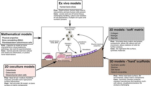
ABSTRACT Bone is the most common site for cancer metastasis. Understanding the interactions within the complex, heterogeneous bone–tumor microenvironment is essential for the development of new therapeutics. Various animal models of tumor‐induced bone disease are routinely used to provide valuable information on the relationship between cancer cells and the skeleton. However, new model systems exist that offer an alternative approach to the use of animals and might more accurately reveal the cellular interactions occurring within the human bone–tumor niche. This review highlights replacement models that mimic the bone microenvironment and where cancer metastases and tumor growth might be assessed alongside bone turnover. Such culture models include the use of calcified regions of animal tissue and scaffolds made from bone mineral hydroxyapatite, synthetic polymers that can be manipulated during manufacture to create structures resembling trabecular bone surfaces, gel composites that can be modified for stiffness and porosity to resemble conditions in the tumor–bone microenvironment. Possibly the most accurate model system involves the use of fresh human bone samples, which can be cultured ex vivo in the presence of human tumor cells and demonstrate similar cancer cell–bone cell interactions as described in vivo. In addition, the use of mathematical modeling and computational biology approaches provide an alternative to preliminary animal testing. The use of such models offers the capacity to mimic significant elements of the human bone–tumor environment, and complement, refine, or replace the use of preclinical models. © 2020 The Authors. JBMR Plus published by Wiley Periodicals, Inc. on behalf of American Society for Bone and Mineral Research.
Osteoprotegerin in Bone Metastases: Mathematical Solution to the Puzzle
Bone is a common site for cancer metastasis. To create space for their growth, cancer cells stimulate bone resorbing osteoclasts. Cytokine RANKL is a key osteoclast activator, while osteoprotegerin (OPG) is a RANKL decoy receptor and an inhibitor of osteoclastogenesis. Consistently, systemic application of OPG decreases metastatic tumor burden in bone. However, OPG produced locally by cancer cells was shown to enhance osteolysis and tumor growth. We propose that OPG produced by cancer cells causes a local reduction in RANKL levels, inducing a steeper RANKL gradient away from the tumor and towards the bone tissue, resulting in faster resorption and tumor expansion. We tested this hypothesis using a mathematical model of nonlinear partial differential equations describing the spatial dynamics of OPG, RANKL, PTHrP, osteoclasts, tumor and bone mass. We demonstrate that at lower expression rates, tumor-derived OPG enhances the chemotactic RANKL gradient and osteolysis, whereas at higher expression rates OPG broadly inhibits RANKL and decreases osteolysis and tumor burden. Moreover, tumor expression of a soluble mediator inducing RANKL in the host tissue, such as PTHrP, is important for correct orientation of the RANKL gradient. A meta-analysis of OPG, RANKL and PTHrP expression in normal prostate, carcinoma and metastatic tissues demonstrated an increase in expression of OPG, but not RANKL, in metastatic prostate cancer, and positive correlation between OPG and PTHrP in metastatic prostate cancer. The proposed mechanism highlights the importance of the spatial distribution of receptors, decoys and ligands, and can be applied to other systems involving regulation of spatially anisotropic processes.
Mathematical Model of Bone Remodeling Captures the Antiresorptive and Anabolic Actions of Various Therapies
A better understanding of the molecular pathways regulating the bone remodeling process should help in the development of new antiresorptive regulators and anabolic regulators, that is, regulators of bone resorption and of bone formation. Understanding the mechanisms by which parathyroid hormone (PTH) influences bone formation and how it switches from anabolic to catabolic action is important for treating osteoporosis (Poole and Reeve in Curr Opin Pharmacol 5:612–617, 2005). In this paper we describe a mathematical model of bone remodeling that incorporates, extends, and integrates several models of particular aspects of this biochemical system (Cabal et al. in J Bone Miner Res 28(8):1830–1836, 2013; Lemaire et al. in J Theor Biol 229:293–309, 2004; Peterson and Riggs in Bone 46:49–63, 2010; Raposo et al. in J Clin Endocrinol Metab 87(9):4330–4340, 2002; Ross et al. in J Disc Cont Dyn Sys Series B 17(6):2185–2200, 2012). We plan to use this model as a bone homeostasis platform to develop anabolic and antiresorptive compounds. The model will allow us to test hypotheses about the dynamics of compounds and to test the potential benefits of combination therapies. At the core of the model is the idealized account of osteoclast and osteoblast signaling given by Lemaire et al. (J Theor Biol 229:293–309, 2004). We have relaxed some of their assumptions about the roles of osteoprotegerin, transforming growth factor $$\upbeta $$, and receptor activator of nuclear factor $$\upkappa $$B ligand; we have devised more detailed models of the interactions of these species. We have incorporated a model of the effect of calcium sensing receptor antagonists on remodeling (Cabal et al. in J Bone Miner Res 28(8):1830–1836, 2013). We have also incorporated a basic model of the effects of vitamin D on calcium homeostasis. We have included a simple model of the mechanism proposed by Bellido et al. (2003), Ross et al. (J Disc Cont Dyn Sys Series B 17(6):2185–2200, 2012), of the influence of PTH on osteoblast apoptosis, a mechanism that accounts for the anabolic response to pulsatile PTH administration. Finally, we have devised a simple model of the administration and effects of bisphosphonates. The biomarkers in the model are procollagen type 1 amino-terminal propeptide and C-terminal telopeptide. Bone mineral density is the model’s principal endpoint.

Mathematical Modeling of Spatio-Temporal Dynamics of a Single Bone Multicellular Unit
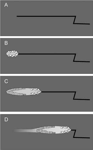
Abstract During bone remodeling, bone-resorbing osteoclasts and bone-forming osteoblasts are organized in bone multicellular units (BMUs), which travel at a rate of 20–40 μm/d for 6–12 mo, maintaining a cylindrical structure. However, the interplay of local BMU geometry with biochemical regulation is poorly understood. We developed a mathematical model of BMU describing changes in time and space of the concentrations of proresorptive cytokine RANKL and its inhibitor osteoprotegerin (OPG), in osteoclast and osteoblast numbers, and in bone mass. We assumed that osteocytes surrounding a microfracture produce RANKL, which attracted osteoclasts. OPG and RANKL were produced by osteoblasts and diffused through bone, RANKL was eliminated by binding to OPG and RANK. Osteoblasts were coupled to osteoclasts through paracrine factors. The evolution of the BMU arising from this model was studied using numerical simulations. Our model recapitulated the spatio-temporal dynamics observed in vivo in a cross-section of bone. In response to a RANKL field, osteoclasts moved as a well-confined cutting cone. The coupling of osteoclasts to osteoblasts allowed for sufficient recruitment of osteoblasts to the resorbed surfaces. The RANKL field was the highest at the microfracture in front of the BMU, whereas the OPG field peaked at the back of the BMU, resulting in the formation of a RANKL/OPG gradient, which strongly affected the rate of BMU progression and its size. Thus, the spatial organization of a BMU provides important constraints on the roles of RANKL and OPG as well as possibly other regulators in determining the outcome of remodeling in the BMU.
Pharmacometrics and systems pharmacology for metabolic bone diseases
Mathematical modelling and simulation (M&S) of drug concentrations, pharmacologic effects and the (patho)physiologic systems within which they interact can be powerful tools for the preclinical, translational and clinical development of drugs. Indeed, the Prescription Drug User Fee Act (PDUFA VI), incorporated as part of the FDA Reauthorization Act of 2017 (FDARA), highlights the goal of advancing model‐informed drug development (MIDD). MIDD can benefit development across many drug classes, including for metabolic bone diseases such as osteoporosis, cancer‐related and numerous rare metabolic bone diseases; conditions characterized by significant morbidity and mortality. A drought looms in terms of the availability of new drugs to better treat these devastating diseases. This review provides an overview of several M&S approaches ranging from simple pharmacokinetic to integrated pharmacometric and systems pharmacology modelling. Examples are included to illustrate the use of these approaches during the development of several drugs for metabolic bone diseases such as bisphosphonates, denosumab, teriparatide and sclerostin inhibitors (romosozumab and blosozumab).

The Cellular Dynamics of Bone Remodeling: A Mathematical Model
The mechanical properties of vertebrate bone are largely determined by a process which involves the complex interplay of three different cell types. This process is called bone remodeling and occurs asynchronously at multiple sites in the mature skeleton. The cells involved are bone resorbing osteoclasts, bone matrix producing osteoblasts, and mechanosensing osteocytes. These cells communicate with each other by means of autocrine and paracrine signaling factors and operate in complex entities, the so-called bone multicellular units (BMUs). To investigate the BMU dynamics in silico, we develop a novel mathematical model resulting in a system of nonlinear partial differential equations (PDEs) with time delays. The model describes the osteoblast and osteoclast populations together with the dynamics of the key messenger molecule RANKL and its decoy receptor OPG. Scaling theory is used to address parameter sensitivity and predict the emergence of pathological remodeling regimes. The model is studied numerically in one and two space dimensions using finite difference schemes in space and explicit delay equation solvers in time. The computational results are in agreement with in vivo observations and provide new insights into the role of the RANKL/OPG pathway in the spatial regulation of bone remodeling.

Mechanobiological osteocyte feedback drives mechanostat regulation of bone in a multiscale computational model
Significant progress has been made to identify the cells and signaling molecules involved in the mechanobiological regulation of bone remodeling. It is now well accepted that osteocytes act as mechanosensory cells in bone expressing several signaling molecules such as nitric oxide (NO) and sclerostin (Scl) which are able to control bone remodeling responses. In this paper, we present a comprehensive multiscale computational model of bone remodeling which incorporates biochemical osteocyte feedback. The mechanostat theory is quantitatively incorporated into the model using mechanical feedback to control expression levels of NO and Scl. The catabolic signaling pathway RANK–RANKL–OPG is co-regulated via (continuous) PTH and NO, while the anabolic Wnt signaling pathway is described via competitive binding reactions between Wnt, Scl and the Wnt receptors LRP5/6. Using this novel model of bone remodeling, we investigate the effects of changes in the mechanical loading and hormonal environment on bone balance. Our numerical simulations show that we can calibrate the mechanostat anabolic and catabolic regulatory mechanisms so that they are mutually exclusive. This is consistent with previous models that use a Wolff-type law to regulate bone resorption and formation separately. Furthermore, mechanical feedback provides an effective mechanism to obtain physiological bone loss responses due to mechanical disuse and/or osteoporosis.
