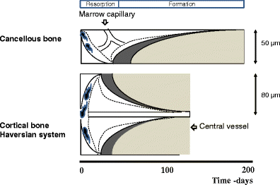
Bone remodeling is a tightly regulated process securing repair of microdamage (targeted remodeling) and replacement of old bone with new bone through sequential osteoclastic resorption and osteoblastic bone formation. The rate of remodeling is regulated by a wide variety of calcitropic hormones (PTH, thyroid hormone, sex steroids etc.). In recent years we have come to appreciate that bone remodeling proceeds in a specialized vascular structure,—the Bone Remodeling Compartment (BRC). The outer lining of this compartment is made up of flattened cells, displaying all the characteristics of lining cells in bone including expression of OPG and RANKL. Reduced bone turnover leads to a decrease in the number of BRCs, while increased turnover causes an increase in the number of BRCs. The secretion of regulatory factors inside a confined space separated from the bone marrow would facilitate local regulation of the remodeling process without interference from growth factors secreted by blood cells in the marrow space. The BRC also creates an environment where cells inside the structure are exposed to denuded bone, which may enable direct cellular interactions with integrins and other matrix factors known to regulate osteoclast/osteoblast activity. However, the denuded bone surface inside the BRC also constitutes an ideal environment for the seeding of bone metastases, known to have high affinity for bone matrix. Circulating osteoclast- and osteoblast precursor cells have been demonstrated in peripheral blood. The dominant pathway regulating osteoclast recruitment is the RANKL/OPG system, while many different factors (RUNX, Osterix) are involved in osteoblast differentiation. Both pathways are modulated by calcitropic hormones.
Osteonal and hemi‐osteonal remodeling: The spatial and temporal framework for signal traffic in adult human bone
Abstract The bone replacement process in the adult skeleton is known as remodeling. When bone is removed by osteoclasts, new bone is laid down by osteoblasts in the same place, because the load bearing requirement is unchanged. Bone is usually replaced because it is too old to carry out its function, which is mainly mechanical in cortical bone and mainly support for homeostasis and hematopoiesis in cancellous bone. Remodeling always begins on a quiescent bone surface, separated from the marrow by flat lining cells that are one of the two modes of terminal differentiation of osteoblasts. Lining cells are gatekeepers, able to be informed of the need for remodeling, and to either execute or mediate all four components of its activation‐selection and preparation of the site, recruitment of mononuclear preosteoclasts, budding of new capillaries, and attraction of preosteoclasts to the chosen site where they fuse into multinucleated osteoclasts. In cortical bone, osteonal remodeling is carried out by a complex and unique structure, the basic multicellular unit (BMU) that comprises a cutting cone of osteoclasts in front, a closing cone lined by osteoblasts following behind, and connective tissue, blood vessels and nerves filling the cavity. The BMU maintains its size, shape and internal organization for many months as it travels through bone in a controlled direction. Individual osteoclast nuclei are short‐lived, turning over about 8% per d, replaced by new preosteoclasts that originated in the bone marrow and travel in the circulation to the site of resorption. Refilling of bone at each successive cross‐sectional location is accomplished by a team of osteoblasts, probably originating from precursors within the local connective tissue, all assembled within a narrow window of time, at the right location, and in the right orientation to the surface. Each osteoblast team forms bone most rapidly at its onset and slows down progressively. Some of the osteoblasts are buried as osteocytes, some die, and the remainder gradually assume the shape of lining cells. Cancellous bone is more accessible to study than cortical bone, but is geometrically complex. Although remodeling conforms to the same sequence of surface activation, resorption and formation, its three‐dimensional organization is difficult to visualize from two‐dimensional histologic sections. But the average sizes of resorption sites, formation sites, and completed structural units increase progressively, as they do in cortical bone, indicating that the cancellous BMU travels across the surface digging a trench rather than a tunnel, but maintaining its size, shape and individual identity by the continuous recruitment of new cells, just as in cortical bone, a process that can be visualized as hemiosteonal remodeling. The conclusion that all remodeling is carried out by individual BMUs has important implications for bone biology, since many questions about how BMUs operate cannot be answered by studying either intact organisms or isolated cell systems. Many different steps in remodeling and many factors that influence each step have been identified, but very little is known about how the process is regulated in vivo to achieve its biologic purposes; most factors studied to date are likely permissive rather than regulatory in nature. Based on the proposed conceptual model of the BMU, much in vitro experimentation is relevant to the growth, modeling and repair of bone, but not to its remodeling in the adult skeleton. Further progress in the understanding of in vivo physiology will require the characterization of gene expression in individual cells to be related to the spatial and temporal organization of the BMU. This is likely to be possible only for osteonal remodeling in cortical bone in which, because of its geometric simplicity, individual BMUs can consistently be observed in two‐dimensional, longitudinal sections. © 1994 Wiley‐Liss, Inc.

Biology of Bone Tissue: Structure, Function, and Factors That Influence Bone Cells
Bone tissue is continuously remodeled through the concerted actions of bone cells, which include bone resorption by osteoclasts and bone formation by osteoblasts, whereas osteocytes act as mechanosensors and orchestrators of the bone remodeling process. This process is under the control of local (e.g., growth factors and cytokines) and systemic (e.g., calcitonin and estrogens) factors that all together contribute for bone homeostasis. An imbalance between bone resorption and formation can result in bone diseases including osteoporosis. Recently, it has been recognized that, during bone remodeling, there are an intricate communication among bone cells. For instance, the coupling from bone resorption to bone formation is achieved by interaction between osteoclasts and osteoblasts. Moreover, osteocytes produce factors that influence osteoblast and osteoclast activities, whereas osteocyte apoptosis is followed by osteoclastic bone resorption. The increasing knowledge about the structure and functions of bone cells contributed to a better understanding of bone biology. It has been suggested that there is a complex communication between bone cells and other organs, indicating the dynamic nature of bone tissue. In this review, we discuss the current data about the structure and functions of bone cells and the factors that influence bone remodeling.

Bone remodeling: A tissue-level process emerging from cell-level molecular algorithms
The human skeleton undergoes constant remodeling throughout the lifetime. Processes occurring on microscopic and molecular scales degrade bone and replace it with new, fully functional tissue. Multiple bone remodeling events occur simultaneously, continuously and independently throughout the body, so that the entire skeleton is completely renewed about every ten years.Bone remodeling is performed by groups of cells called Bone Multicellular Units (BMU). BMUs consist of different cell types, some specialized in the resorption of old bone, others encharged with producing new bone to replace the former. These processes are tightly regulated so that the amount of new bone produced is in perfect equilibrium with that of old bone removed, thus maintaining bone microscopic structure.To date, many regulatory molecules involved in bone remodeling have been identified, but the precise mechanism of BMU operation remains to be fully elucidated. Given the complexity of the signaling pathways already known, one may question whether such complexity is an inherent requirement of the process or whether some subset of the multiple constituents could fulfill the essential role, leaving functional redundancy to serve an alternative safety role. We propose in this work a minimal model of BMU function that involves a limited number of signals able to account for fully functional BMU operation. Our main assumptions were i) at any given time, any cell within a BMU can select only one among a limited choice of decisions, i.e. divide, die, migrate or differentiate, ii) this decision is irreversibly determined by depletion of an appropriate internal inhibitor and iii) the dynamics of any such inhibitor are coupled to that of specific external mediators, such as hormones, cytokines, growth factors. It was thus shown that efficient BMU operation manifests as an emergent process, which results from the individual and collective decisions taken by cells within the BMU unit in the absence of any external planning.
The Role of Osteocytes in Targeted Bone Remodeling: A Mathematical Model
Until recently many studies of bone remodeling at the cellular level have focused on the behavior of mature osteoblasts and osteoclasts, and their respective precursor cells, with the role of osteocytes and bone lining cells left largely unexplored. This is particularly true with respect to the mathematical modeling of bone remodeling. However, there is increasing evidence that osteocytes play important roles in the cycle of targeted bone remodeling, in serving as a significant source of RANKL to support osteoclastogenesis, and in secreting the bone formation inhibitor sclerostin. Moreover, there is also increasing interest in sclerostin, an osteocyte-secreted bone formation inhibitor, and its role in regulating local response to changes in the bone microenvironment. Here we develop a cell population model of bone remodeling that includes the role of osteocytes, sclerostin, and allows for the possibility of RANKL expression by osteocyte cell populations. We have aimed to give a simple, yet still tractable, model that remains faithful to the underlying system based on the known literature. This model extends and complements many of the existing mathematical models for bone remodeling, but can be used to explore aspects of the process of bone remodeling that were previously beyond the scope of prior modeling work. Through numerical simulations we demonstrate that our model can be used to explore theoretically many of the qualitative features of the role of osteocytes in bone biology as presented in recent literature.
A review of mathematical modeling of bone remodeling from a systems biology perspective
Bone remodeling is an essential, delicately balanced physiological process of coordinated activity of bone cells that remove and deposit new bone tissue in the adult skeleton. Due to the complex nature of this process, many mathematical models of bone remodeling have been developed. Each of these models has unique features, but they have underlying patterns. In this review, the authors highlight the important aspects frequently found in mathematical models for bone remodeling and discuss how and why these aspects are included when considering the physiology of the bone basic multicellular unit, which is the term used for the collection of cells responsible for bone remodeling. The review also emphasizes the view of bone remodeling from a systems biology perspective. Understanding the systemic mechanisms involved in remodeling will help provide information on bone pathology associated with aging, endocrine disorders, cancers, and inflammatory conditions and enhance systems pharmacology. Furthermore, some features of the bone remodeling cycle and interactions with other organ systems that have not yet been modeled mathematically are discussed as promising future directions in the field.

The bone remodelling cycle
The bone remodelling cycle replaces old and damaged bone and is a highly regulated, lifelong process essential for preserving bone integrity and maintaining mineral homeostasis. During the bone remodelling cycle, osteoclastic resorption is tightly coupled to osteoblastic bone formation. The remodelling cycle occurs within the basic multicellular unit and comprises five co-ordinated steps; activation, resorption, reversal, formation and termination. These steps occur simultaneously but asynchronously at multiple different locations within the skeleton. Study of rare human bone disease and animal models have helped to elucidate the cellular and molecular mechanisms that regulate the bone remodelling cycle. The key signalling pathways controlling osteoclastic bone resorption and osteoblastic bone formation are receptor activator of nuclear factor-κB (RANK)/RANK ligand/osteoprotegerin and canonical Wnt signalling. Cytokines, growth factors and prostaglandins act as paracrine regulators of the cycle, whereas endocrine regulators include parathyroid hormone, vitamin D, calcitonin, growth hormone, glucocorticoids, sex hormones, and thyroid hormone. Disruption of the bone remodelling cycle and any resulting imbalance between bone resorption and formation leads to metabolic bone disease, most commonly osteoporosis. The advances in understanding the cellular and molecular mechanisms underlying bone remodelling have also provided targets for pharmacological interventions which include antiresorptive and anabolic therapies. This review will describe the remodelling process and its regulation, discuss osteoporosis and summarize the commonest pharmacological interventions used in its management.

Physiological Bone Remodeling: Systemic Regulation and Growth Factor Involvement
Bone remodeling is essential for adult bone homeostasis. It comprises two phases: bone formation and resorption. The balance between the two phases is crucial for sustaining bone mass and systemic mineral homeostasis. This review highlights recent work on physiological bone remodeling and discusses our knowledge of how systemic and growth factors regulate this process.

A review of mathematical modeling of bone remodeling from a systems biology perspective
Bone remodeling is an essential physiological process in the adult skeleton. Due to the complex nature of this process, many mathematical models of bone remodeling have been developed. Each of these models has unique features, but they have underlying patterns. In this review, the authors highlight the important aspects frequently found in mathematical models for bone remodeling and discuss how and why these aspects are included when considering the physiology of the bone basic multicellular unit, which is the term used for the collection of cells responsible for bone remodeling. The review also emphasizes the view of bone remodeling from a systems biology perspective. Understanding the systemic mechanisms involved in remodeling will help provide information on bone pathology associated with aging, endocrine disorders, cancers, and inflammatory conditions and enhance systems pharmacology. Furthermore, some features of the bone remodeling cycle and interactions with other organ systems that have not yet been modeled mathematically are discussed as promising future directions in the field.
Osteoclasts Provide Coupling Signals to Osteoblast Lineage Cells Through Multiple Mechanisms
Bone remodeling is essential for the repair and replacement of damaged and old bone. The major principle underlying this process is that osteoclast-mediated resorption of a quantum of bone is followed by osteoblast precursor recruitment; these cells differentiate to matrix-producing osteoblasts, which form new bone to replace what was resorbed. Evidence from osteopetrotic syndromes indicate that osteoclasts not only resorb bone, but also provide signals to promote bone formation. Osteoclasts act upon osteoblast lineage cells throughout their differentiation by facilitating growth factor release from resorbed matrix, producing secreted proteins and microvesicles, and expressing membrane-bound factors. These multiple mechanisms mediate the coupling of bone formation to resorption in remodeling. Additional interactions of osteoclasts with osteoblast lineage cells, including interactions with canopy and reversal cells, are required to achieve coordination between bone formation and resorption during bone remodeling.

Mathematical Model of Bone Remodeling Captures the Antiresorptive and Anabolic Actions of Various Therapies
A better understanding of the molecular pathways regulating the bone remodeling process should help in the development of new antiresorptive regulators and anabolic regulators, that is, regulators of bone resorption and of bone formation. Understanding the mechanisms by which parathyroid hormone (PTH) influences bone formation and how it switches from anabolic to catabolic action is important for treating osteoporosis (Poole and Reeve in Curr Opin Pharmacol 5:612–617, 2005). In this paper we describe a mathematical model of bone remodeling that incorporates, extends, and integrates several models of particular aspects of this biochemical system (Cabal et al. in J Bone Miner Res 28(8):1830–1836, 2013; Lemaire et al. in J Theor Biol 229:293–309, 2004; Peterson and Riggs in Bone 46:49–63, 2010; Raposo et al. in J Clin Endocrinol Metab 87(9):4330–4340, 2002; Ross et al. in J Disc Cont Dyn Sys Series B 17(6):2185–2200, 2012). We plan to use this model as a bone homeostasis platform to develop anabolic and antiresorptive compounds. The model will allow us to test hypotheses about the dynamics of compounds and to test the potential benefits of combination therapies. At the core of the model is the idealized account of osteoclast and osteoblast signaling given by Lemaire et al. (J Theor Biol 229:293–309, 2004). We have relaxed some of their assumptions about the roles of osteoprotegerin, transforming growth factor $$\upbeta $$, and receptor activator of nuclear factor $$\upkappa $$B ligand; we have devised more detailed models of the interactions of these species. We have incorporated a model of the effect of calcium sensing receptor antagonists on remodeling (Cabal et al. in J Bone Miner Res 28(8):1830–1836, 2013). We have also incorporated a basic model of the effects of vitamin D on calcium homeostasis. We have included a simple model of the mechanism proposed by Bellido et al. (2003), Ross et al. (J Disc Cont Dyn Sys Series B 17(6):2185–2200, 2012), of the influence of PTH on osteoblast apoptosis, a mechanism that accounts for the anabolic response to pulsatile PTH administration. Finally, we have devised a simple model of the administration and effects of bisphosphonates. The biomarkers in the model are procollagen type 1 amino-terminal propeptide and C-terminal telopeptide. Bone mineral density is the model’s principal endpoint.

A mechano-chemo-biological model for bone remodeling with a new mechano-chemo-transduction approach
Bone remodeling is a fundamental biological process that develops in bone tissue along its whole lifetime. It refers to a continuous bone transformation with new bone formation and old bone resorption that changes the internal microstructure and composition of the tissue. The main objectives of bone remodeling are: repair of the internal microcracks; adaptation of the macroscopic stiffness and strength to the actual changing mechanical demands; and control of the calcium homeostasis. Understanding this process and predicting its evolution is critical to reduce the effects of long-term disuse as happens during periods of reduced mobility. It is also important in the design of bone implants to avoid long-term stress shielding. Many mathematical models have been proposed from the earliest purely phenomenological to the latest that include biological knowledge. However, there still exists a lack of connection between the mechanical driving force and the biochemical and cell processes it triggers. Here, and following previous works that model independently the mechanobiological and biochemical processes in bone remodeling, we present a more complete model, useful for both cortical and trabecular bone, that uses a new mechanotransduction approach based on the effect of strains onto the bonding–unbonding rate of RANK/RANKL/OPG receptor–ligand reactions. We compare the results of this model with previous ones, showing a good agreement in similar conditions. We also apply it to realistic situations such as a femoral bone after implantation of a hip prosthesis, getting similar results to the clinical ones in the final bone density distribution. Finally, we extend this approach to the anisotropic case, getting not only the mean density, but also the directional homogenization of the microstructure. This biochemical approach permits, not only to predict the bone evolution under changes in the mechanical loads, but also, to consider anabolic and catabolic drugs to control bone density, such as those used in osteoporosis.

Bone Remodeling in Post-menopausal Osteoporosis
Bone mass in the skeleton is dependent on the coordinated activities of bone-forming osteoblasts and bone-resorbing osteoclasts in discrete bone multi-cellular units. Remodeling of bone in these units is important not only for maintaining bone mass, but also to repair microdamage, to prevent accumulation of too much old bone, and for mineral homeostasis. The activities of osteoblasts and osteoclasts are controlled by a variety of hormones and cytokines, as well as by mechanical loading. Most importantly, sex hormones are very crucial for keeping bone mass in balance, and the lack of either estrogen or testosterone leads to decreased bone mass and increased risk for osteoporosis. The prevalence of osteoporotic fractures is increasing dramatically in the Western part of the world and is a major health problem in many countries. In the present review, the cellular and molecular mechanisms controlling bone remodeling and the influence of sex hormones on these processes are summarized. In a separate paper in this issue, the pathogenesis of post-menopausal osteoporosis will be compared with that of inflammation-induced bone remodeling, including the evidence for and against the hypothesis that concomitant post-menopausal osteoporotic disease influences the progression of periodontal disease.
Mechanobiological osteocyte feedback drives mechanostat regulation of bone in a multiscale computational model
Significant progress has been made to identify the cells and signaling molecules involved in the mechanobiological regulation of bone remodeling. It is now well accepted that osteocytes act as mechanosensory cells in bone expressing several signaling molecules such as nitric oxide (NO) and sclerostin (Scl) which are able to control bone remodeling responses. In this paper, we present a comprehensive multiscale computational model of bone remodeling which incorporates biochemical osteocyte feedback. The mechanostat theory is quantitatively incorporated into the model using mechanical feedback to control expression levels of NO and Scl. The catabolic signaling pathway RANK–RANKL–OPG is co-regulated via (continuous) PTH and NO, while the anabolic Wnt signaling pathway is described via competitive binding reactions between Wnt, Scl and the Wnt receptors LRP5/6. Using this novel model of bone remodeling, we investigate the effects of changes in the mechanical loading and hormonal environment on bone balance. Our numerical simulations show that we can calibrate the mechanostat anabolic and catabolic regulatory mechanisms so that they are mutually exclusive. This is consistent with previous models that use a Wolff-type law to regulate bone resorption and formation separately. Furthermore, mechanical feedback provides an effective mechanism to obtain physiological bone loss responses due to mechanical disuse and/or osteoporosis.

Regulation of Bone Remodeling by Parathyroid Hormone
Parathyroid hormone (PTH) exerts profound effects on skeletal homeostasis through multiple cellular and molecular mechanisms. Continuous hyperparathyroidism causes net loss of bone mass, despite accelerating bone formation by osteoblasts. Intermittent treatment with PTH analogs represents the only Food and Drug Administration (FDA)-approved bone anabolic osteoporosis treatment strategy. Functional PTH receptors are present on cells of the osteoblast lineage, ranging from early skeletal stem cells to matrix-embedded osteocytes. In addition, bone remodeling by osteoclasts liberates latent growth factors present within bone matrix. Here, we will provide an overview of the multiple cellular and molecular mechanisms through which PTH influences bone homeostasis. Notably, net skeletal effects of continuous versus intermittent can differ significantly. Where possible, we will highlight mechanisms through which continuous hyperparathyroidism leads to bone loss, and through which intermittent hyperparathyroidism boosts bone mass. Given the therapeutic usage of intermittent PTH (iPTH) treatment for osteoporosis, particular attention will be paid toward mechanisms underlying the bone anabolic effects of once daily PTH administration.
Osteoclast differentiation and activation
Osteoclasts are specialized cells derived from the monocyte/macrophage haematopoietic lineage that develop and adhere to bone matrix, then secrete acid and lytic enzymes that degrade it in a specialized, extracellular compartment. Discovery of the RANK signalling pathway in the osteoclast has provided insight into the mechanisms of osteoclastogenesis and activation of bone resorption, and how hormonal signals impact bone structure and mass. Further study of this pathway is providing the molecular basis for developing therapeutics to treat osteoporosis and other diseases of bone loss.
Osteocyte-Related Cytokines Regulate Osteoclast Formation and Bone Resorption
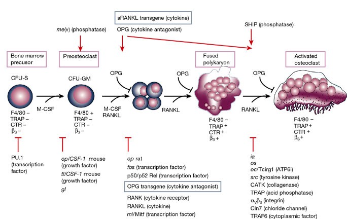
The process of bone remodeling is the result of the regulated balance between bone cell populations, namely bone-forming osteoblasts, bone-resorbing osteoclasts, and the osteocyte, the mechanosensory cell type. Osteoclasts derived from the hematopoietic stem cell lineage are the principal cells involved in bone resorption. In osteolytic diseases such as rheumatoid arthritis, periodontitis, and osteoporosis, the balance is lost and changes in favor of bone resorption. Therefore, it is vital to elucidate the mechanisms of osteoclast formation and bone resorption. It has been reported that osteocytes express Receptor activator of nuclear factor κΒ ligand (RANKL), an essential factor for osteoclast formation. RANKL secreted by osteocytes is the most important factor for physiologically supported osteoclast formation in the developing skeleton and in pathological bone resorption such as experimental periodontal bone loss. TNF-α directly enhances RANKL expression in osteocytes and promotes osteoclast formation. Moreover, TNF-α enhances sclerostin expression in osteocytes, which also increases osteoclast formation. These findings suggest that osteocyte-related cytokines act directly to enhance osteoclast formation and bone resorption. In this review, we outline the most recent knowledge concerning bone resorption-related cytokines and discuss the osteocyte as the master regulator of bone resorption and effector in osteoclast formation.