


Matatall et al. show that chronic infection causes pancytopenia and hematopoietic stem cell (HSC) depletion in mice. HSCs are lost through impaired self-renewal and increased terminal differentiation, which can be triggered by induction of the interferon gamma-responsive transcription factor BATF2. This study elucidates mechanisms underlying bone marrow failure during chronic infections.
A T Cell View of the Bone Marrow
The majority of T cells present in the bone marrow (BM) represent an activated/memory phenotype and most of these, if not all, are circulating T cells. Their lodging in the bone marrow keeps them activated, turning the bone marrow microenvironment into a “memory reservoir”. This article will focus on how T cell activation in the marrow results in both direct and indirect effect on the hematopoiesis. The hematopoietic stem cell niche will be presented, with its main components and organization, along with the role-played by T-lymphocytes in basal and pathologic conditions and their effect on the bone remodeling process. Also discussed herein will be how “normal” bone mass peak is achieved only in the presence of an intact adaptive immune system, with T and B cells playing critical roles in this process. Our main hypothesis is that the partnership between T cells and cells of the bone marrow microenvironment orchestrates numerous processes regulating immunity, hematopoiesis and bone remodeling.

Osteoimmunology: shared mechanisms and crosstalk between the immune and bone systems
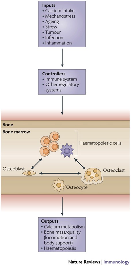
Osteoimmunology is an interdisciplinary field, that covers the shared mechanisms and interactions between bone cells and immune cellsReceptor activator of nuclear factor-κB (NF-κB) ligand (RANKL) is an osteoclast-differentiation factor that links the activated immune system and bone loss. In addition, abnormal bone homeostasis has been observed in various mice deficient in immunomodulatory molecules.Osteoclast differentiation is dependent on the transcription factor nuclear factor of activated T cells, cytoplasmic 1 (NFATc1), which is induced and activated by RANKL and its co-stimulatory (immunoglobulin-like) receptors.Interleukin-17 (IL-17)-producing T helper cells (TH17 cells) are the key T-cell subset that links T-cell activation and bone destruction in autoimmune arthritis.Bone cells are involved in the maintenance and mobilization of haematopoietic stem cells.Osteoimmunology is becoming increasingly important for understanding the pathogenesis of, and developing new therapeutic strategies for, diseases that affect both systems.
Why Does Inflammation Seem to Underlie All Sickness?
It’s a culprit in diseases ranging from arthritis to depression

Updates on Osteoimmunology: What's New on the Cross-Talk Between Bone and Immune System
The term osteoimmunology was coined many years ago to describe the research field that deals with the cross-regulation between bone cells and the immune system. As a matter of fact, many factors that are classically considered immune-related, such as interleukins (i.e. IL-6, -11,-17 and -23), tumor necrosis factor (TNF)-α, receptor-activator of nuclear factor kappa B (RANK) and its ligand (RANKL), nuclear factor of activated t-cell, cytoplasmatic-1 (NFATc1) and others have all been found to be crucial in osteoclast and osteoblast biology. Conversely, bone cells, which we used to think would only regulate each other and take care of remodeling bone, actually regulate immune cells, by creating the so-called “endosteal niche”. Both osteoblasts and osteoclasts participate to this niche, either by favoring engraftment, or mobilization of hematopoietic stem cells (HSCs). In this review, we will describe the main milestones at the base of the osteoimmunology and present the key cellular players of the bone-immune system cross-talk, including HSCs, osteoblasts, osteoclasts, bone marrow macrophages, osteomacs, T- and B-lymphocytes, dendritic cells and neutrophils. We will also briefly describe some pathological conditions in which the bone-immune system cross-talk plays a crucial role, with the final aim to portray the state of the art in the mechanisms regulating the bone-immune system interplay, and some of the latest molecular players in the field. This is important to encourage investigation in this field, to identify new targets in the treatment of bone and immune diseases.

A unified network systems approach uncovers a core program underlying T follicular helper cell differentiation
Characterizing multi-scale processes underlying immune-state progression is critical for defining their function. This becomes pertinent for functionally diverse and plastic immune cells, such as T follicular helper (Tfh) cells. Here, we adopt a multi-scale network-systems approach that incorporates both regulatory and protein-protein interactions. This approach integrates diverse data types, captures regulation across levels of immune system organization, and recapitulates known Tfh differentiation drivers. Further, we present CoreNet, a core Tfh gene set that is conserved between humans and mice, across tissue types and disease contexts, and is consistent across data modalities. Using CoreNet, we implicate NR3C1 and interleukin (IL)-12 in the regulation of Tfh differentiation. Notably, IL-12 is permissive for differentiation of Tfh precursors but blocks differentiation into germinal center Tfh cells. Overall, this work elucidates networks with unexplored roles governing Tfh differentiation across species and tissues, while providing a generalizable framework. CoreNet is accessible through an interactive web server: https://pitt-csi.shinyapps.io/tfhcorenet/.
OSTEOIMMUNOLOGY: Interplay Between the Immune System and Bone Metabolism
Studies of bone and the immune system have converged in recent years under the banner of osteoimmunology. The immune system is spawned in the bone marrow reservoir, and investigators now recognize that important niches also exist there for memory lymphocytes. At the same time, various factors produced during immune responses are capable of profoundly affecting regulation of bone. Mechanisms have evolved to prevent excessive interference by the immune system with bone homeostasis, yet pathologic bone loss is a common sequela associated with autoimmunity and cancer. There are also developmental links, or parallels, between bone and the immune system. Cells that regulate bone turnover share a common precursor with inflammatory immune cells and may restrict themselves anatomically, in part by utilizing a signaling network analogous to lymphocyte costimulation. Efforts are currently under way to further characterize how these two organ systems overlap and to develop therapeutic strategies that benefit from this understanding.

Osteoimmunology: The Nexus between bone and immune system
Osteoimmunology is an interdisciplinary research field which combines the existing fields of osteology (bone biology) and immunology under one umbrella. The observation that contributed enormously to the emergence of osteoimmunology as an independent field of investigation was the enhanced bone loss in various inflammatory bone diseases such as rheumatoid arthritis, osteoporosis and periodontitis. T helper cells (Th1, Th2, Treg and Th17) along with various other immune cells (B cells, DC, macrophages etc.) are actively involved in bone homeostasis. The present review thus provides an overview of the nexus between these two prominent systems (Bone and Immune system) of an organism, which reside in a common niche (bone marrow) and thus cross-communicate to modulate their respective development. Investigations in the field of osteoimmunology thus promise the advent of new era in the field with novel therapeutics for bone loss in various inflammatory conditions. A molecular insight into the field of osteoimmunology can lead to novel approaches for the prevention and treatment of diverse inflammatory conditions such as osteoporosis, rheumatoid arthritis and osteoarthritis.
<p>The Rising Era of “Immunoporosis”: Role of Immune System in the Pathophysiology of Osteoporosis</p>
We exhaustively revisit the characteristics, mechanism of action, and function of both innate and adaptive immune cells .

Immunoporosis: Role of Innate Immune Cells in Osteoporosis
Osteoporosis or porous bone disorder is the result of an imbalance in an otherwise highly balanced physiological process known as ‘bone remodeling’. The immune system is intricately involved in bone physiology as well as pathologies. Inflammatory diseases are often correlated with osteoporosis. Inflammatory mediators such as reactive oxygen species (ROS), and pro-inflammatory cytokines and chemokines directly or indirectly act on the bone cells and play a role in the pathogenesis of osteoporosis. Recently, Srivastava et al. (Srivastava RK, Dar HY, Mishra PK. Immunoporosis: Immunology of Osteoporosis-Role of T Cells. Frontiers in immunology. 2018;9:657) have coined the term “immunoporosis” to emphasize the role of immune cells in the pathology of osteoporosis. Accumulated pieces of evidence suggest both innate and adaptive immune cells contribute to osteoporosis. However, innate cells are the major effectors of inflammation. They sense various triggers to inflammation such as pathogen-associated molecular patterns (PAMPS), damage-associated molecular patterns (DAMPS), cellular stress, etc., thus producing pro-inflammatory mediators that play a critical role in the pathogenesis of osteoporosis. In this review, we have discussed the role of the innate immune cells in great detail and divided these cells into different sections in a systemic manner. In the beginning, we talked about cells of the myeloid lineage, including macrophages, monocytes, and dendritic cells. This group of cells explicitly influences the skeletal system by the action of production of pro-inflammatory cytokines and can transdifferentiate into osteoclast. Other cells of the myeloid lineage, such as neutrophils, eosinophils, and mast cells, largely impact osteoporosis via the production of pro-inflammatory cytokines. Further, we talked about the cells of the lymphoid lineage, including natural killer cells and innate lymphoid cells, which share innate-like properties and play a role in osteoporosis. In addition to various innate immune cells, we also discussed the impact of classical pro-inflammatory cytokines on osteoporosis. We also highlighted the studies regarding the impact of physiological and metabolic changes in the body, which results in chronic inflammatory conditions such as ageing, ultimately triggering osteoporosis.

Severe osteopetrosis, defective interleukin‐1 signalling and lymph node organogenesis in <i>TRAF6</i> ‐deficient mice
Background TRAF6, a member of the tumour necrosis factor receptor‐associated factor family, was first identified as a transducer of CD40 and interleukin‐1 receptor (IL‐1R) signals based on the interaction of TRAF6 with the cytoplasmic tail of CD40 and with the IL‐1R associated kinase in vitro . However, the functions of TRAF6 in vivo remain unidentified. Results We show that TRAF6 −/− mice exhibit severe osteopetrosis and are defective in osteoclast formation. In vitro culture experiments revealed that osteoclast precursor cells derived from TRAF6 −/− mice are unable to differentiate to functional osteoclasts in response to osteoclast differentiation factor (ODF). In bone marrow of TRAF6 −/− mice, the number of sIgM + B220 + immature B cells is markedly reduced while the ratio of proB to preB cells is not affected. In contrast, development of thymocytes is not affected. Furthermore, TRAF6 −/− mice are defective in lymph node organogenesis and IL‐1 signalling in thymocytes. Conclusions The results identify TRAF6 as an essential component of ODF signalling pathway, and also show that TRAF6 plays pivotal roles in immune and inflammatory systems in vivo .

Osteoimmunology: A Current Update of the Interplay Between Bone and the Immune System
Immunology, already a discipline in its own right, has become a major part of many different medical fields. However, its relationship to orthopedics and trauma surgery has unfortunately, and perhaps unjustly, been developing rather slowly. Discover-ies in recent years have emphasized the immense breadth of communication and connection between both systems and, importantly, the highly promising therapeu-tic opportunities.Recent discoveries of factors originally assigned to the immune system have now also been shown to have a significant impact on bone health and disease, which has greatly changed how we approach treatment of bone pathologies. In case of bone fracture, immune cells, especially macrophages, are present through-out the whole healing process, assure defense against pathogens and discharge a complex variety of effectors to regulate bone modelling. In rheumatoid arthritis and osteoporosis, the immune system contributes to the formation of the pathological and chronic conditions. Fascinatingly, prosthesis failure is not at all solely a me-chanical problem of improper strain but works in conjunction with an active contri-bution of the immune system as a reaction to irritant debris from material wear.Unravelling conjoined mechanisms of the immune and osseous systems heralds therapeutic possibilities for ailments of both. Contemplation of the bone as merely an unchanging support pillar is outdated and obsolete. Instead it is manda-tory that this highly diverse network be incorporated in our understanding of the immune system and hematopoiesis.

Osteopetrosis in mice lacking NF-κB1 and NF-κB2
The nfkbl and nfkb2 genes encode closely related products regulating immune and inflammatory responses1–3. Their role during development and differentiation remains unclear. The generation of nfkb1 null mice (p50−/−) resulted in altered immune responses, but had no effect on development4. Similarly, nfkb2 knockout mice (p52−/−) did not show developmental defects (J.C. et al., manuscript submitted). We have investigated the potential for in vivo compensatory functions of these genes by generating double-knockout mice. The surprising result was that the animals developed osteopetrosis because of a defect in osteoclast differentiation, suggesting redundant functions of NF-κB1 and NF-κB2 proteins in the development of this cell lineage. The osteopetrotic phenotype was rescued by bone marrow transplantation, indicating that the hematopoietic component was impaired. These results define a new mouse osteopetrotic mutant and implicate NF-κB proteins in bone development, raising new directions in the treatment of bone disorders.
Immunoporosis: Immunology of Osteoporosis—Role of T Cells
The role of immune system in various bone pathologies such as osteoporosis, osteoarthritis and rheumatoid arthritis is now well established. This had led to the emergence of a modern field of systems biology called as osteoimmunology, an integrated research between fields of immunology and bone biology under one umbrella. Osteoporosis is one of the most common inflammatory bone-loss conditions with more than 200 million individuals affected worldwide. T helper cells along with various other immune cells are major players involved in bone homeostasis. In the present review, we specifically discuss the role of various defined T lymphocyte subsets (Th cells comprising Th1, Th2, Th9, Th17, Th22, regulatory T cells, follicular helper T cells, natural killer T cells, γδ T cells and CD8+ T cells) in the pathophysiology of osteoporosis. The study of the specific role of immune system in osteoporosis has now been proposed by our group as “Immunoporosis: The immunology of osteoporosis” with special emphasis on the role of various subsets of T lymphocytes. The establishment of this new field had been the need of the hour due to the emergence of novel roles of various T cell lymphocytes in accelerated bone loss observed during osteoporosis. Activated T cells either directly or indirectly through the secretion of various cytokines and factors modulate bone health and thereby regulate bone remodelling. Various studies have summarized the role of inflammation in pathogenesis of osteoporosis, but very few reports had delineated the precise role of various T cell subsets in the pathobiology of osteoporosis. The present review thus for the first time clearly highlights and summarizes the role of various T lymphocytes in the development and pathophysiology of osteoporosis, giving birth to a new field of biology termed as “Immunoporosis”. This novel field will thus provide an overview of the nexus between the cellular components of both bone and immune systems, responsible for the observed bone loss in osteoporosis. A molecular insight into the upcoming and novel field of immunoporosis would thus lead to development of innovative approaches for the prevention and treatment of osteoporosis.

Bone versus immune system
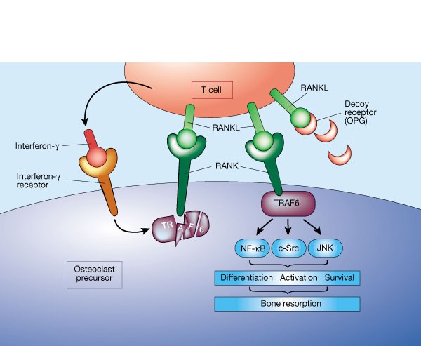
A molecule on activated T cells triggers bone loss by switching on bone-resorbing cells. Fortunately, it seems that this mechanism is kept in check by another molecule, secreted by the T cells.
Osteoporosis in Rheumatoid Arthritis: Dangerous Liaisons
Osteoporosis has been classically considered a comorbidity of rheumatoid arthritis (RA). However, recent advances in the pathogenesis of osteoporosis in RA have shown a close interplay between cells of the immune system and those involved in bone remodeling, introducing new actors into the classic route in which osteoclast activation is related to the RANK/RANKL/OPG pathway. In fact, the inflammatory state in early stages of RA, mediated by interleukin (IL)-1, IL-6, IL-8 and tumour necrosis factor (TNF)-α has the ability to activate and differentiate osteoclasts not only through their relationship with RANKL, but also through the Wnt/DKK1/sclerostin pathway, leading to bone loss. The role of synovial fibroblasts and activated T lymphocytes in the expression of the RANKL system and its connection to bone destruction is also depicted. In addition, autoantibodies such as rheumatoid factor and anti-citrullinated protein antibodies are other pathogenic mechanisms for the development of bone erosions and systemic osteoporosis in RA, even before the onset of arthritis. The aim of this review is to unravel the relationship between different factors involved in the development of osteoporosis in RA patients, both the classic factors and the most novel, based on the relationship of autoantibodies with bone remodeling. Furthermore, we propose that bone mineral density measured by different techniques may be helpful as a biomarker of severity in early arthritis patients.

Comparing phenotypic manifolds with Kompot: Detecting differential abundance and gene expression at single-cell resolution
Single-cell studies are frequently designed to compare across conditions such as health and disease. However, existing computational approaches typically rely on grouping cells into discrete populations before making comparisons, which can limit resolution for detecting state-dependent changes. Here, we introduce Kompot, a statistical framework for comparative analysis of multi-condition single-cell data. Kompot quantifies both differential abundance, capturing how cells redistribute across the phenotypic space, and differential expression, identifying condition-specific transcriptional changes that may be localized, heterogeneous, or oppositely regulated across states. By modeling cell density and gene expression as continuous functions over a shared cell-state representation, Kompot enables single-cell–resolution inference with principled uncertainty estimates, without requiring predefined clusters or cell types. Applying Kompot to aging murine bone marrow, we identified a continuum of shifts in hematopoietic stem cell and mature cell states, transcriptional remodeling of monocytes independent of compositional changes, and divergent regulation of oxidative stress response genes across cell types. We demonstrate the utility of Kompot in disease settings by identifying cell-state and gene expression changes associated with improved efficacy of combinatorial immunotherapy in melanoma. Additionally, Kompot enables multi-sample comparative analysis by accounting for sample-to-sample heterogeneity. By capturing both global and cell-state–specific effects of perturbation, the Kompot framework is broadly applicable to dissecting condition-specific effects in complex single-cell landscapes. ### Competing Interest Statement The authors have declared no competing interest. National Institutes of Health, R35GM147125, R01CA292932, T32GM136534, S10OD028685 The Mark Foundation for Cancer Research, https://ror.org/00v7th354, Endeavor Award Edward P. Evans Foundation, https://ror.org/03h22gm35, Discovery Research Grant Brotman Baty Institute, https://ror.org/03jxvbk42, Pilot Award
