


Molecular Physiology; Immunology; Cell Biology;
Role of T cells in ovariectomy induced bone loss—revisited
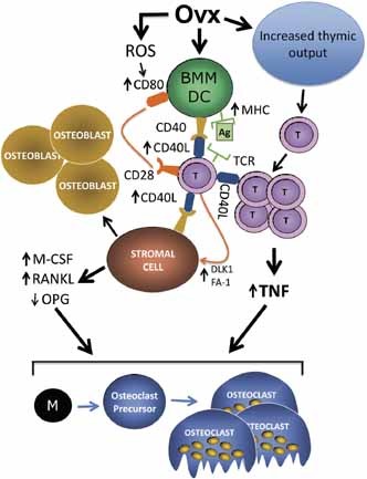
More than a decade has passed since the publication of the first report implicating T cells in the bone loss induced by ovariectomy (ovx).1 Since then ther
Osteoimmunology: shared mechanisms and crosstalk between the immune and bone systems
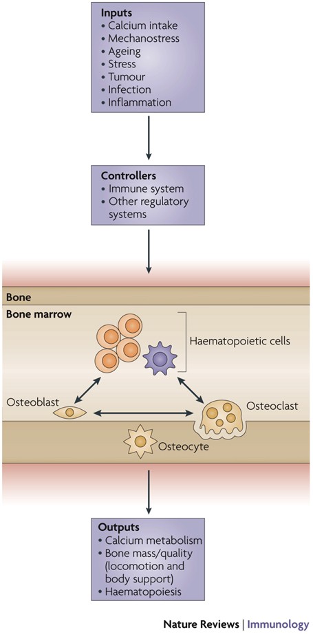
Osteoimmunology is an interdisciplinary field, that covers the shared mechanisms and interactions between bone cells and immune cellsReceptor activator of nuclear factor-κB (NF-κB) ligand (RANKL) is an osteoclast-differentiation factor that links the activated immune system and bone loss. In addition, abnormal bone homeostasis has been observed in various mice deficient in immunomodulatory molecules.Osteoclast differentiation is dependent on the transcription factor nuclear factor of activated T cells, cytoplasmic 1 (NFATc1), which is induced and activated by RANKL and its co-stimulatory (immunoglobulin-like) receptors.Interleukin-17 (IL-17)-producing T helper cells (TH17 cells) are the key T-cell subset that links T-cell activation and bone destruction in autoimmune arthritis.Bone cells are involved in the maintenance and mobilization of haematopoietic stem cells.Osteoimmunology is becoming increasingly important for understanding the pathogenesis of, and developing new therapeutic strategies for, diseases that affect both systems.
Bone versus immune system
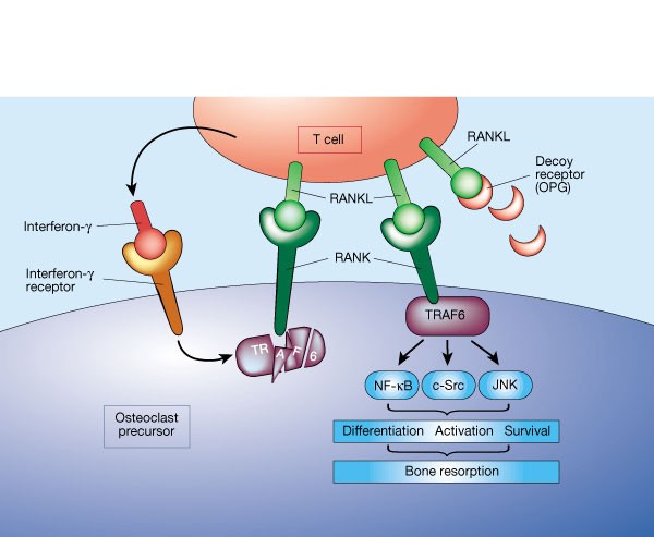
A molecule on activated T cells triggers bone loss by switching on bone-resorbing cells. Fortunately, it seems that this mechanism is kept in check by another molecule, secreted by the T cells.
Critical Role of Estrogens on Bone Homeostasis in Both Male and Female: From Physiology to Medical Implications
Bone is a multi-skilled tissue, protecting major organs, regulating calcium phosphate balance and producing hormones. Its development during childhood determines height and stature as well as resistance against fracture in advanced age. Estrogens are key regulators of bone turnover in both females and males. These hormones play a major role in longitudinal and width growth throughout puberty as well as in the regulation of bone turnover. In women, estrogen deficiency is one of the major causes of postmenopausal osteoporosis. In this review, we will summarize the main clinical and experimental studies reporting the effects of estrogens not only in females but also in males, during different life stages. Effects of estrogens on bone involve either Estrogen Receptor (ER)α or ERβ depending on the type of bone (femur, vertebrae, tibia, mandible), the compartment (trabecular or cortical), cell types involved (osteoclasts, osteoblasts and osteocytes) and sex. Finally, we will discuss new ongoing strategies to increase the benefit/risk ratio of the hormonal treatment of menopause.
Regulation of osteoblastogenesis and bone mass by Wnt10b
Wnts comprise a family of secreted signaling proteins that regulate diverse developmental processes. Activation of Wnt signaling by Wnt10b inhibits differentiation of preadipocytes and blocks adipose tissue development; however, the effect of Wnt10b on other mesenchymal lineages has not been defined. To explore the physiological role of Wnt signaling in bone development, we analyzed FABP4-Wnt10b mice, which express the Wnt10b transgene in marrow. Femurs from FABP4-Wnt10b mice have almost four times as much bone in the distal metaphyses and are mechanically stronger. These mice maintain elevated bone mass at least through 23 months of age. In addition, FABP4-Wnt10b mice are protected from the bone loss characteristic of estrogen deficiency. We used pharmacological and genetic approaches to demonstrate that canonical Wnt signaling stimulates osteoblastogenesis and inhibits adipogenesis of bipotential mesenchymal precursors. Wnt10b shifts cell fate toward the osteoblast lineage by induction of the osteoblastogenic transcription factors Runx2, Dlx5, and osterix and suppression of the adipogenic transcription factors C/EBPα and PPARγ. One mechanism whereby Wnt10b promotes osteoblastogenesis is suppression of PPARγ expression. Finally, Wnt10b-/- mice have decreased trabecular bone and serum osteocalcin, confirming that Wnt10b is an endogenous regulator of bone formation.

Estrogen and the skeleton
Estrogen is the major hormonal regulator of bone metabolism in women and men. Therefore, there is considerable interest in unraveling the pathways by which estrogen exerts its protective effects on bone. Although the major consequence of the loss of estrogen is an increase in bone resorption, estrogen deficiency is associated with a gap between bone resorption and formation, indicating that estrogen is also important for maintaining bone formation at the cellular level. Direct estrogen effects on osteocytes, osteoclasts, and osteoblasts lead to inhibition of bone remodeling, decreased bone resorption, and maintenance of bone formation, respectively.

Immunoporosis: Immunology of Osteoporosis—Role of T Cells
The role of immune system in various bone pathologies such as osteoporosis, osteoarthritis and rheumatoid arthritis is now well established. This had led to the emergence of a modern field of systems biology called as osteoimmunology, an integrated research between fields of immunology and bone biology under one umbrella. Osteoporosis is one of the most common inflammatory bone-loss conditions with more than 200 million individuals affected worldwide. T helper cells along with various other immune cells are major players involved in bone homeostasis. In the present review, we specifically discuss the role of various defined T lymphocyte subsets (Th cells comprising Th1, Th2, Th9, Th17, Th22, regulatory T cells, follicular helper T cells, natural killer T cells, γδ T cells and CD8+ T cells) in the pathophysiology of osteoporosis. The study of the specific role of immune system in osteoporosis has now been proposed by our group as “Immunoporosis: The immunology of osteoporosis” with special emphasis on the role of various subsets of T lymphocytes. The establishment of this new field had been the need of the hour due to the emergence of novel roles of various T cell lymphocytes in accelerated bone loss observed during osteoporosis. Activated T cells either directly or indirectly through the secretion of various cytokines and factors modulate bone health and thereby regulate bone remodelling. Various studies have summarized the role of inflammation in pathogenesis of osteoporosis, but very few reports had delineated the precise role of various T cell subsets in the pathobiology of osteoporosis. The present review thus for the first time clearly highlights and summarizes the role of various T lymphocytes in the development and pathophysiology of osteoporosis, giving birth to a new field of biology termed as “Immunoporosis”. This novel field will thus provide an overview of the nexus between the cellular components of both bone and immune systems, responsible for the observed bone loss in osteoporosis. A molecular insight into the upcoming and novel field of immunoporosis would thus lead to development of innovative approaches for the prevention and treatment of osteoporosis.

Estrogens and Androgens in Skeletal Physiology and Pathophysiology
Estrogens and androgens influence the growth and maintenance of the mammalian skeleton and are responsible for its sexual dimorphism. Estrogen deficiency at menopause or loss of both estrogens and androgens in elderly men contribute to the development of osteoporosis, one of the most common and impactful metabolic diseases of old age. In the last 20 years, basic and clinical research advances, genetic insights from humans and rodents, and newer imaging technologies have changed considerably the landscape of our understanding of bone biology as well as the relationship between sex steroids and the physiology and pathophysiology of bone metabolism. Together with the appreciation of the side effects of estrogen-related therapies on breast cancer and cardiovascular diseases, these advances have also drastically altered the treatment of osteoporosis. In this article, we provide a comprehensive review of the molecular and cellular mechanisms of action of estrogens and androgens on bone, their influences on skeletal homeostasis during growth and adulthood, the pathogenetic mechanisms of the adverse effects of their deficiency on the female and male skeleton, as well as the role of natural and synthetic estrogenic or androgenic compounds in the pharmacotherapy of osteoporosis. We highlight latest advances on the crosstalk between hormonal and mechanical signals, the relevance of the antioxidant properties of estrogens and androgens, the difference of their cellular targets in different bone envelopes, the role of estrogen deficiency in male osteoporosis, and the contribution of estrogen or androgen deficiency to the monomorphic effects of aging on skeletal involution.

The Microbial Metabolite Butyrate Stimulates Bone Formation via T Regulatory Cell-Mediated Regulation of WNT10B Expression
Tyagi et al. show that oral supplementation with the widely used probiotic, Lactobacillus rhamnosus GG (LGG), increases bone mass in mice by increasing the serum levels of the short chain fatty acid butyrate. LGG or butyrate increase the frequency of regulatory T (Treg) cells in the intestine and in the bone marrow. Treg cells stimulate CD8+ T cells to secrete the Wnt ligand Wnt10b, which stimulates bone formation by activating Wnt signaling in osteoblasts. Therefore, LGG and butyrate may represent new interventions for the prevention and treatment of osteoporosis.

Osteoimmunology: The Nexus between bone and immune system
Osteoimmunology is an interdisciplinary research field which combines the existing fields of osteology (bone biology) and immunology under one umbrella. The observation that contributed enormously to the emergence of osteoimmunology as an independent field of investigation was the enhanced bone loss in various inflammatory bone diseases such as rheumatoid arthritis, osteoporosis and periodontitis. T helper cells (Th1, Th2, Treg and Th17) along with various other immune cells (B cells, DC, macrophages etc.) are actively involved in bone homeostasis. The present review thus provides an overview of the nexus between these two prominent systems (Bone and Immune system) of an organism, which reside in a common niche (bone marrow) and thus cross-communicate to modulate their respective development. Investigations in the field of osteoimmunology thus promise the advent of new era in the field with novel therapeutics for bone loss in various inflammatory conditions. A molecular insight into the field of osteoimmunology can lead to novel approaches for the prevention and treatment of diverse inflammatory conditions such as osteoporosis, rheumatoid arthritis and osteoarthritis.
Physiological and pathophysiological bone turnover — role of the immune system
Receptor activator of NF-κB ligand (RANKL) and osteoprotegerin (OPG) are key downstream effectors of bone resorption; tumour necrosis factor (TNF) might synergize with RANKL to superinduce osteoclastic bone resorptionB cells, regulated by T cells, are a key source of basal OPG whereas activated T cells and B cells are key sources of TNF and RANKL in inflammatory conditionsShort-term antiresorptive therapies might safely prevent bone loss associated with combination antiretroviral therapyAnergic and parathyroid-hormone-treated T cells secrete the protein Wnt-10b, which promotes bone formationNovel therapeutic strategies targeting the immune system might promote bone formation and decrease bone resorption to manage osteoporotic bone loss and prevent fracture
Wnt Pathway in Bone Repair and Regeneration – What Do We Know So Far
Wnt signaling plays a central regulatory role across a remarkably diverse range of functions during embryonic development, including those involved in the formation of bone and cartilage. Wnt signaling continues to play a critical role in adult osteogenic differentiation of mesenchymal stem cells. Disruptions in this highly-conserved and complex system leads to various pathological conditions, including impaired bone healing, autoimmune diseases and malignant degeneration. For reconstructive surgeons, critically sized skeletal defects represent a major challenge. These are frequently associated with significant morbidity in both the recipient and donor sites. The Wnt pathway is an attractive therapeutic target with the potential to directly modulate stem cells responsible for skeletal tissue regeneration and promote bone growth, suggesting that Wnt factors could be used to stimulate bone healing after trauma. This review summarizes our current understanding of the essential role of the Wnt pathway in bone regeneration and repair.

Wnt10b Increases Postnatal Bone Formation by Enhancing Osteoblast Differentiation
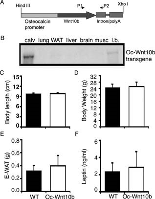
Abstract Overexpression of Wnt10b from the osteocalcin promoter in transgenic mice increases postnatal bone mass. Increases in osteoblast perimeter, mineralizing surface, and bone formation rate without detectable changes in pre-osteoblast proliferation, osteoblast apoptosis, or osteoclast number and activity suggest that, in this animal model, Wnt10b primarily increases bone mass by stimulating osteoblastogenesis. Introduction: Wnt signaling regulates many aspects of development including postnatal accrual of bone. Potential mechanisms for how Wnt signaling increases bone mass include regulation of osteoblast and/or osteoclast number and activity. To help differentiate between these possibilities, we studied mice in which Wnt10b is expressed specifically in osteoblast lineage cells or in mice devoid of Wnt10b. Materials and Methods: Transgenic mice, in which mouse Wnt10b is expressed from the human osteocalcin promoter (Oc-Wnt10b), were generated in C57BL/6 mice. Transgene expression was evaluated by RNase protection assay. Quantitative assessment of bone variables was done by radiography, μCT, and static and dynamic histomorphometry. Mechanisms of bone homeostasis were evaluated with assays for BrdU, TUNEL, and TRACP5b activity, as well as serum levels of C-terminal telopeptide of type I collagen (CTX). The endogenous role of Wnt10b in bone was assessed by dynamic histomorphometry in Wnt10b−/− mice. Results: Oc-Wnt10b mice have increased mandibular bone and impaired eruption of incisors during postnatal development. Analyses of femoral distal metaphyses show significantly higher BMD, bone volume fraction, and trabecular number. Increased bone formation is caused by increases in number of osteoblasts per bone surface, rate of mineral apposition, and percent mineralizing surface. Although number of osteoclasts per bone surface is not altered, Oc-Wnt10b mice have increased total osteoclast activity because of higher bone mass. In Wnt10b−/− mice, changes in mineralizing variables and osteoblast perimeter in femoral distal metaphyses were not observed; however, bone formation rate is reduced because of decreased total bone volume and trabecular number. Conclusions: High bone mass in Oc-Wnt10b mice is primarily caused by increased osteoblastogenesis, with a minor contribution from elevated mineralizing activity of osteoblasts.
Osteoimmunology: The Conceptual Framework Unifying the Immune and Skeletal Systems
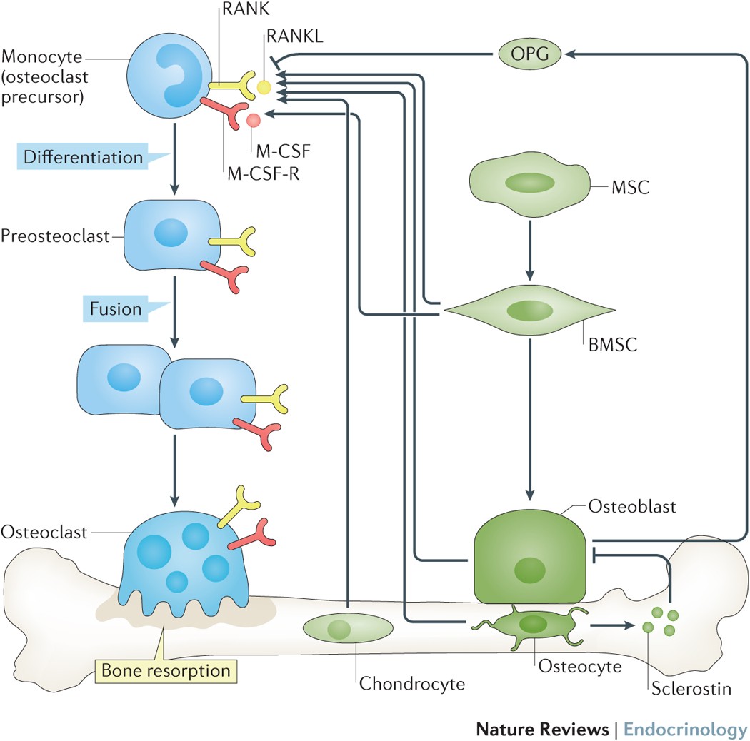
The immune and skeletal systems share a variety of molecules, including cytokines, chemokines, hormones, receptors, and transcription factors. Bone cells interact with immune cells under physiological and pathological conditions. Osteoimmunology was created as a new interdisciplinary field in large part to highlight the shared molecules and reciprocal interactions between the two systems in both heath and disease. Receptor activator of NF-κB ligand (RANKL) plays an essential role not only in the development of immune organs and bones, but also in autoimmune diseases affecting bone, thus effectively comprising the molecule that links the two systems. Here we review the function, gene regulation, and signal transduction of osteoimmune molecules, including RANKL, in the context of osteoclastogenesis as well as multiple other regulatory functions. Osteoimmunology has become indispensable for understanding the pathogenesis of a number of diseases such as rheumatoid arthritis (RA). We review the various osteoimmune pathologies, including the bone destruction in RA, in which pathogenic helper T cell subsets [such as IL-17-expressing helper T (Th17) cells] induce bone erosion through aberrant RANKL expression. We also focus on cellular interactions and the identification of the communication factors in the bone marrow, discussing the contribution of bone cells to the maintenance and regulation of hematopoietic stem and progenitors cells. Thus the time has come for a basic reappraisal of the framework for understanding both the immune and bone systems. The concept of a unified osteoimmune system will be absolutely indispensable for basic and translational approaches to diseases related to bone and/or the immune system.

Bone Remodeling in Post-menopausal Osteoporosis
Bone mass in the skeleton is dependent on the coordinated activities of bone-forming osteoblasts and bone-resorbing osteoclasts in discrete bone multi-cellular units. Remodeling of bone in these units is important not only for maintaining bone mass, but also to repair microdamage, to prevent accumulation of too much old bone, and for mineral homeostasis. The activities of osteoblasts and osteoclasts are controlled by a variety of hormones and cytokines, as well as by mechanical loading. Most importantly, sex hormones are very crucial for keeping bone mass in balance, and the lack of either estrogen or testosterone leads to decreased bone mass and increased risk for osteoporosis. The prevalence of osteoporotic fractures is increasing dramatically in the Western part of the world and is a major health problem in many countries. In the present review, the cellular and molecular mechanisms controlling bone remodeling and the influence of sex hormones on these processes are summarized. In a separate paper in this issue, the pathogenesis of post-menopausal osteoporosis will be compared with that of inflammation-induced bone remodeling, including the evidence for and against the hypothesis that concomitant post-menopausal osteoporotic disease influences the progression of periodontal disease.

The Regulation of Bone Metabolism and Disorders by Wnt Signaling
Wnt, a secreted glycoprotein, has an approximate molecular weight of 40 kDa, and it is a cytokine involved in various biological phenomena including ontogeny, morphogenesis, carcinogenesis, and maintenance of stem cells. The Wnt signaling pathway can be classified into two main pathways: canonical and non-canonical. Of these, the canonical Wnt signaling pathway promotes osteogenesis. Sclerostin produced by osteocytes is an inhibitor of this pathway, thereby inhibiting osteogenesis. Recently, osteoporosis treatment using an anti-sclerostin therapy has been introduced. In this review, the basics of Wnt signaling, its role in bone metabolism and its involvement in skeletal disorders have been covered. Furthermore, the clinical significance and future scopes of Wnt signaling in osteoporosis, osteoarthritis, rheumatoid arthritis and neoplasia are discussed.