


Butyrate, a short-chain fatty acid produced by the gut microbiota, has pivotal roles in the regulation of the immune system. Recent studies have revealed that butyrate increases the differentiation of peripheral regulatory T cells in the gut–bone axis and promotes osteoblasts’ bone forming activity. However, the mechanism of the therapeutic benefit of butyrate in bone remodeling remains incompletely understood. Here, we develop a multicompartment mathematical model to quantitatively predict the contribution of butyrate on the expansion of regulatory T cells in the gut, blood, and bone compartments. We investigate the interplay between regulatory T cell-derived TGF-β and CD8+ T cell-derived Wnt-10b with changes in gut butyrate concentration. In addition, we connect our model to a detailed model of bone metabolism to study the impacts of butyrate and Wnt-10b on trabecular bone volume. Our results indicate both direct and indirect immune-mediated impacts of butyrate on bone metabolism.
The Microbial Metabolite Butyrate Stimulates Bone Formation via T Regulatory Cell-Mediated Regulation of WNT10B Expression
Tyagi et al. show that oral supplementation with the widely used probiotic, Lactobacillus rhamnosus GG (LGG), increases bone mass in mice by increasing the serum levels of the short chain fatty acid butyrate. LGG or butyrate increase the frequency of regulatory T (Treg) cells in the intestine and in the bone marrow. Treg cells stimulate CD8+ T cells to secrete the Wnt ligand Wnt10b, which stimulates bone formation by activating Wnt signaling in osteoblasts. Therefore, LGG and butyrate may represent new interventions for the prevention and treatment of osteoporosis.

The gut microbiota regulates bone mass in mice
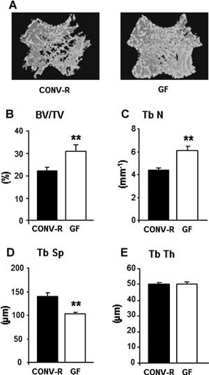
Abstract The gut microbiota modulates host metabolism and development of immune status. Here we show that the gut microbiota is also a major regulator of bone mass in mice. Germ-free (GF) mice exhibit increased bone mass associated with reduced number of osteoclasts per bone surface compared with conventionally raised (CONV-R) mice. Colonization of GF mice with a normal gut microbiota normalizes bone mass. Furthermore, GF mice have decreased frequency of CD4+ T cells and CD11b+/GR 1 osteoclast precursor cells in bone marrow, which could be normalized by colonization. GF mice exhibited reduced expression of inflammatory cytokines in bone and bone marrow compared with CONV-R mice. In summary, the gut microbiota regulates bone mass in mice, and we provide evidence for a mechanism involving altered immune status in bone and thereby affected osteoclast-mediated bone resorption. Further studies are required to evaluate the gut microbiota as a novel therapeutic target for osteoporosis. © 2012 American Society for Bone and Mineral Research.
Immunoporosis: Immunology of Osteoporosis—Role of T Cells
The role of immune system in various bone pathologies such as osteoporosis, osteoarthritis and rheumatoid arthritis is now well established. This had led to the emergence of a modern field of systems biology called as osteoimmunology, an integrated research between fields of immunology and bone biology under one umbrella. Osteoporosis is one of the most common inflammatory bone-loss conditions with more than 200 million individuals affected worldwide. T helper cells along with various other immune cells are major players involved in bone homeostasis. In the present review, we specifically discuss the role of various defined T lymphocyte subsets (Th cells comprising Th1, Th2, Th9, Th17, Th22, regulatory T cells, follicular helper T cells, natural killer T cells, γδ T cells and CD8+ T cells) in the pathophysiology of osteoporosis. The study of the specific role of immune system in osteoporosis has now been proposed by our group as “Immunoporosis: The immunology of osteoporosis” with special emphasis on the role of various subsets of T lymphocytes. The establishment of this new field had been the need of the hour due to the emergence of novel roles of various T cell lymphocytes in accelerated bone loss observed during osteoporosis. Activated T cells either directly or indirectly through the secretion of various cytokines and factors modulate bone health and thereby regulate bone remodelling. Various studies have summarized the role of inflammation in pathogenesis of osteoporosis, but very few reports had delineated the precise role of various T cell subsets in the pathobiology of osteoporosis. The present review thus for the first time clearly highlights and summarizes the role of various T lymphocytes in the development and pathophysiology of osteoporosis, giving birth to a new field of biology termed as “Immunoporosis”. This novel field will thus provide an overview of the nexus between the cellular components of both bone and immune systems, responsible for the observed bone loss in osteoporosis. A molecular insight into the upcoming and novel field of immunoporosis would thus lead to development of innovative approaches for the prevention and treatment of osteoporosis.

Osteoimmunology: The Conceptual Framework Unifying the Immune and Skeletal Systems
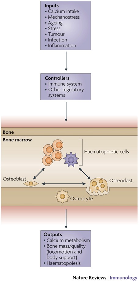
The immune and skeletal systems share a variety of molecules, including cytokines, chemokines, hormones, receptors, and transcription factors. Bone cells interact with immune cells under physiological and pathological conditions. Osteoimmunology was created as a new interdisciplinary field in large part to highlight the shared molecules and reciprocal interactions between the two systems in both heath and disease. Receptor activator of NF-κB ligand (RANKL) plays an essential role not only in the development of immune organs and bones, but also in autoimmune diseases affecting bone, thus effectively comprising the molecule that links the two systems. Here we review the function, gene regulation, and signal transduction of osteoimmune molecules, including RANKL, in the context of osteoclastogenesis as well as multiple other regulatory functions. Osteoimmunology has become indispensable for understanding the pathogenesis of a number of diseases such as rheumatoid arthritis (RA). We review the various osteoimmune pathologies, including the bone destruction in RA, in which pathogenic helper T cell subsets [such as IL-17-expressing helper T (Th17) cells] induce bone erosion through aberrant RANKL expression. We also focus on cellular interactions and the identification of the communication factors in the bone marrow, discussing the contribution of bone cells to the maintenance and regulation of hematopoietic stem and progenitors cells. Thus the time has come for a basic reappraisal of the framework for understanding both the immune and bone systems. The concept of a unified osteoimmune system will be absolutely indispensable for basic and translational approaches to diseases related to bone and/or the immune system.

Osteoimmunology: The Nexus between bone and immune system
Osteoimmunology is an interdisciplinary research field which combines the existing fields of osteology (bone biology) and immunology under one umbrella. The observation that contributed enormously to the emergence of osteoimmunology as an independent field of investigation was the enhanced bone loss in various inflammatory bone diseases such as rheumatoid arthritis, osteoporosis and periodontitis. T helper cells (Th1, Th2, Treg and Th17) along with various other immune cells (B cells, DC, macrophages etc.) are actively involved in bone homeostasis. The present review thus provides an overview of the nexus between these two prominent systems (Bone and Immune system) of an organism, which reside in a common niche (bone marrow) and thus cross-communicate to modulate their respective development. Investigations in the field of osteoimmunology thus promise the advent of new era in the field with novel therapeutics for bone loss in various inflammatory conditions. A molecular insight into the field of osteoimmunology can lead to novel approaches for the prevention and treatment of diverse inflammatory conditions such as osteoporosis, rheumatoid arthritis and osteoarthritis.
Osteoimmunology: shared mechanisms and crosstalk between the immune and bone systems
Osteoimmunology is an interdisciplinary field, that covers the shared mechanisms and interactions between bone cells and immune cellsReceptor activator of nuclear factor-κB (NF-κB) ligand (RANKL) is an osteoclast-differentiation factor that links the activated immune system and bone loss. In addition, abnormal bone homeostasis has been observed in various mice deficient in immunomodulatory molecules.Osteoclast differentiation is dependent on the transcription factor nuclear factor of activated T cells, cytoplasmic 1 (NFATc1), which is induced and activated by RANKL and its co-stimulatory (immunoglobulin-like) receptors.Interleukin-17 (IL-17)-producing T helper cells (TH17 cells) are the key T-cell subset that links T-cell activation and bone destruction in autoimmune arthritis.Bone cells are involved in the maintenance and mobilization of haematopoietic stem cells.Osteoimmunology is becoming increasingly important for understanding the pathogenesis of, and developing new therapeutic strategies for, diseases that affect both systems.
A review of mathematical modeling of bone remodeling from a systems biology perspective
Bone remodeling is an essential physiological process in the adult skeleton. Due to the complex nature of this process, many mathematical models of bone remodeling have been developed. Each of these models has unique features, but they have underlying patterns. In this review, the authors highlight the important aspects frequently found in mathematical models for bone remodeling and discuss how and why these aspects are included when considering the physiology of the bone basic multicellular unit, which is the term used for the collection of cells responsible for bone remodeling. The review also emphasizes the view of bone remodeling from a systems biology perspective. Understanding the systemic mechanisms involved in remodeling will help provide information on bone pathology associated with aging, endocrine disorders, cancers, and inflammatory conditions and enhance systems pharmacology. Furthermore, some features of the bone remodeling cycle and interactions with other organ systems that have not yet been modeled mathematically are discussed as promising future directions in the field.
OSTEOIMMUNOLOGY: Interplay Between the Immune System and Bone Metabolism
Studies of bone and the immune system have converged in recent years under the banner of osteoimmunology. The immune system is spawned in the bone marrow reservoir, and investigators now recognize that important niches also exist there for memory lymphocytes. At the same time, various factors produced during immune responses are capable of profoundly affecting regulation of bone. Mechanisms have evolved to prevent excessive interference by the immune system with bone homeostasis, yet pathologic bone loss is a common sequela associated with autoimmunity and cancer. There are also developmental links, or parallels, between bone and the immune system. Cells that regulate bone turnover share a common precursor with inflammatory immune cells and may restrict themselves anatomically, in part by utilizing a signaling network analogous to lymphocyte costimulation. Efforts are currently under way to further characterize how these two organ systems overlap and to develop therapeutic strategies that benefit from this understanding.

A review of mathematical modeling of bone remodeling from a systems biology perspective
Bone remodeling is an essential, delicately balanced physiological process of coordinated activity of bone cells that remove and deposit new bone tissue in the adult skeleton. Due to the complex nature of this process, many mathematical models of bone remodeling have been developed. Each of these models has unique features, but they have underlying patterns. In this review, the authors highlight the important aspects frequently found in mathematical models for bone remodeling and discuss how and why these aspects are included when considering the physiology of the bone basic multicellular unit, which is the term used for the collection of cells responsible for bone remodeling. The review also emphasizes the view of bone remodeling from a systems biology perspective. Understanding the systemic mechanisms involved in remodeling will help provide information on bone pathology associated with aging, endocrine disorders, cancers, and inflammatory conditions and enhance systems pharmacology. Furthermore, some features of the bone remodeling cycle and interactions with other organ systems that have not yet been modeled mathematically are discussed as promising future directions in the field.

Transforming Growth Factor-β1 to the Bone
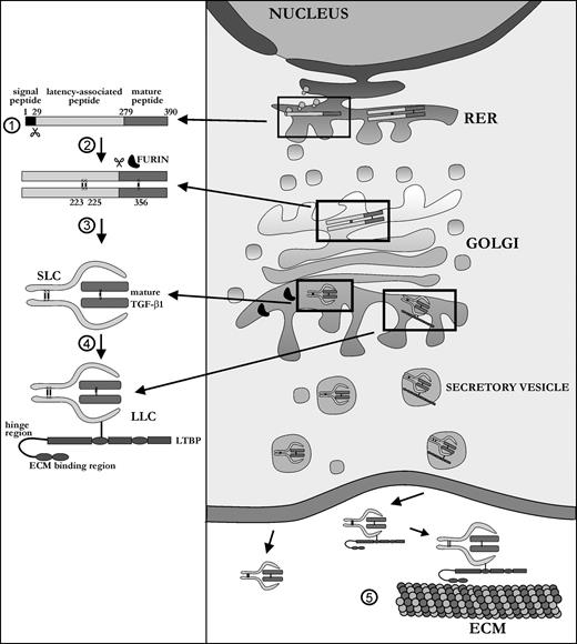
TGF-β1 is a ubiquitous growth factor that is implicated in the control of proliferation, migration, differentiation, and survival of many different cell types. It influences such diverse processes as embryogenesis, angiogenesis, inflammation, and wound healing. In skeletal tissue, TGF-β1 plays a major role in development and maintenance, affecting both cartilage and bone metabolism, the latter being the subject of this review. Because it affects both cells of the osteoblast and osteoclast lineage, TGF-β1 is one of the most important factors in the bone environment, helping to retain the balance between the dynamic processes of bone resorption and bone formation. Many seemingly contradictory reports have been published on the exact functioning of TGF-β1 in the bone milieu. This review provides an overall picture of the bone-specific actions of TGF-β1 and reconciles experimental discrepancies that have been reported for this multifunctional cytokine.
Updates on Osteoimmunology: What's New on the Cross-Talk Between Bone and Immune System
The term osteoimmunology was coined many years ago to describe the research field that deals with the cross-regulation between bone cells and the immune system. As a matter of fact, many factors that are classically considered immune-related, such as interleukins (i.e. IL-6, -11,-17 and -23), tumor necrosis factor (TNF)-α, receptor-activator of nuclear factor kappa B (RANK) and its ligand (RANKL), nuclear factor of activated t-cell, cytoplasmatic-1 (NFATc1) and others have all been found to be crucial in osteoclast and osteoblast biology. Conversely, bone cells, which we used to think would only regulate each other and take care of remodeling bone, actually regulate immune cells, by creating the so-called “endosteal niche”. Both osteoblasts and osteoclasts participate to this niche, either by favoring engraftment, or mobilization of hematopoietic stem cells (HSCs). In this review, we will describe the main milestones at the base of the osteoimmunology and present the key cellular players of the bone-immune system cross-talk, including HSCs, osteoblasts, osteoclasts, bone marrow macrophages, osteomacs, T- and B-lymphocytes, dendritic cells and neutrophils. We will also briefly describe some pathological conditions in which the bone-immune system cross-talk plays a crucial role, with the final aim to portray the state of the art in the mechanisms regulating the bone-immune system interplay, and some of the latest molecular players in the field. This is important to encourage investigation in this field, to identify new targets in the treatment of bone and immune diseases.

Physiological and pathophysiological bone turnover — role of the immune system
Receptor activator of NF-κB ligand (RANKL) and osteoprotegerin (OPG) are key downstream effectors of bone resorption; tumour necrosis factor (TNF) might synergize with RANKL to superinduce osteoclastic bone resorptionB cells, regulated by T cells, are a key source of basal OPG whereas activated T cells and B cells are key sources of TNF and RANKL in inflammatory conditionsShort-term antiresorptive therapies might safely prevent bone loss associated with combination antiretroviral therapyAnergic and parathyroid-hormone-treated T cells secrete the protein Wnt-10b, which promotes bone formationNovel therapeutic strategies targeting the immune system might promote bone formation and decrease bone resorption to manage osteoporotic bone loss and prevent fracture
Gut microbiota and bone metabolism
Abstract Osteoporosis is the most common metabolic skeletal disease. It is characterized by the deterioration of the skeletal microarchitecture and bone loss, leading to ostealgia, and even bone fractures. Accumulating evidence has indicated that there is an inextricable relationship between the gut microbiota (GM) and bone homeostasis involving host–microbiota crosstalk. Any perturbation of the GM can play an initiating and reinforcing role in disrupting the bone remodeling balance during the development of osteoporosis. Although the GM is known to influence bone metabolism, the mechanisms associated with these effects remain unclear. Herein, we review the current knowledge of how the GM affects bone metabolism in health and disease, summarize the correlation between pathogen‐associated molecular patterns of GM structural components and bone metabolism, and discuss the potential mechanisms underlying how GM metabolites regulate bone turnover. Deciphering the complicated relationship between the GM and bone health will provide new insights into the prevention and treatment of osteoporosis.

Loss of Bone and Wnt10b Expression in Male Type 1 Diabetic Mice Is Blocked by the Probiotic Lactobacillus reuteri
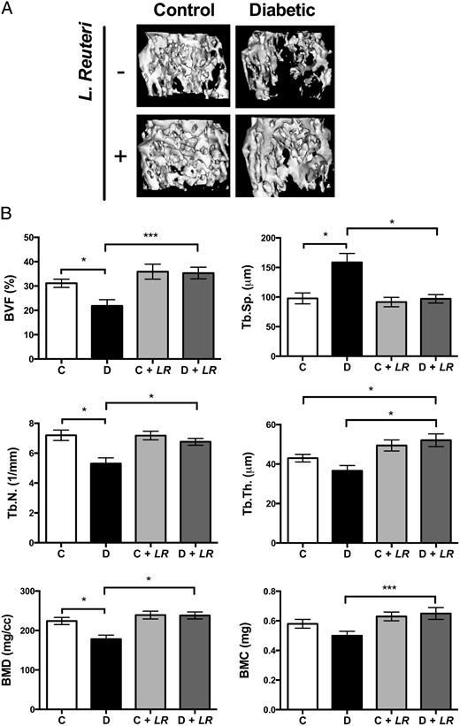
Type 1 diabetes (T1D)–induced osteoporosis is characterized by a predominant suppression of osteoblast number and activity, as well as increased bone marrow adiposity but no change in osteoclast activity. The fundamental mechanisms and alternative anabolic treatments (with few side effects) for T1D bone loss remain undetermined. Recent studies by our laboratory and others indicate that probiotics can benefit bone health. Here, we demonstrate that Lactobacillus reuteri, a probiotic with anti-inflammatory and bone health properties, prevents T1D-induced bone loss and marrow adiposity in mice. We further found that L. reuteri treatment prevented the suppression of Wnt10b in T1D bone. Consistent with a role for attenuated bone Wnt10b expression in T1D osteoporosis, we observed that bone-specific Wnt10b transgenic mice are protected from T1D bone loss. To examine the mechanisms of this protection, we focused on TNF-α, a cytokine up-regulated in T1D that causes suppression of osteoblast Wnt10b expression in vitro. Addition of L. reuteri prevented TNF-α–mediated suppression of Wnt10b and osteoblast maturation markers. Taken together, our findings reveal a mechanism by which T1D causes bone loss and open new avenues for use of probiotics to benefit the bone.
The Role of Osteocytes in Targeted Bone Remodeling: A Mathematical Model
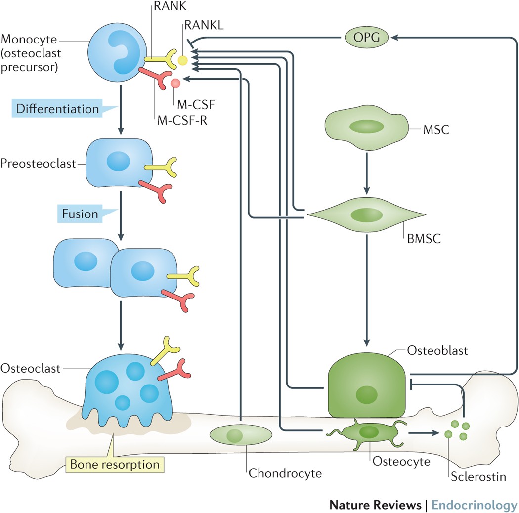
Until recently many studies of bone remodeling at the cellular level have focused on the behavior of mature osteoblasts and osteoclasts, and their respective precursor cells, with the role of osteocytes and bone lining cells left largely unexplored. This is particularly true with respect to the mathematical modeling of bone remodeling. However, there is increasing evidence that osteocytes play important roles in the cycle of targeted bone remodeling, in serving as a significant source of RANKL to support osteoclastogenesis, and in secreting the bone formation inhibitor sclerostin. Moreover, there is also increasing interest in sclerostin, an osteocyte-secreted bone formation inhibitor, and its role in regulating local response to changes in the bone microenvironment. Here we develop a cell population model of bone remodeling that includes the role of osteocytes, sclerostin, and allows for the possibility of RANKL expression by osteocyte cell populations. We have aimed to give a simple, yet still tractable, model that remains faithful to the underlying system based on the known literature. This model extends and complements many of the existing mathematical models for bone remodeling, but can be used to explore aspects of the process of bone remodeling that were previously beyond the scope of prior modeling work. Through numerical simulations we demonstrate that our model can be used to explore theoretically many of the qualitative features of the role of osteocytes in bone biology as presented in recent literature.
Molecular Signaling Pathways and Essential Metabolic Elements in Bone Remodeling: An Implication of Therapeutic Targets for Bone Diseases
Bone is one of the dynamic tissues in the human body that undergoes continuous remodelling through subsequent actions of bone cells, osteoclasts, and osteoblasts. Several signal transduction pathways are involved in the transition of mesenchymal stem cells into osteoblasts. These primarily include Runx2, ATF4, Wnt signaling and sympathetic signalling. The differentiation of osteoclasts is controlled by M-CSF, RANKL, and costimulatory signalling. It is well known that bone remodelling is regulated through receptor activator of nuclear factor-kappa B ligand followed by the binding to RANK, which eventually induces the differentiation of osteoclasts. The resorbing osteoclasts secrete TRAP, cathepsin K, MMP-9 and gelatinase to digest the proteinaceous matrix of type I collagen and form a saucer-shaped lacuna along with resorption tunnels in the trabecular bone. Osteoblasts secrete a soluble decoy receptor, osteoprotegerin that prevents the binding of RANK/RANKL and thus moderating osteoclastogenesis. Moreover, bone homeostasis is also regulated by several growth factors, cytokines, calciotropic hormones, parathyroid hormone and sex steroids. The current review presents a correlation of the probable molecular targets underlying the regulation of bone mass and the role of essential metabolic elements in bone remodelling. Targeting these signaling pathways may help design newer therapies for treating bone diseases.
