


In mice, provision of butyrate—a short-chain fatty acid produced by commensal microorganisms during starch fermentation—facilitates extrathymic generation and differentiation of Foxp3+ regulatory T cells, demonstrating that metabolic by-products are sensed by cells of the immune system and affect the balance between pro- and anti-inflammatory cells.
Commensal microbe-derived butyrate induces the differentiation of colonic regulatory T cells
The gut microbial metabolite butyrate is shown to induce the differentiation of colonic T regulatory cells in mice and to ameliorate the development of colitis; it also increases histone H3 acetylation at the Foxp3 promoter.

The Microbial Metabolites, Short-Chain Fatty Acids, Regulate Colonic T<sub>reg</sub> Cell Homeostasis
Protecting the Guts Regulatory T cells (T regs ) in the gut are important sentinels in maintaining the peace between our gut and its trillions of resident bacteria and have been shown to be regulated by specific strains of bacteria in mouse models. Smith et al. (p. 569 , published online 4 July; see the Perspective by Bollrath and Powrie ) asked whether metabolite(s) generated by resident bacterial species may regulate T regs in the gut. Indeed, short-chain fatty acids (SCFAs), bacterial fermentation products of dietary fibers produced by a range of bacteria, restored colonic T reg numbers in mice devoid of a gut microbiota and increased T reg numbers in colonized mice. The effects of SCFAs on T regs were mediated through GPCR43, a receptor for SCFAs, which is expressed on colonic T regs . Mice fed SCFAs were protected against experimentally induced colitis in a manner that was dependent on GPR43-expressing T regs . , Bacterial fermentation products regulate the number and function of regulatory T cells in the mouse colon. [Also see Perspective by Bollrath and Powrie ] , Regulatory T cells (T regs ) that express the transcription factor Foxp3 are critical for regulating intestinal inflammation. Candidate microbe approaches have identified bacterial species and strain-specific molecules that can affect intestinal immune responses, including species that modulate T reg responses. Because neither all humans nor mice harbor the same bacterial strains, we posited that more prevalent factors exist that regulate the number and function of colonic T regs . We determined that short-chain fatty acids, gut microbiota–derived bacterial fermentation products, regulate the size and function of the colonic T reg pool and protect against colitis in a Ffar2 -dependent manner in mice. Our study reveals that a class of abundant microbial metabolites underlies adaptive immune microbiota coadaptation and promotes colonic homeostasis and health.

Activation of Gpr109a, Receptor for Niacin and the Commensal Metabolite Butyrate, Suppresses Colonic Inflammation and Carcinogenesis
Commensal gut microflora and dietary fiber protect against colonic inflammation and colon cancer through unknown targets. Butyrate, a bacterial product from fermentation of dietary fiber in the colon, has been implicated in this process. GPR109A (encoded by Niacr1) is a receptor for butyrate in the colon. GPR109A is also a receptor for niacin, which is also produced by gut microbiota and suppresses intestinal inflammation. Here we showed that Gpr109a signaling promoted anti-inflammatory properties in colonic macrophages and dendritic cells and enabled them to induce differentiation of Treg cells and IL-10-producing T cells.

The contribution of gut bacterial metabolites in the human immune signaling pathway of non-communicable diseases
The interaction disorder between gut microbiota and its host has been documented in different non-communicable diseases (NCDs) such as metabolic syndrome, neurodegenerative disease, and autoimmune ...

The Microbial Metabolite Butyrate Stimulates Bone Formation via T Regulatory Cell-Mediated Regulation of WNT10B Expression
Tyagi et al. show that oral supplementation with the widely used probiotic, Lactobacillus rhamnosus GG (LGG), increases bone mass in mice by increasing the serum levels of the short chain fatty acid butyrate. LGG or butyrate increase the frequency of regulatory T (Treg) cells in the intestine and in the bone marrow. Treg cells stimulate CD8+ T cells to secrete the Wnt ligand Wnt10b, which stimulates bone formation by activating Wnt signaling in osteoblasts. Therefore, LGG and butyrate may represent new interventions for the prevention and treatment of osteoporosis.

Regulation of Intestinal Barrier Function by Microbial Metabolites
The human gastrointestinal tract (GI) harbors a diverse population of microbial life that continually shapes host pathophysiological responses. Despite readily available abundant metagenomic data, the functional dynamics of gut microbiota remain to be explored in various health and disease conditions. Microbiota generate a variety of metabolites from dietary products that influence host health and pathophysiological functions. Since gut microbial metabolites are produced in close proximity to gut epithelium, presumably they have significant impact on gut barrier function and immune responses.

Mathematical Modeling of the Gut–Bone Axis and Implications of Butyrate Treatment on Osteoimmunology
Butyrate, a short-chain fatty acid produced by the gut microbiota, has pivotal roles in the regulation of the immune system. Recent studies have revealed that butyrate increases the differentiation of peripheral regulatory T cells in the gut–bone axis and promotes osteoblasts’ bone forming activity. However, the mechanism of the therapeutic benefit of butyrate in bone remodeling remains incompletely understood. Here, we develop a multicompartment mathematical model to quantitatively predict the contribution of butyrate on the expansion of regulatory T cells in the gut, blood, and bone compartments. We investigate the interplay between regulatory T cell-derived TGF-β and CD8+ T cell-derived Wnt-10b with changes in gut butyrate concentration. In addition, we connect our model to a detailed model of bone metabolism to study the impacts of butyrate and Wnt-10b on trabecular bone volume. Our results indicate both direct and indirect immune-mediated impacts of butyrate on bone metabolism.

From the gut to the lung: microbiota-associated metabolites as regulators of respiratory immunometabolism
The development and progression of respiratory diseases are influenced by both the local pulmonary microenvironment and the intestinal ecosystem. Research on the gut-lung axis has shown that diverse small-molecule metabolites produced or modified by the gut microbiota can cross the intestinal barrier and enter the systemic circulation, where they may modulate immune-cell response thresholds, functional polarisation, and inflammatory dynamics in the distal lung. The local respiratory microbiota may also contribute to the pulmonary metabolic microenvironment, although its role in metabolite production and immune regulation remains less clearly defined.

Dietary Fiber and Bacterial SCFA Enhance Oral Tolerance and Protect against Food Allergy through Diverse Cellular Pathways
Tan et al. examine the beneficial roles of dietary fiber in peanut allergy using mice. The authors find that this effect involves reshaping of the gut microbiota as well as increased levels of short-chain fatty acids and activity of their receptors GPR43 and GPR109a. High-fiber feeding also increased tolerogenic CD103+ DCs activity, leading to increased Treg cell differentiation.

The gut microbiota regulates bone mass in mice
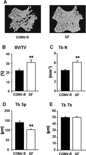
Abstract The gut microbiota modulates host metabolism and development of immune status. Here we show that the gut microbiota is also a major regulator of bone mass in mice. Germ-free (GF) mice exhibit increased bone mass associated with reduced number of osteoclasts per bone surface compared with conventionally raised (CONV-R) mice. Colonization of GF mice with a normal gut microbiota normalizes bone mass. Furthermore, GF mice have decreased frequency of CD4+ T cells and CD11b+/GR 1 osteoclast precursor cells in bone marrow, which could be normalized by colonization. GF mice exhibited reduced expression of inflammatory cytokines in bone and bone marrow compared with CONV-R mice. In summary, the gut microbiota regulates bone mass in mice, and we provide evidence for a mechanism involving altered immune status in bone and thereby affected osteoclast-mediated bone resorption. Further studies are required to evaluate the gut microbiota as a novel therapeutic target for osteoporosis. © 2012 American Society for Bone and Mineral Research.
Interactions between gut microbiota and skeletal muscle
The gut microbiota is now recognized as a major contributor to the host’s nutrition, metabolism, immunity, and neurological functions. Imbalanced microbiota (ie, dysbiosis) is linked to undernutrition-induced stunting, inflammatory and metabolic diseases, and cancers. Skeletal muscle also takes part in the interorgan crosstalk regulating substrate metabolism, immunity, and health. Here, we review the reciprocal influence of gut microbiota and skeletal muscle in relation to juvenile growth, performance, aging, and chronic diseases. Several routes involving the vascular system and organs such as the liver and adipose tissue connect the gut microbiota and skeletal muscle, with effects on fitness and health. Therapeutic perspectives arise from the health benefits observed with changes in gut microbiota and muscle activity, further encouraging multimodal therapeutic strategies.

Gut microbiota and bone metabolism
Abstract Osteoporosis is the most common metabolic skeletal disease. It is characterized by the deterioration of the skeletal microarchitecture and bone loss, leading to ostealgia, and even bone fractures. Accumulating evidence has indicated that there is an inextricable relationship between the gut microbiota (GM) and bone homeostasis involving host–microbiota crosstalk. Any perturbation of the GM can play an initiating and reinforcing role in disrupting the bone remodeling balance during the development of osteoporosis. Although the GM is known to influence bone metabolism, the mechanisms associated with these effects remain unclear. Herein, we review the current knowledge of how the GM affects bone metabolism in health and disease, summarize the correlation between pathogen‐associated molecular patterns of GM structural components and bone metabolism, and discuss the potential mechanisms underlying how GM metabolites regulate bone turnover. Deciphering the complicated relationship between the GM and bone health will provide new insights into the prevention and treatment of osteoporosis.

Loss of Bone and Wnt10b Expression in Male Type 1 Diabetic Mice Is Blocked by the Probiotic Lactobacillus reuteri
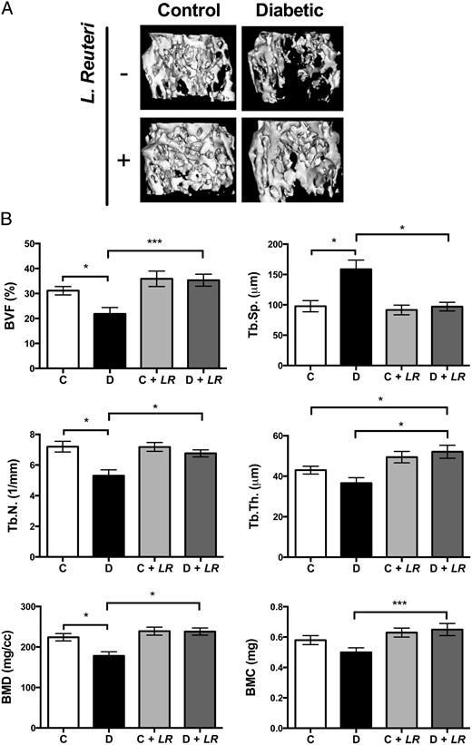
Type 1 diabetes (T1D)–induced osteoporosis is characterized by a predominant suppression of osteoblast number and activity, as well as increased bone marrow adiposity but no change in osteoclast activity. The fundamental mechanisms and alternative anabolic treatments (with few side effects) for T1D bone loss remain undetermined. Recent studies by our laboratory and others indicate that probiotics can benefit bone health. Here, we demonstrate that Lactobacillus reuteri, a probiotic with anti-inflammatory and bone health properties, prevents T1D-induced bone loss and marrow adiposity in mice. We further found that L. reuteri treatment prevented the suppression of Wnt10b in T1D bone. Consistent with a role for attenuated bone Wnt10b expression in T1D osteoporosis, we observed that bone-specific Wnt10b transgenic mice are protected from T1D bone loss. To examine the mechanisms of this protection, we focused on TNF-α, a cytokine up-regulated in T1D that causes suppression of osteoblast Wnt10b expression in vitro. Addition of L. reuteri prevented TNF-α–mediated suppression of Wnt10b and osteoblast maturation markers. Taken together, our findings reveal a mechanism by which T1D causes bone loss and open new avenues for use of probiotics to benefit the bone.
Specific inulin‐type fructan fibers protect against autoimmune diabetes by modulating gut immunity, barrier function, and microbiota homeostasis
Scope Dietary fibers capable of modifying gut barrier and microbiota homeostasis affect the progression of type 1 diabetes (T1D). Here, we aim to compare modulatory effects of inulin‐type fructans (ITFs), natural soluble dietary fibers with different degrees of fermentability from chicory root, on T1D development in nonobese diabetic mice. Methods and results Female nonobese diabetic mice were weaned to long‐ and short‐chain ITFs [ITF(l) and ITF(s), 5%] supplemented diet up to 24 weeks. T1D incidence, pancreatic‐gut immune responses, gut barrier function, and microbiota composition were analyzed. ITF(l) but not ITF(s) supplementation dampened the incidence of T1D. ITF(l) promoted modulatory T‐cell responses, as evidenced by increased CD25 + Foxp3 + CD4 + regulatory T cells, decreased IL17A + CD4 + Th17 cells, and modulated cytokine production profile in the pancreas, spleen, and colon. Furthermore, ITF(l) suppressed NOD like receptor protein 3 caspase‐1‐p20‐IL‐1β inflammasome in the colon. Expression of barrier reinforcing tight junction proteins occludin and claudin‐2, antimicrobial peptides β‐defensin‐1, and cathelicidin‐related antimicrobial peptide as well as short‐chain fatty acid production were enhanced by ITF(l). Next‐generation sequencing analysis revealed that ITF(l) enhanced Firmicutes/Bacteroidetes ratio to an antidiabetogenic balance and enriched modulatory Ruminococcaceae and Lactobacilli . Conclusion Our data demonstrate that ITF(l) but not ITF(s) delays the development of T1D via modulation of gut‐pancreatic immunity, barrier function, and microbiota homeostasis.

The gut microbiota in osteoarthritis: where do we stand and what can we do?
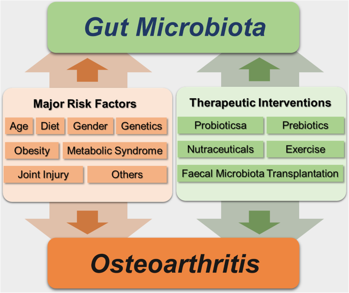
Osteoarthritis (OA) is one of the most frequent musculoskeletal diseases characterized by degeneration of articular cartilage, subchondral bone remodeling, and synovial membrane inflammation, which is a leading cause of global disability, morbidity, and decreased quality of life. Interpreting the potential mechanisms of OA pathogenesis is essential for developing novel prevention and disease-modifying therapeutic interventions. Gut microbiota is responsible for a series of metabolic, immunological, and structural and neurological functions, potentially elucidating the heterogeneity of OA phenotypes and individual features. In this narrative review, we summarized research evidence supporting the hypothesis of a “gut-joint axis” and the interaction between gut microbiota and the OA-relevant factors, including age, gender, genetics, metabolism, central nervous system, and joint injury, elucidating the underlying mechanisms of this intricate interaction. In the context, we also speculated the promising manipulation of gut microbiota in OA management, such as exercise and fecal microbiota transplantation (FMT), highlighting the clinical values of gut microbiota. Additionally, future research directions, such as more convincing studies by the interventions of gut microbiota, the gene regulation of host contributing to or attributed to the specific phenotypes of gut microbiota related to OA, and the relevance of distinct cell subgroups to gut microbiota, are expected. Moreover, gut microbiota is also the potential biomarker related to inflammation and gut dysbiosis that is able to predict OA progression and monitor the efficacy of therapeutic intervention.
The gut-bone axis: how bacterial metabolites bridge the distance
1Department of Internal Medicine 3, Rheumatology and Immunology, Friedrich-Alexander University Erlangen-Nürnberg (FAU) and Universitätsklinikum Erlangen, Erlangen, Germany.