

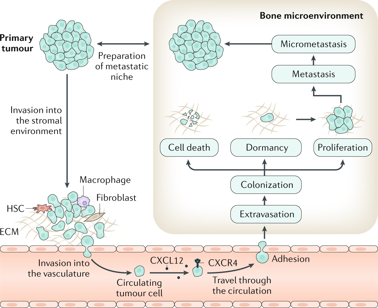
Bone is the most frequent site for metastasis for many cancers, notably for tumours originating in the breast and the prostate. Tumour cells can escape from the primary tumour site and colonize the bone microenvironment. Within the bone, these disseminated tumour cells, as well as those arising in the context of multiple myeloma, may assume a state of dormancy, remaining quiescent for years before resuming proliferation and causing overt metastasis, which causes bone destruction via activation of osteoclast-mediated osteolysis. This structural damage can lead to considerable morbidity, including pain, fractures and impaired quality of life. Although treatment of bone metastases and myeloma bone disease is rarely curative, disease control is often possible for many years through the use of systemic anticancer treatments on a background of multidisciplinary supportive care. This care should include bone-targeted agents to inhibit tumour-associated osteolysis and prevent skeletal morbidity as well as use of appropriate local treatments such as radiation therapy, orthopaedic surgery and specialist palliative care to minimize the impact of metastatic bone disease on physical functioning. In this Primer, we provide an overview of the clinical features, the pathophysiology and the specific treatment approaches to prevent and treat bone metastases from solid tumours as well as myeloma bone disease.
Mathematical modelling of the pathogenesis of multiple myeloma‐induced bone disease
SUMMARY Multiple myeloma (MM) is the second most common haematological malignancy and results in destructive bone lesions. The interaction between MM cells and the bone microenvironment plays an important role in the development of the tumour cells and MM‐induced bone disease and forms a ‘vicious cycle’ of tumour development and bone destruction, intensified by suppression of osteoblast activity and promotion of osteoclast activity. In this paper, a mathematical model is proposed to simulate how the interaction between MM cells and the bone microenvironment facilitates the development of the tumour cells and the resultant bone destruction. It includes both the roles of inhibited osteoblast activity and stimulated osteoclast activity. The model is able to mimic the temporal variation of bone cell concentrations and resultant bone volume after the invasion and then removal of the tumour cells and explains why MM‐induced bone lesions rarely heal even after the complete removal of MM cells. The behaviour of the model compares well with published experimental data. The model serves as a first step to understand the development of MM‐induced bone disease and could be applied further to evaluate the current therapies against MM‐induced bone disease and even suggests new potential therapeutic targets. © 2014 The Authors. International Journal for Numerical Methods in Biomedical Engineering published by John Wiley & Sons Ltd

Systems Modeling of Bortezomib and Dexamethasone Combinatorial Effects on Bone Homeostasis in Multiple Myeloma Patients
Osteolytic bone disease is one of the most debilitating manifestations of multiple myeloma (MM). Bortezomib is a proteasome inhibitor that shows both anticancer and bone anabolic properties and is being evaluated for its positive effects in MM patients with skeletal complications. Dexamethasone is a potent corticosteroid that is often given in combination with bortezomib for its antineoplastic effects; however, bone loss and osteoporosis are major adverse effects of long-term steroid-based therapies.

Denosumab Compared With Zoledronic Acid for the Treatment of Bone Metastases in Patients With Advanced Breast Cancer: A Randomized, Double-Blind Study
Purpose This randomized study compared denosumab, a fully human monoclonal antibody against receptor activator of nuclear factor κ B (RANK) ligand, with zoledronic acid in delaying or preventing skeletal-related events (SREs) in patients with breast cancer with bone metastases. Patients and Methods Patients were randomly assigned to receive either subcutaneous denosumab 120 mg and intravenous placebo (n = 1,026) or intravenous zoledronic acid 4 mg adjusted for creatinine clearance and subcutaneous placebo (n = 1,020) every 4 weeks. All patients were strongly recommended to take daily calcium and vitamin D supplements. The primary end point was time to first on-study SRE (defined as pathologic fracture, radiation or surgery to bone, or spinal cord compression). Results Denosumab was superior to zoledronic acid in delaying time to first on-study SRE (hazard ratio, 0.82; 95% CI, 0.71 to 0.95; P = .01 superiority) and time to first and subsequent (multiple) on-study SREs (rate ratio, 0.77; 95% CI, 0.66 to 0.89; P = .001). Reduction in bone turnover markers was greater with denosumab. Overall survival, disease progression, and rates of adverse events (AEs) and serious AEs were similar between groups. An excess of renal AEs and acute-phase reactions occurred with zoledronic acid; hypocalcemia occurred more frequently with denosumab. Osteonecrosis of the jaw occurred infrequently (2.0%, denosumab; 1.4%, zoledronic acid; P = .39). Conclusion Denosumab was superior to zoledronic acid in delaying or preventing SREs in patients with breast cancer metastatic to bone and was generally well tolerated. With the convenience of a subcutaneous injection and no requirement for renal monitoring, denosumab represents a potential treatment option for patients with bone metastases.

Osteoprotegerin in Bone Metastases: Mathematical Solution to the Puzzle
Bone is a common site for cancer metastasis. To create space for their growth, cancer cells stimulate bone resorbing osteoclasts. Cytokine RANKL is a key osteoclast activator, while osteoprotegerin (OPG) is a RANKL decoy receptor and an inhibitor of osteoclastogenesis. Consistently, systemic application of OPG decreases metastatic tumor burden in bone. However, OPG produced locally by cancer cells was shown to enhance osteolysis and tumor growth. We propose that OPG produced by cancer cells causes a local reduction in RANKL levels, inducing a steeper RANKL gradient away from the tumor and towards the bone tissue, resulting in faster resorption and tumor expansion. We tested this hypothesis using a mathematical model of nonlinear partial differential equations describing the spatial dynamics of OPG, RANKL, PTHrP, osteoclasts, tumor and bone mass. We demonstrate that at lower expression rates, tumor-derived OPG enhances the chemotactic RANKL gradient and osteolysis, whereas at higher expression rates OPG broadly inhibits RANKL and decreases osteolysis and tumor burden. Moreover, tumor expression of a soluble mediator inducing RANKL in the host tissue, such as PTHrP, is important for correct orientation of the RANKL gradient. A meta-analysis of OPG, RANKL and PTHrP expression in normal prostate, carcinoma and metastatic tissues demonstrated an increase in expression of OPG, but not RANKL, in metastatic prostate cancer, and positive correlation between OPG and PTHrP in metastatic prostate cancer. The proposed mechanism highlights the importance of the spatial distribution of receptors, decoys and ligands, and can be applied to other systems involving regulation of spatially anisotropic processes.
Modeling the Human Bone–Tumor Niche: Reducing and Replacing the Need for Animal Data
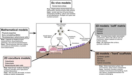
ABSTRACT Bone is the most common site for cancer metastasis. Understanding the interactions within the complex, heterogeneous bone–tumor microenvironment is essential for the development of new therapeutics. Various animal models of tumor‐induced bone disease are routinely used to provide valuable information on the relationship between cancer cells and the skeleton. However, new model systems exist that offer an alternative approach to the use of animals and might more accurately reveal the cellular interactions occurring within the human bone–tumor niche. This review highlights replacement models that mimic the bone microenvironment and where cancer metastases and tumor growth might be assessed alongside bone turnover. Such culture models include the use of calcified regions of animal tissue and scaffolds made from bone mineral hydroxyapatite, synthetic polymers that can be manipulated during manufacture to create structures resembling trabecular bone surfaces, gel composites that can be modified for stiffness and porosity to resemble conditions in the tumor–bone microenvironment. Possibly the most accurate model system involves the use of fresh human bone samples, which can be cultured ex vivo in the presence of human tumor cells and demonstrate similar cancer cell–bone cell interactions as described in vivo. In addition, the use of mathematical modeling and computational biology approaches provide an alternative to preliminary animal testing. The use of such models offers the capacity to mimic significant elements of the human bone–tumor environment, and complement, refine, or replace the use of preclinical models. © 2020 The Authors. JBMR Plus published by Wiley Periodicals, Inc. on behalf of American Society for Bone and Mineral Research.
Randomized, Double-Blind Study of Denosumab Versus Zoledronic Acid in the Treatment of Bone Metastases in Patients With Advanced Cancer (Excluding Breast and Prostate Cancer) or Multiple Myeloma
Purpose This study compared denosumab, a fully human monoclonal anti-receptor activator of nuclear factor kappa-B ligand antibody, with zoledronic acid (ZA) for delaying or preventing skeletal-related events (SRE) in patients with advanced cancer and bone metastases (excluding breast and prostate) or myeloma. Patients and Methods Eligible patients were randomly assigned in a double-blind, double-dummy design to receive monthly subcutaneous denosumab 120 mg (n = 886) or intravenous ZA 4 mg (dose adjusted for renal impairment; n = 890). Daily supplemental calcium and vitamin D were strongly recommended. The primary end point was time to first on-study SRE (pathologic fracture, radiation or surgery to bone, or spinal cord compression). Results Denosumab was noninferior to ZA in delaying time to first on-study SRE (hazard ratio, 0.84; 95% CI, 0.71 to 0.98; P = .0007). Although directionally favorable, denosumab was not statistically superior to ZA in delaying time to first on-study SRE (P = .03 unadjusted; P = .06 adjusted for multiplicity) or time to first-and-subsequent (multiple) SRE (rate ratio, 0.90; 95% CI, 0.77 to 1.04; P = .14). Overall survival and disease progression were similar between groups. Hypocalcemia occurred more frequently with denosumab. Osteonecrosis of the jaw occurred at similarly low rates in both groups. Acute-phase reactions after the first dose occurred more frequently with ZA, as did renal adverse events and elevations in serum creatinine based on National Cancer Institute Common Toxicity Criteria for Adverse Events grading. Conclusion Denosumab was noninferior (trending to superiority) to ZA in preventing or delaying first on-study SRE in patients with advanced cancer metastatic to bone or myeloma. Denosumab represents a potential novel treatment option with the convenience of subcutaneous administration and no requirement for renal monitoring or dose adjustment.

An Integrated Computational Model of the Bone Microenvironment in Bone-Metastatic Prostate Cancer
Abstract Bone metastasis will impact most men with advanced prostate cancer. The vicious cycle of bone degradation and formation driven by metastatic prostate cells in bone yields factors that drive cancer growth. Mechanistic insights into this vicious cycle have suggested new therapeutic opportunities, but complex temporal and cellular interactions in the bone microenvironment make drug development challenging. We have integrated biologic and computational approaches to generate a hybrid cellular automata model of normal bone matrix homeostasis and the prostate cancer-bone microenvironment. The model accurately reproduces the basic multicellular unit bone coupling process, such that introduction of a single prostate cancer cell yields a vicious cycle similar in cellular composition and pathophysiology to models of prostate-to-bone metastasis. Notably, the model revealed distinct phases of osteolytic and osteogenic activity, a critical role for mesenchymal stromal cells in osteogenesis, and temporal changes in cellular composition. To evaluate the robustness of the model, we assessed the effect of established bisphosphonate and anti-RANKL therapies on bone metastases. At approximately 100% efficacy, bisphosphonates inhibited cancer progression while, in contrast with clinical observations in humans, anti-RANKL therapy fully eradicated metastases. Reducing anti-RANKL yielded clinically similar results, suggesting that better targeting or dosing could improve patient survival. Our work establishes a computational model that can be tailored for rapid assessment of experimental therapies and delivery of precision medicine to patients with prostate cancer with bone metastases. Cancer Res; 74(9); 2391–401. ©2014 AACR.
Bisphosphonates: Mechanism of Action and Role in Clinical Practice
Bisphosphonates are primary agents in the current pharmacological arsenal against osteoclast-mediated bone loss due to osteoporosis, Paget disease of bone, malignancies metastatic to bone, multiple myeloma, and hypercalcemia of malignancy. In addition to currently approved uses, bisphosphonates are commonly prescribed for prevention and treatment of a variety of other skeletal conditions, such as low bone density and osteogenesis imperfecta. However, the recent recognition that bisphosphonate use is associated with pathologic conditions including osteonecrosis of the jaw has sharpened the level of scrutiny of the current widespread use of bisphosphonate therapy.

Dynamic Biochemical and Cellular Models of Bone Physiology: Integrating Remodeling Processes, Tumor Growth, and Therapy
Bone is an activeCoelho, Rui M. Neto, Joana P. connective tissue composed of different types of cells. The dynamic behavior of bone remodeling processes is typically represented throughValério, Duarte differential equations, which represent the physiological phenomena occurring in this organ. These models takeVinga, Susana into account the tight biochemical regulation between osteoclasts and osteoblasts and have also been enriched with variables and parameters related to bone pathologies and treatment. This chapter reviews some of the more recent models describing bone physiology, focusing on those that include the main cellular processes, along the biochemical control, and also the pharmacokinetic/pharmacodynamic (PK/PD) of the most common treatments for diseases such as cancer. These models are then compared in terms of the simulations obtained and, finally, some highlights on integrating them with the biomechanical component of the system which will be given. These models are expected to provide a valuable insight into this complex system and to support the development of clinical decision systems for bone pathologies.

Current Status of the Diagnosis and Management of Osteoporosis
Osteoporosis has been defined as the silent disease of the 21st century, becoming a public health risk due to its severity, chronicity and progression and affecting mainly postmenopausal women and older adults. Osteoporosis is characterized by an imbalance between bone resorption and bone production. It is diagnosed through different methods such as bone densitometry and dual X-rays. The treatment of this pathology focuses on different aspects. On the one hand, pharmacological treatments are characterized by the use of anti-resorptive drugs, as well as emerging regenerative medicine treatments such as cell therapies and the use of bioactive hydrogels. On the other hand, non-pharmacological treatments are associated with lifestyle habits that should be incorporated, such as physical activity, diet and the cessation of harmful habits such as a high consumption of alcohol or smoking. This review seeks to provide an overview of the theoretical basis in relation to bone biology, the existing methods for diagnosis and the treatments of osteoporosis, including the development of new strategies.

The Effect of Inflammation on Bone
Bone remodeling is the continual process to renew the adult skeleton through the sequential action of osteoblasts and osteoclasts. Nuclear factor RANK, an osteoclast receptor, and its ligand RANKL, expressed on the surface of osteoblasts, result in coordinated control of bone remodeling. Inflammation, a feature of illness and injury, plays a distinct role in skewing this process toward resorption. It does so via the interaction of inflammatory mediators and their related peptides with osteoblasts and osteoclasts, to alter the expression of RANK and RANKL. Such chemical mediators include TNFa, glucocorticoids, histamine, bradykinin, PGE2, systemic RANKL from immune cells, and interleukins 1 and 6. A thorough understanding of bone response to injury and disease, and ability to detect such biomarkers, as well as imaging to identify early structural and mechanical property changes in bone architecture, is important in improving management and outcomes of bone related pathology. While gut health and vitamin and mineral availability appear vitally important, there are very few intervention strategies currently available to favorably alter bone pathology induced by inflammation. Further research is required in this field to advance early detection and treatments.

The Development of Molecular Biology of Osteoporosis
Osteoporosis is one of the major bone disorders that affects both women and men, and causes bone deterioration and bone strength. Bone remodeling maintains bone mass and mineral homeostasis through the balanced action of osteoblasts and osteoclasts, which are responsible for bone formation and bone resorption, respectively. The imbalance in bone remodeling is known to be the main cause of osteoporosis. The imbalance can be the result of the action of various molecules produced by one bone cell that acts on other bone cells and influence cell activity. The understanding of the effect of these molecules on bone can help identify new targets and therapeutics to prevent and treat bone disorders. In this article, we have focused on molecules that are produced by osteoblasts, osteocytes, and osteoclasts and their mechanism of action on these cells. We have also summarized the different pharmacological osteoporosis treatments that target different molecular aspects of these bone cells to minimize osteoporosis.
Biology of Bone Tissue: Structure, Function, and Factors That Influence Bone Cells
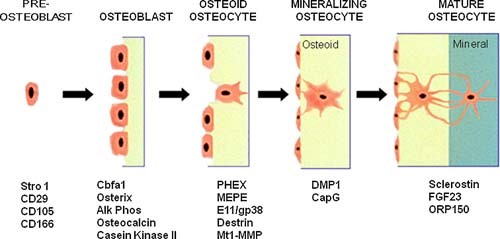
Bone tissue is continuously remodeled through the concerted actions of bone cells, which include bone resorption by osteoclasts and bone formation by osteoblasts, whereas osteocytes act as mechanosensors and orchestrators of the bone remodeling process. This process is under the control of local (e.g., growth factors and cytokines) and systemic (e.g., calcitonin and estrogens) factors that all together contribute for bone homeostasis. An imbalance between bone resorption and formation can result in bone diseases including osteoporosis. Recently, it has been recognized that, during bone remodeling, there are an intricate communication among bone cells. For instance, the coupling from bone resorption to bone formation is achieved by interaction between osteoclasts and osteoblasts. Moreover, osteocytes produce factors that influence osteoblast and osteoclast activities, whereas osteocyte apoptosis is followed by osteoclastic bone resorption. The increasing knowledge about the structure and functions of bone cells contributed to a better understanding of bone biology. It has been suggested that there is a complex communication between bone cells and other organs, indicating the dynamic nature of bone tissue. In this review, we discuss the current data about the structure and functions of bone cells and the factors that influence bone remodeling.

The amazing osteocyte
Abstract The last decade has provided a virtual explosion of data on the molecular biology and function of osteocytes. Far from being the “passive placeholder in bone,” this cell has been found to have numerous functions, such as acting as an orchestrator of bone remodeling through regulation of both osteoclast and osteoblast activity and also functioning as an endocrine cell. The osteocyte is a source of soluble factors not only to target cells on the bone surface but also to target distant organs, such as kidney, muscle, and other tissues. This cell plays a role in both phosphate metabolism and calcium availability and can remodel its perilacunar matrix. Osteocytes compose 90% to 95% of all bone cells in adult bone and are the longest lived bone cell, up to decades within their mineralized environment. As we age, these cells die, leaving behind empty lacunae that frequently micropetrose. In aged bone such as osteonecrotic bone, empty lacunae are associated with reduced remodeling. Inflammatory factors such as tumor necrosis factor and glucocorticoids used to treat inflammatory disease induce osteocyte cell death, but by different mechanisms with potentially different outcomes. Therefore, healthy, viable osteocytes are necessary for proper functionality of bone and other organs. © 2011 American Society for Bone and Mineral Research.
Long-term use of denosumab and its association with skeletal-related events and osteonecrosis of the jaw
Denosumab, an inhibitor of receptor activator of nuclear factor kappa-B ligand, reduces skeletal-related events (SREs) and is approved for solid tumors with bone metastases. We studied long-term denosumab efficacy and safety because real-world data is scarce. This single-arm, single-center retrospective study included denosumab-treated breast cancer patients with bone metastases. Kaplan–Meier survival curves assessed exposure, SREs, osteonecrosis of the jaw (ONJ), and death. 132 patients were enrolled. The median denosumab exposure was 28.3 months (range 1.0–84.9). In the first year, 11.1% experienced SREs. This increased to 18.6% in the second, 21% in the third, and 35.1% in the fourth year and beyond. The median time to first on-study SRE has not been reached. 10 denosumab users (7.6%) developed ONJ. ONJ incidence was 0.9% in the first year, 6.2% in the second, 13.6% in the third, and 16.2% in subsequent years. The median time to first on-study ONJ has not been reached yet. Seven patients resumed denosumab after careful management of ONJ. Our data suggest that long-term treatment with denosumab may further prevent or postpone SREs at the cost of an increased risk of ONJ. The majority of patients who resumed denosumab did not experience a recurrence of ONJ.

OSTEOIMMUNOLOGY: Interplay Between the Immune System and Bone Metabolism
Studies of bone and the immune system have converged in recent years under the banner of osteoimmunology. The immune system is spawned in the bone marrow reservoir, and investigators now recognize that important niches also exist there for memory lymphocytes. At the same time, various factors produced during immune responses are capable of profoundly affecting regulation of bone. Mechanisms have evolved to prevent excessive interference by the immune system with bone homeostasis, yet pathologic bone loss is a common sequela associated with autoimmunity and cancer. There are also developmental links, or parallels, between bone and the immune system. Cells that regulate bone turnover share a common precursor with inflammatory immune cells and may restrict themselves anatomically, in part by utilizing a signaling network analogous to lymphocyte costimulation. Efforts are currently under way to further characterize how these two organ systems overlap and to develop therapeutic strategies that benefit from this understanding.
