

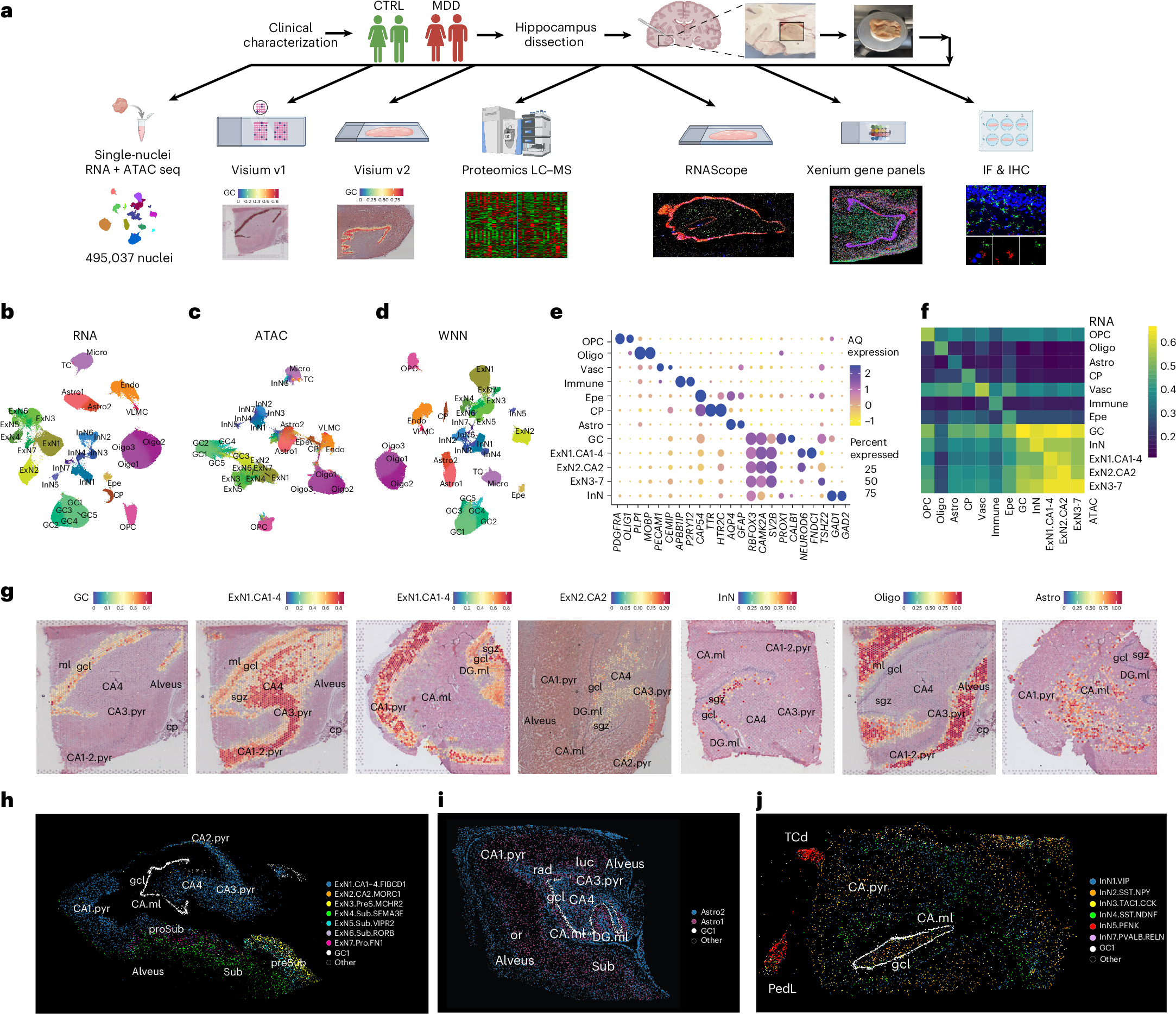
Major depressive disorder (MDD) is associated with reduced hippocampal volume, altered connectivity and negative memory bias, suggesting disrupted hippocampal plasticity. Dysregulated adult hippocampal neurogenesis is a potential contributor, but its relevance in humans and role in MDD remain unclear. Here we investigated the molecular basis of hippocampal dysfunction in nonmedicated individuals with MDD by integrating analyses of neurogenic trajectories, cell-type- and subfield-specific gene expression, chromatin accessibility and protein expression. We identify a neurogenic lineage in the adult human hippocampal subgranular zone and provide evidence for a stalled neurogenic process in MDD, associated with transcriptional regulation, stress-related reprogramming and interferon signaling across developmental stages. Excitatory and inhibitory neurons show dysregulation of transcription factor networks affecting cell states. Cellular stress, excitatory–inhibitory imbalance, impaired synaptic plasticity, reduced metabolic capacity and immune activation, underlie impaired neurogenesis and reduced hippocampus circuit plasticity. Findings indicate genetic and epigenetic regulation of gene expression in MDD, and overlapping pathogenetic mechanisms with autoimmune, neurodevelopmental and neurodegenerative diseases. This work provides a new understanding of the pathogenesis of hippocampus-dependent cognitive symptoms in MDD and suggests potential therapeutic targets.
Dysregulated adult hippocampal neurogenesis in major depressive disorder
Major depressive disorder (MDD) is associated with reduced hippocampal volume, altered connectivity and negative memory bias, suggesting disrupted hippocampal plasticity. Dysregulated adult hippocampal neurogenesis is a potential contributor, but its relevance in humans and role in MDD remain unclear. Here we investigated the molecular basis of hippocampal dysfunction in nonmedicated individuals with MDD by integrating analyses of neurogenic trajectories, cell-type- and subfield-specific gene expression, chromatin accessibility and protein expression. We identify a neurogenic lineage in the adult human hippocampal subgranular zone and provide evidence for a stalled neurogenic process in MDD, associated with transcriptional regulation, stress-related reprogramming and interferon signaling across developmental stages. Excitatory and inhibitory neurons show dysregulation of transcription factor networks affecting cell states. Cellular stress, excitatory–inhibitory imbalance, impaired synaptic plasticity, reduced metabolic capacity and immune activation, underlie impaired neurogenesis and reduced hippocampus circuit plasticity. Findings indicate genetic and epigenetic regulation of gene expression in MDD, and overlapping pathogenetic mechanisms with autoimmune, neurodevelopmental and neurodegenerative diseases. This work provides a new understanding of the pathogenesis of hippocampus-dependent cognitive symptoms in MDD and suggests potential therapeutic targets.
Depression doesn't shrink the brain like we thought it did | New Scientist
For the past decade, depression has been linked to a shrunken hippocampus, which has a vital role in memory. But a large study using up-to-date scanning techniques suggests this may not be the case

Single-cell multiregion dissection of Alzheimer’s disease
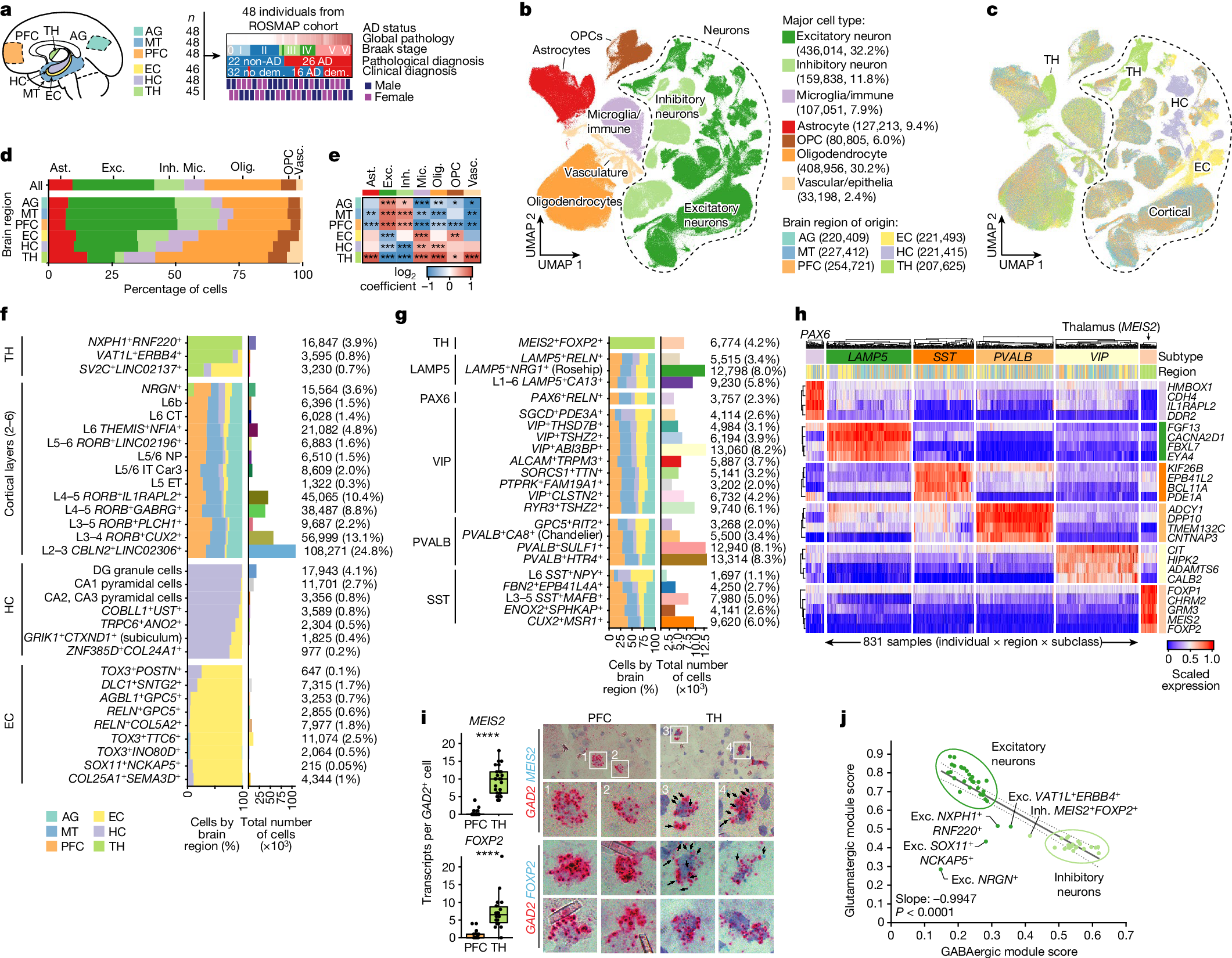
Alzheimer’s disease is the leading cause of dementia worldwide, but the cellular pathways that underlie its pathological progression across brain regions remain poorly understood1–3. Here we report a single-cell transcriptomic atlas of six different brain regions in the aged human brain, covering 1.3 million cells from 283 post-mortem human brain samples across 48 individuals with and without Alzheimer’s disease. We identify 76 cell types, including region-specific subtypes of astrocytes and excitatory neurons and an inhibitory interneuron population unique to the thalamus and distinct from canonical inhibitory subclasses. We identify vulnerable populations of excitatory and inhibitory neurons that are depleted in specific brain regions in Alzheimer’s disease, and provide evidence that the Reelin signalling pathway is involved in modulating the vulnerability of these neurons. We develop a scalable method for discovering gene modules, which we use to identify cell-type-specific and region-specific modules that are altered in Alzheimer’s disease and to annotate transcriptomic differences associated with diverse pathological variables. We identify an astrocyte program that is associated with cognitive resilience to Alzheimer’s disease pathology, tying choline metabolism and polyamine biosynthesis in astrocytes to preserved cognitive function late in life. Together, our study develops a regional atlas of the ageing human brain and provides insights into cellular vulnerability, response and resilience to Alzheimer’s disease pathology.
The neuroimaging correlates of depression established across six large-scale population datasets
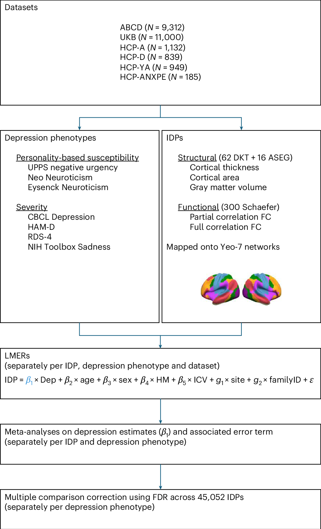
Depression has been linked to reduced size of subcortical regions and abnormal functional connectivity in frontal and default mode networks. However, recent meta-analyses have failed to identify significant converging correlates of depression across the literature such that a conclusive mapping of the neuroimaging correlates of depression remains elusive. We leveraged 23,417 participants across 6 population datasets to comprehensively establish the neuroimaging correlates of depression. We found reductions in gray matter volume/cortical surface area associated with depression in the frontal cortex, anterior cingulate and insula, confirming previous studies showing the importance of prefrontal and default mode regions in depression. Our findings demonstrate multiple surprising results, including a lack of depression correlates in subcortical brain regions and significant depression correlates in somatomotor and visual regions. Overall, these results shed new light on key brain regions involved in the pathophysiology of depression, updating our understanding of the neuroimaging correlates of depression symptoms.
The neuroimaging correlates of depression established across six large-scale population datasets
Depression has been linked to reduced size of subcortical regions and abnormal functional connectivity in frontal and default mode networks. However, recent meta-analyses have failed to identify significant converging correlates of depression across the literature such that a conclusive mapping of the neuroimaging correlates of depression remains elusive. We leveraged 23,417 participants across 6 population datasets to comprehensively establish the neuroimaging correlates of depression. We found reductions in gray matter volume/cortical surface area associated with depression in the frontal cortex, anterior cingulate and insula, confirming previous studies showing the importance of prefrontal and default mode regions in depression. Our findings demonstrate multiple surprising results, including a lack of depression correlates in subcortical brain regions and significant depression correlates in somatomotor and visual regions. Overall, these results shed new light on key brain regions involved in the pathophysiology of depression, updating our understanding of the neuroimaging correlates of depression symptoms.
Comparing phenotypic manifolds with Kompot: Detecting differential abundance and gene expression at single-cell resolution
Single-cell studies are frequently designed to compare across conditions such as health and disease. However, existing computational approaches typically rely on grouping cells into discrete populations before making comparisons, which can limit resolution for detecting state-dependent changes. Here, we introduce Kompot, a statistical framework for comparative analysis of multi-condition single-cell data. Kompot quantifies both differential abundance, capturing how cells redistribute across the phenotypic space, and differential expression, identifying condition-specific transcriptional changes that may be localized, heterogeneous, or oppositely regulated across states. By modeling cell density and gene expression as continuous functions over a shared cell-state representation, Kompot enables single-cell–resolution inference with principled uncertainty estimates, without requiring predefined clusters or cell types. Applying Kompot to aging murine bone marrow, we identified a continuum of shifts in hematopoietic stem cell and mature cell states, transcriptional remodeling of monocytes independent of compositional changes, and divergent regulation of oxidative stress response genes across cell types. We demonstrate the utility of Kompot in disease settings by identifying cell-state and gene expression changes associated with improved efficacy of combinatorial immunotherapy in melanoma. Additionally, Kompot enables multi-sample comparative analysis by accounting for sample-to-sample heterogeneity. By capturing both global and cell-state–specific effects of perturbation, the Kompot framework is broadly applicable to dissecting condition-specific effects in complex single-cell landscapes. ### Competing Interest Statement The authors have declared no competing interest. National Institutes of Health, R35GM147125, R01CA292932, T32GM136534, S10OD028685 The Mark Foundation for Cancer Research, https://ror.org/00v7th354, Endeavor Award Edward P. Evans Foundation, https://ror.org/03h22gm35, Discovery Research Grant Brotman Baty Institute, https://ror.org/03jxvbk42, Pilot Award

Dynamic Landscape Analysis of cell fate decisions provides predictive models of neural development from single-cell data
Building a mechanistic understanding of cell fate decisions remains a fundamental goal of developmental biology, with implications for stem cell therapies, regenerative medicine and understanding disease mechanisms. Single-cell transcriptomics provides a detailed picture of the cellular states observed during these decisions, but building dynamic and predictive models from these data remains a challenge. Here, we present dynamic landscape analysis (DLA), an integrative framework that applies dynamical systems theory to identify stable cell states, map transition pathways, and generate a predictive cell fate decision landscape from single-cell data. Applying this framework to vertebrate neural tube development revealed that progenitor specification by Sonic Hedgehog (Shh) can be captured in a landscape with an unexpected topology in which initially divergent lineages converge to the same fate through multiple distinct routes. The model accurately predicted cellular responses and cell fate allocation for unseen dynamic signalling regimes. Cross-species validation using human embryonic organoid data demonstrated conservation of this decision-making architecture. By modelling the dynamic responses that drive cell fate decisions, the DLA framework provides a quantitative and generative framework for extracting mechanistic insights from high-dimensional single-cell data.
Differences in Krox20-Dependent Regulation of Hoxa2 and Hoxb2 during Hindbrain Development
During hindbrain development, segmental regulation of the paralogous Hoxa2 and Hoxb2 genes in rhombomeres (r) 3 and 5 involves Krox20-dependent enhancers that have been conserved during the duplication of the vertebrate Hox clusters from a common ancestor. Examining these evolutionarily related control regions could provide important insight into the degree to which the basic Krox20-dependent mechanisms, cis-regulatory components, and their organization have been conserved. Toward this goal we have performed a detailed functional analysis of a mouse Hoxa2 enhancer capable of directing reporter expression in r3 and r5. The combined activities of five separate cis-regions, in addition to the conserved Krox20 binding sites, are involved in mediating enhancer function. A CTTT (BoxA) motif adjacent to the Krox20 binding sites is important for r3/r5 activity. The BoxA motif is similar to one (Box1) found in the Hoxb2 enhancer and indicates that the close proximity of these Box motifs to Krox20 sites is a common feature of Krox20 targets in vivo. Two other rhombomeric elements (RE1 and RE3) are essential for r3/r5 activity and share common TCT motifs, indicating that they interact with a similar cofactor(s). TCT motifs are also found in the Hoxb2 enhancer, suggesting that they may be another common feature of Krox20-dependent control regions. The two remaining Hoxa2 cis-elements, RE2 and RE4, are not conserved in the Hoxb2 enhancer and define differences in some of components that can contribute to the Krox20-dependent activities of these enhancers. Furthermore, analysis of regulatory activities of these enhancers in a Krox20 mutant background has uncovered differences in their degree of dependence upon Krox20 for segmental expression. Together, this work has revealed a surprising degree of complexity in the number of cis-elements and regulatory components that contribute to segmental expression mediated by Krox20 and sheds light on the diversity and evolution of Krox20 target sites and Hox regulatory elements in vertebrates.
Leading Innovation in Psychiatric Treatment | Alto Neuroscience
We leverage brain biology to develop personalized medicines, helping patients get better faster.

Spatial Transcriptomics As Rasterized Image Tensors (STARIT) characterizes cell states with subcellular molecular heterogeneity
Imaging-based spatially resolved transcriptomics (imSRT) technologies provide high-throughput molecular-resolution spatial characterization of genes within cells. Conventional analysis methods to identify cell-types and states in imSRT data rely on gene count matrices derived from tallying the number of mRNA molecules detected for each gene per segmented cell, thereby overlooking subcellular heterogeneity that can be useful in defining cell states. To take advantage of the molecular-resolution information in imSRT data and potentially identify cell-states based on subcellular heterogeneity, we developed STARIT (Spatial Transcriptomics As Rasterized Image Tensors). STARIT converts transcripts within segmented cells in imSRT data into an image-based tensor representation that can be combined with deep learning computer vision models for downstream analysis. Using simulated and real imSRT data, we demonstrate that STARIT distinguishes transcriptionally distinct cell-types and further separates cell states based on subcellular transcript localization, which conventional gene count analysis fails to capture. By providing a standardized framework to encode subcellular molecular information in imSRT data, STARIT will enable deeper insights into subcellular heterogeneity and enhance the identification and characterization of cell-types and states that are overlooked by gene count representations.
A unified network systems approach uncovers a core program underlying T follicular helper cell differentiation
Characterizing multi-scale processes underlying immune-state progression is critical for defining their function. This becomes pertinent for functionally diverse and plastic immune cells, such as T follicular helper (Tfh) cells. Here, we adopt a multi-scale network-systems approach that incorporates both regulatory and protein-protein interactions. This approach integrates diverse data types, captures regulation across levels of immune system organization, and recapitulates known Tfh differentiation drivers. Further, we present CoreNet, a core Tfh gene set that is conserved between humans and mice, across tissue types and disease contexts, and is consistent across data modalities. Using CoreNet, we implicate NR3C1 and interleukin (IL)-12 in the regulation of Tfh differentiation. Notably, IL-12 is permissive for differentiation of Tfh precursors but blocks differentiation into germinal center Tfh cells. Overall, this work elucidates networks with unexplored roles governing Tfh differentiation across species and tissues, while providing a generalizable framework. CoreNet is accessible through an interactive web server: https://pitt-csi.shinyapps.io/tfhcorenet/.
What stress does to the brain - Neuroscience News
A new study sheds light on how the brain changes as a result of chronic stress. Selective noradrenaline release rewires connectivity patterns between different brain regions. Activity in the amygdala and networks that process sensory stimuli increased.

Motif Neurotech — Therapeutic Brain Computer Interface For Mental Health
We're building devices to help you monitor and regulate your mental health and performance.
.png)
Gene regulatory networks: from correlative models to causal explanations
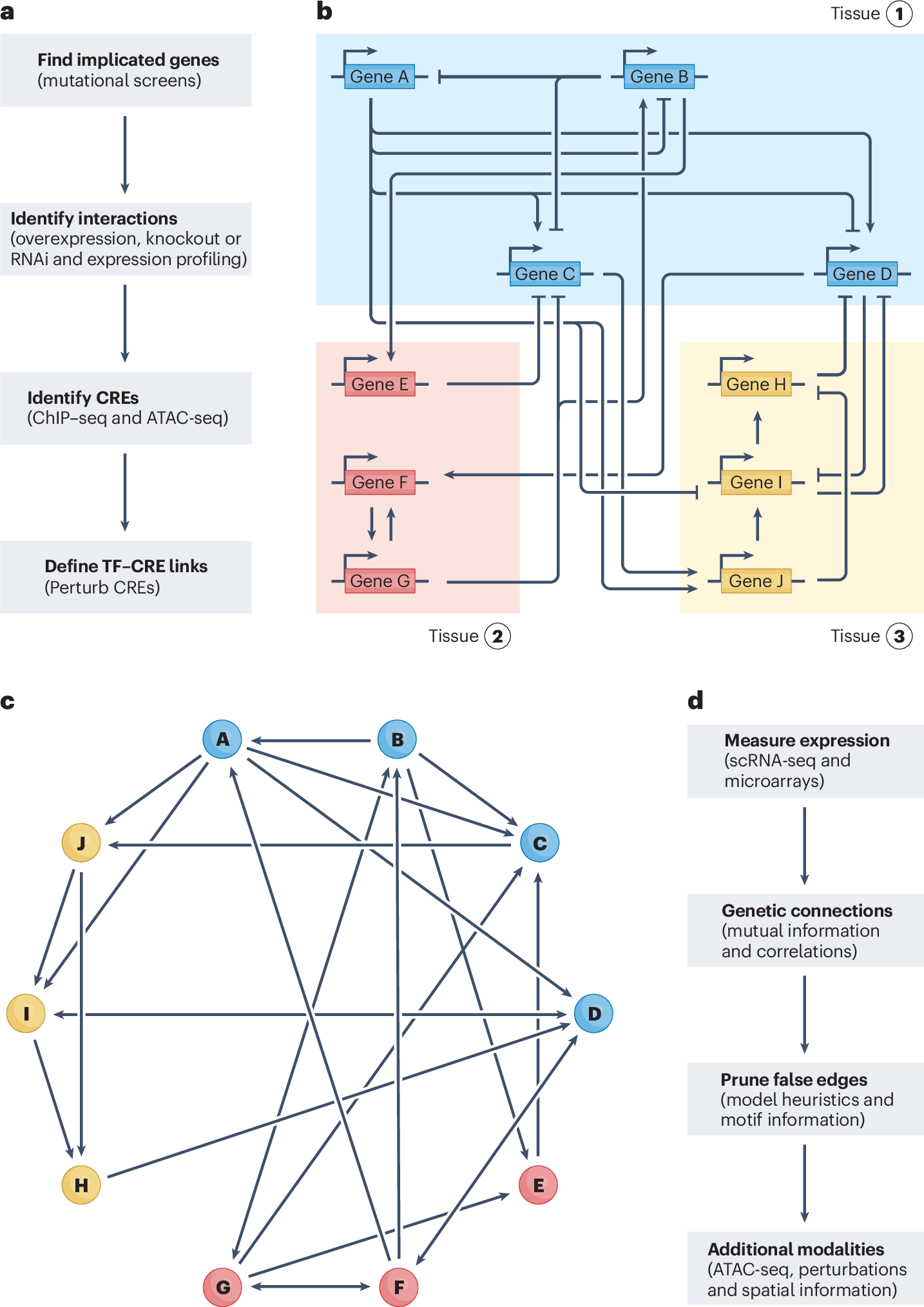
Gene regulatory networks (GRNs) explain how the genome controls cellular behaviour and tissue morphogenesis, serving to connect molecular mechanism to functional output. Single-cell technologies now provide descriptions of these networks with unprecedented detail, but this advance has also revealed gene regulatory systems that are too complex for our existing conceptual frameworks. GRNs, which should provide mechanistic explanations, are increasingly reduced to statistical correlations — ‘hairballs’ that fail to capture molecular causation. Here, we explore why this dilemma exists and propose a path forward. We argue that methods in ‘representation learning’ can be used to model GRNs, without needing to capture every molecular detail. For this framework, we advocate three linked principles: models must be inherently mechanistic, with structures grounded in cellular and evolutionary biology; molecular principles and constraints must be used to reduce the solution space for learning GRN models; and more sophisticated forms of experimental perturbation and synthetic biological engineering are needed to train models and test predictions. By reimagining GRNs through these principles, we can bridge the gap from data abundance to new conceptual understanding.
Embryo-scale reverse genetics at single-cell resolution
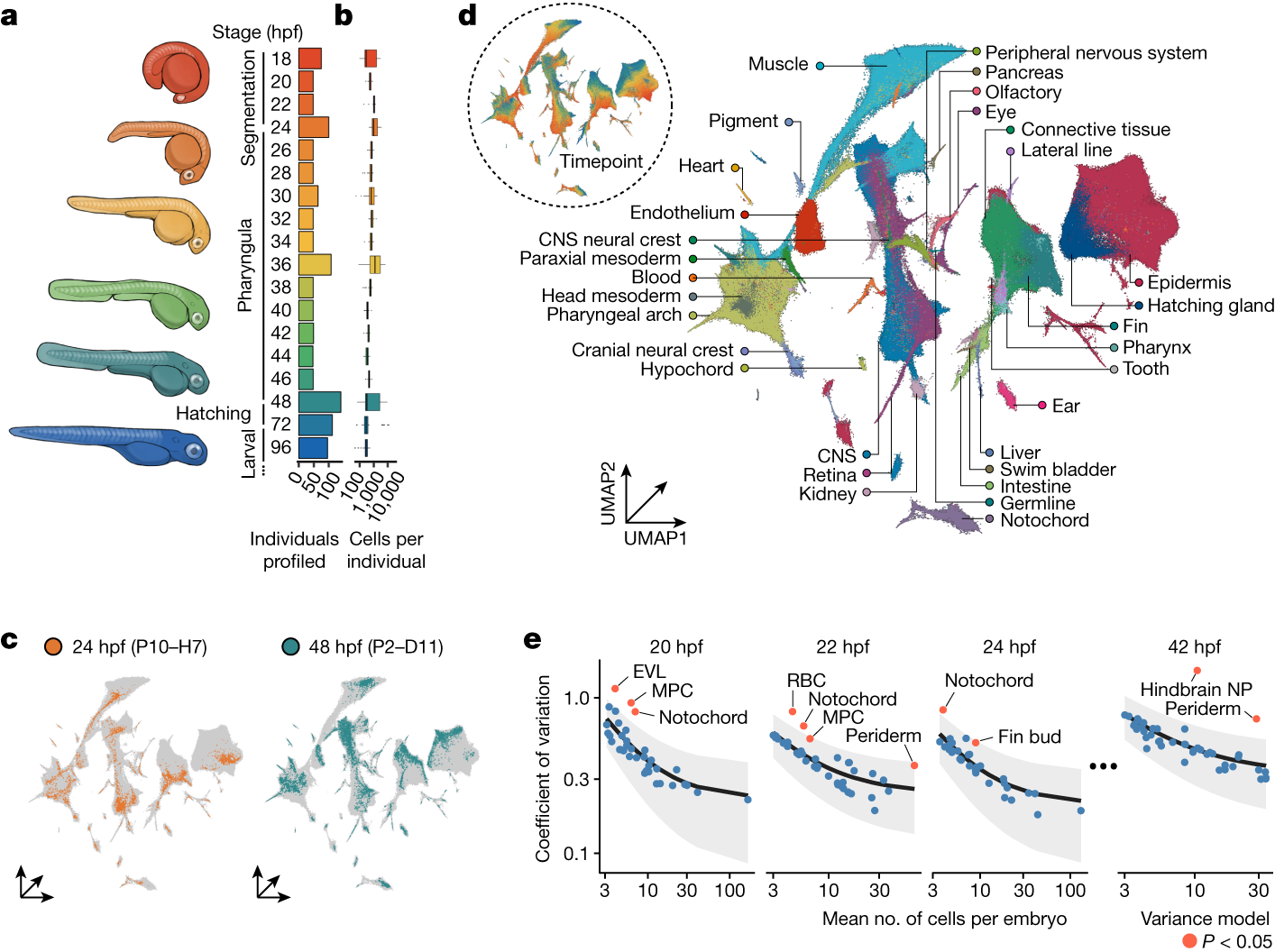
The maturation of single-cell transcriptomic technologies has facilitated the generation of comprehensive cellular atlases from whole embryos1–4. A majority of these data, however, has been collected from wild-type embryos without an appreciation for the latent variation that is present in development. Here we present the ‘zebrafish single-cell atlas of perturbed embryos’: single-cell transcriptomic data from 1,812 individually resolved developing zebrafish embryos, encompassing 19 timepoints, 23 genetic perturbations and a total of 3.2 million cells. The high degree of replication in our study (eight or more embryos per condition) enables us to estimate the variance in cell type abundance organism-wide and to detect perturbation-dependent deviance in cell type composition relative to wild-type embryos. Our approach is sensitive to rare cell types, resolving developmental trajectories and genetic dependencies in the cranial ganglia neurons, a cell population that comprises less than 1% of the embryo. Additionally, time-series profiling of individual mutants identified a group of brachyury-independent cells with strikingly similar transcriptomes to notochord sheath cells, leading to new hypotheses about early origins of the skull. We anticipate that standardized collection of high-resolution, organism-scale single-cell data from large numbers of individual embryos will enable mapping of the genetic dependencies of zebrafish cell types, while also addressing longstanding challenges in developmental genetics, including the cellular and transcriptional plasticity underlying phenotypic diversity across individuals.
BMP–Smad1/9 signaling plays a critical role in regulating zebrafish PGC proliferation
The germ cell fate in zebrafish is determined by germ plasm, whereas mammalian germ cell fate is induced by bone morphogenetic protein (BMP) signaling. It remains elusive whether BMP signaling is implicated in zebrafish germ cell development. Here, we demonstrate that BMP–Smad1/9 signaling plays a critical role in zebrafish primordial germ cell (PGC) maintenance rather than fate determination. BMP inhibition or smad1/9 knockdown reduces PGC numbers. Furthermore, we generated PGC-specific smad1/9 knockouts using a transgenic approach with PGC-specifically expressed Cas9 and ubiquitously expressed guide RNAs. Smad1/9 deficiency in PGCs leads to impaired PGC proliferation and increased apoptosis, consequently reducing PGC numbers. Transcriptome analysis revealed unchanged PGC-specific gene expression, but a marked upregulation of DNA damage response-related genes, which is validated by ectopic ATR–pChk1 activation in PGCs and PGC restoring by ATR inhibition. Collectively, these findings underscore conserved but functionally distinct roles of BMP signaling in vertebrate PGC development.
