

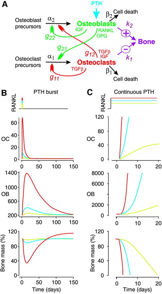
Mathematical Model of Paracrine Interactions between Osteoclasts and Osteoblasts Predicts Anabolic Action of Parathyroid Hormone on Bone
Abstract. To restore falling plasma calcium levels, PTH promotes calcium liberation from bone. PTH targets bone-forming cells, osteoblasts, to increase exp
Regulation of Bone Remodeling by Parathyroid Hormone
Parathyroid hormone (PTH) exerts profound effects on skeletal homeostasis through multiple cellular and molecular mechanisms. Continuous hyperparathyroidism causes net loss of bone mass, despite accelerating bone formation by osteoblasts. Intermittent treatment with PTH analogs represents the only Food and Drug Administration (FDA)-approved bone anabolic osteoporosis treatment strategy. Functional PTH receptors are present on cells of the osteoblast lineage, ranging from early skeletal stem cells to matrix-embedded osteocytes. In addition, bone remodeling by osteoclasts liberates latent growth factors present within bone matrix. Here, we will provide an overview of the multiple cellular and molecular mechanisms through which PTH influences bone homeostasis. Notably, net skeletal effects of continuous versus intermittent can differ significantly. Where possible, we will highlight mechanisms through which continuous hyperparathyroidism leads to bone loss, and through which intermittent hyperparathyroidism boosts bone mass. Given the therapeutic usage of intermittent PTH (iPTH) treatment for osteoporosis, particular attention will be paid toward mechanisms underlying the bone anabolic effects of once daily PTH administration.
Dynamics of bone cell signaling and PTH treatments of osteoporosis
In this paper we analyze and generalize the dynamical system introduced by Lemaire and co-workers [14] as a model of cell signaling in bone remodeling. We show that for large classes of parameter values, including the physically-realistic baseline values, the system has a unique physically relevant equilibrium which is a global attractor. We generalize that model, minimally, to incorporate a mechanism by which parathyroid hormone (PTH) retards osteoblast apoptosis. We show that with this mechanism, which is a simplified version of that proposed by Bellido and co-workers [4], the model exhibits a well-known phenomenon that has puzzled researchers: the system responds catabolically to the continuous administration of PTH andally to appropriately pulsed administration of PTH.
The osteocyte as a signaling cell
Osteocytes, former osteoblasts encapsulated by mineralized bone matrix, are far from being passive and metabolically inactive bone cells. Instead, osteocytes are multifunctional and dynamic cells capable of integrating hormonal and mechanical signals and transmitting them to effector cells in bone and in distant tissues. Osteocytes are a major source of molecules that regulate bone homeostasis by integrating both mechanical cues and hormonal signals that coordinate the differentiation and function of osteoclasts and osteoblasts. Osteocyte function is altered in both rare and common bone diseases, suggesting that osteocyte dysfunction is directly involved in the pathophysiology of several disorders affecting the skeleton. Advances in osteocyte biology initiated the development of novel therapeutics interfering with osteocyte-secreted molecules. Moreover, osteocytes are targets and key distributors of biological signals mediating the beneficial effects of several bone therapeutics used in the clinic. Here we review the most recent discoveries in osteocyte biology demonstrating that osteocytes regulate bone homeostasis and bone marrow fat via paracrine signaling, influence body composition and energy metabolism via endocrine signaling, and contribute to the damaging effects of diabetes mellitus and hematologic and metastatic cancers in the skeleton.

The bone remodelling cycle
The bone remodelling cycle replaces old and damaged bone and is a highly regulated, lifelong process essential for preserving bone integrity and maintaining mineral homeostasis. During the bone remodelling cycle, osteoclastic resorption is tightly coupled to osteoblastic bone formation. The remodelling cycle occurs within the basic multicellular unit and comprises five co-ordinated steps; activation, resorption, reversal, formation and termination. These steps occur simultaneously but asynchronously at multiple different locations within the skeleton. Study of rare human bone disease and animal models have helped to elucidate the cellular and molecular mechanisms that regulate the bone remodelling cycle. The key signalling pathways controlling osteoclastic bone resorption and osteoblastic bone formation are receptor activator of nuclear factor-κB (RANK)/RANK ligand/osteoprotegerin and canonical Wnt signalling. Cytokines, growth factors and prostaglandins act as paracrine regulators of the cycle, whereas endocrine regulators include parathyroid hormone, vitamin D, calcitonin, growth hormone, glucocorticoids, sex hormones, and thyroid hormone. Disruption of the bone remodelling cycle and any resulting imbalance between bone resorption and formation leads to metabolic bone disease, most commonly osteoporosis. The advances in understanding the cellular and molecular mechanisms underlying bone remodelling have also provided targets for pharmacological interventions which include antiresorptive and anabolic therapies. This review will describe the remodelling process and its regulation, discuss osteoporosis and summarize the commonest pharmacological interventions used in its management.

Parathyroid hormone temporal effects on bone formation and resorption
Parathyroid hormone (PTH) paradoxically causes net bone loss (resorption) when administered in a continuous fashion, and net bone formation (deposition) when administered intermittently. Currently no pharmacological formulations are available to promote bone formation, as needed for the treatment of osteoporosis. The paradoxical behavior of PTH confuses endocrinologists, thus, a model bone resorption or deposition dependent on the timing of PTH administration would de-mystify this behavior and provide the basis for logical drug formulation. We developed a mathematical model that accounts for net bone loss with continuous PTH administration and net bone formation with intermittent PTH administration, based on the differential effects of PTH on the osteoblastic and osteoclastic populations of cells.

Biology of Bone Tissue: Structure, Function, and Factors That Influence Bone Cells
Bone tissue is continuously remodeled through the concerted actions of bone cells, which include bone resorption by osteoclasts and bone formation by osteoblasts, whereas osteocytes act as mechanosensors and orchestrators of the bone remodeling process. This process is under the control of local (e.g., growth factors and cytokines) and systemic (e.g., calcitonin and estrogens) factors that all together contribute for bone homeostasis. An imbalance between bone resorption and formation can result in bone diseases including osteoporosis. Recently, it has been recognized that, during bone remodeling, there are an intricate communication among bone cells. For instance, the coupling from bone resorption to bone formation is achieved by interaction between osteoclasts and osteoblasts. Moreover, osteocytes produce factors that influence osteoblast and osteoclast activities, whereas osteocyte apoptosis is followed by osteoclastic bone resorption. The increasing knowledge about the structure and functions of bone cells contributed to a better understanding of bone biology. It has been suggested that there is a complex communication between bone cells and other organs, indicating the dynamic nature of bone tissue. In this review, we discuss the current data about the structure and functions of bone cells and the factors that influence bone remodeling.

Simulated Interventions to Ameliorate Age-Related Bone Loss Indicate the Importance of Timing
Bone remodeling is the continuous process of bone resorption by osteoclasts and bone formation by osteoblasts, in order to maintain homeostasis. The activity of osteoclasts and osteoblasts is regulated by a network of signaling pathways, including Wnt, parathyroid hormone (PTH), RANKL/OPG and TGF-β, in response to stimuli such as mechanical loading. During aging there is a gradual loss of bone mass due to dysregulation of signaling pathways. This may be due to a decline in physical activity with age and/or changes in hormones and other signaling molecules. In particular, hormones such as PTH have a circadian rhythm which may be disrupted in aging. Due to the complexity of the molecular and cellular networks involved in bone remodeling, several mathematical models have been proposed to aid understanding of the processes involved. However, to date there are no models which explicitly consider the effects of mechanical loading, the circadian rhythm of PTH and the dynamics of signaling molecules on bone remodeling. Therefore, we have constructed a network model of the system using a modular approach which will allow further modifications as required in future research. The model was used to simulate the effects of mechanical loading and also the effects of different interventions such as continuous or intermittent administration of PTH. Our model predicts that the absence of regular mechanical loading and/or an impaired PTH circadian rhythm leads to a gradual decrease in bone mass over time which can be restored by simulated interventions and that the effectiveness of some interventions may depend on their timing.

Mathematical Model of Bone Remodeling Captures the Antiresorptive and Anabolic Actions of Various Therapies
A better understanding of the molecular pathways regulating the bone remodeling process should help in the development of new antiresorptive regulators and anabolic regulators, that is, regulators of bone resorption and of bone formation. Understanding the mechanisms by which parathyroid hormone (PTH) influences bone formation and how it switches from anabolic to catabolic action is important for treating osteoporosis (Poole and Reeve in Curr Opin Pharmacol 5:612–617, 2005). In this paper we describe a mathematical model of bone remodeling that incorporates, extends, and integrates several models of particular aspects of this biochemical system (Cabal et al. in J Bone Miner Res 28(8):1830–1836, 2013; Lemaire et al. in J Theor Biol 229:293–309, 2004; Peterson and Riggs in Bone 46:49–63, 2010; Raposo et al. in J Clin Endocrinol Metab 87(9):4330–4340, 2002; Ross et al. in J Disc Cont Dyn Sys Series B 17(6):2185–2200, 2012). We plan to use this model as a bone homeostasis platform to develop anabolic and antiresorptive compounds. The model will allow us to test hypotheses about the dynamics of compounds and to test the potential benefits of combination therapies. At the core of the model is the idealized account of osteoclast and osteoblast signaling given by Lemaire et al. (J Theor Biol 229:293–309, 2004). We have relaxed some of their assumptions about the roles of osteoprotegerin, transforming growth factor $$\upbeta $$, and receptor activator of nuclear factor $$\upkappa $$B ligand; we have devised more detailed models of the interactions of these species. We have incorporated a model of the effect of calcium sensing receptor antagonists on remodeling (Cabal et al. in J Bone Miner Res 28(8):1830–1836, 2013). We have also incorporated a basic model of the effects of vitamin D on calcium homeostasis. We have included a simple model of the mechanism proposed by Bellido et al. (2003), Ross et al. (J Disc Cont Dyn Sys Series B 17(6):2185–2200, 2012), of the influence of PTH on osteoblast apoptosis, a mechanism that accounts for the anabolic response to pulsatile PTH administration. Finally, we have devised a simple model of the administration and effects of bisphosphonates. The biomarkers in the model are procollagen type 1 amino-terminal propeptide and C-terminal telopeptide. Bone mineral density is the model’s principal endpoint.

Osteoclast differentiation and activation
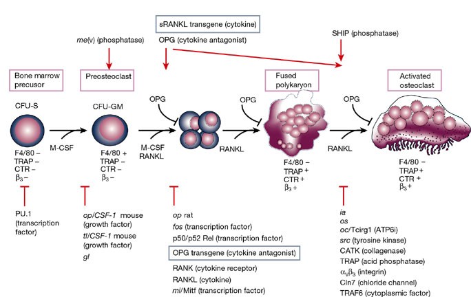
Osteoclasts are specialized cells derived from the monocyte/macrophage haematopoietic lineage that develop and adhere to bone matrix, then secrete acid and lytic enzymes that degrade it in a specialized, extracellular compartment. Discovery of the RANK signalling pathway in the osteoclast has provided insight into the mechanisms of osteoclastogenesis and activation of bone resorption, and how hormonal signals impact bone structure and mass. Further study of this pathway is providing the molecular basis for developing therapeutics to treat osteoporosis and other diseases of bone loss.
Physiological and pathophysiological bone turnover — role of the immune system
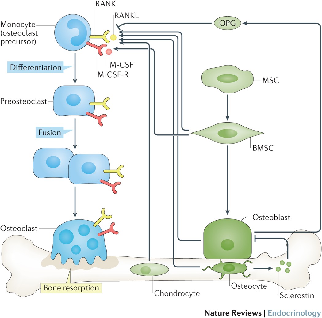
Receptor activator of NF-κB ligand (RANKL) and osteoprotegerin (OPG) are key downstream effectors of bone resorption; tumour necrosis factor (TNF) might synergize with RANKL to superinduce osteoclastic bone resorptionB cells, regulated by T cells, are a key source of basal OPG whereas activated T cells and B cells are key sources of TNF and RANKL in inflammatory conditionsShort-term antiresorptive therapies might safely prevent bone loss associated with combination antiretroviral therapyAnergic and parathyroid-hormone-treated T cells secrete the protein Wnt-10b, which promotes bone formationNovel therapeutic strategies targeting the immune system might promote bone formation and decrease bone resorption to manage osteoporotic bone loss and prevent fracture
Complex Dynamics of Osteoclast Formation and Death in Long-Term Cultures
BackgroundOsteoclasts, cells responsible for bone resorption, contribute to the development of degenerative, metabolic and neoplastic bone diseases, which are often characterized by persistent changes in bone microenvironment. We aimed to investigate the dynamics of osteoclast formation and death in cultures that considerably exceeded the length of standard protocol and to design a mathematical model describing osteoclastogenesis.Methodology/Principal FindingsRAW 264.7 monocytic cells fuse to form multinucleated osteoclasts upon treatment with pro-resorptive cytokine RANKL. We have found that in long-term experiments (15–26 days), the dynamics of changes in osteoclast numbers was remarkably complex and qualitatively variable in different experiments. Whereas 19 of 46 experiments exhibited single peak of osteoclast formation, in 27 experiments we observed development of successive waves of osteoclast formation and death. Periodic changes in osteoclast numbers were confirmed in long-term cultures of mouse bone marrow cells treated with M-CSF and RANKL. Because the dynamics of changes in osteoclast numbers was found to be largely independent of monocytes, a two-species model of ordinary differential equations describing the changes in osteoclasts and monocytes was ineffective in recapitulating the oscillations in osteoclast numbers. Following experimental observation that medium collected from mature osteoclasts inhibited osteoclastogenesis in fresh cultures, we introduced a third variable, factor f, to describe osteoclast-derived inhibitor. This model allowed us to simulate the oscillatory changes in osteoclasts, which were coupled to oscillatory changes in the factor f, whereas monocytes changed exponentially. Importantly, to achieve the experimentally observed oscillations with increasing amplitude, we also had to assume that osteoclast presence stimulates osteoclast formation.Conclusions/SignificanceThis study identifies the critical role for osteoclast autocrine regulation in controlling long-term dynamic of osteoclast formation and death and describes the complementary roles for negative and positive feedback mediators in determining the sharp dynamics of activation and inactivation of osteoclasts.
Osteocyte-Related Cytokines Regulate Osteoclast Formation and Bone Resorption
The process of bone remodeling is the result of the regulated balance between bone cell populations, namely bone-forming osteoblasts, bone-resorbing osteoclasts, and the osteocyte, the mechanosensory cell type. Osteoclasts derived from the hematopoietic stem cell lineage are the principal cells involved in bone resorption. In osteolytic diseases such as rheumatoid arthritis, periodontitis, and osteoporosis, the balance is lost and changes in favor of bone resorption. Therefore, it is vital to elucidate the mechanisms of osteoclast formation and bone resorption. It has been reported that osteocytes express Receptor activator of nuclear factor κΒ ligand (RANKL), an essential factor for osteoclast formation. RANKL secreted by osteocytes is the most important factor for physiologically supported osteoclast formation in the developing skeleton and in pathological bone resorption such as experimental periodontal bone loss. TNF-α directly enhances RANKL expression in osteocytes and promotes osteoclast formation. Moreover, TNF-α enhances sclerostin expression in osteocytes, which also increases osteoclast formation. These findings suggest that osteocyte-related cytokines act directly to enhance osteoclast formation and bone resorption. In this review, we outline the most recent knowledge concerning bone resorption-related cytokines and discuss the osteocyte as the master regulator of bone resorption and effector in osteoclast formation.

Parathyroid Hormone and Teriparatide for the Treatment of Osteoporosis: A Review of the Evidence and Suggested Guidelines for Its Use
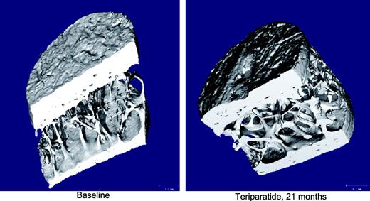
All therapies currently recommended for the management of osteoporosis act mainly to inhibit bone resorption and reduce bone remodeling. PTH and its analog
Bone Remodeling in Post-menopausal Osteoporosis
Bone mass in the skeleton is dependent on the coordinated activities of bone-forming osteoblasts and bone-resorbing osteoclasts in discrete bone multi-cellular units. Remodeling of bone in these units is important not only for maintaining bone mass, but also to repair microdamage, to prevent accumulation of too much old bone, and for mineral homeostasis. The activities of osteoblasts and osteoclasts are controlled by a variety of hormones and cytokines, as well as by mechanical loading. Most importantly, sex hormones are very crucial for keeping bone mass in balance, and the lack of either estrogen or testosterone leads to decreased bone mass and increased risk for osteoporosis. The prevalence of osteoporotic fractures is increasing dramatically in the Western part of the world and is a major health problem in many countries. In the present review, the cellular and molecular mechanisms controlling bone remodeling and the influence of sex hormones on these processes are summarized. In a separate paper in this issue, the pathogenesis of post-menopausal osteoporosis will be compared with that of inflammation-induced bone remodeling, including the evidence for and against the hypothesis that concomitant post-menopausal osteoporotic disease influences the progression of periodontal disease.
Application of a mechanism-based disease systems model for osteoporosis to clinical data
A recently proposed mechanism-based disease systems model for osteoporosis (Schmidt et al., J Pharmacokinet Pharmacodyn 38:873–900, 2011) was applied to clinical data from post-menopausal women (n = 767) receiving various doses of the selective estrogen receptor modulator tibolone. Plasma bone-specific alkaline phosphatase activity and urinary N-telopeptide were used as biomarkers reflecting the activity of osteoblasts (bone forming cells) and osteoclasts (bone removing cells), respectively. These data were analyzed in conjunction with data on osteocalcin and on bone mineral density (BMD) (both lumbar spine and total hip), which reflect the activity of both cell types. While the dynamics of bone turnover markers changes rapidly, closely following changes in the activity of bone cells, changes in BMD are slower and have their own dynamics. Application of the mechanism-based disease systems model to the clinical data allowed for an adequate description of the data and yielded parameter estimates that are consistent with physiological values reported in the literature (Lemaire et al., J Theor Biol 229:293–309, 2004). The fitted model enabled characterization of (i) the critical time scales involved in disease progression, (ii) the dynamics of the system during onset and offset of the therapeutic intervention, and (iii) the distinction between responders and low-responders to tibolone treatment.


TRANCE (tumor necrosis factor–related activation-induced cytokine) is a recently described member of the tumor necrosis factor superfamily that stimulates