ABSTRACT Cellular senescence is a fundamental biological process contributing to aging, often accompanied by extensive chromatin remodeling. Dynamic alterations of three‐dimensional (3D) genomic spatial structure, driven by chromatin reorganization, play a critical role in cell fate determination, but their relevance in therapy‐induced senescence (TIS) remains underexplored. Here, we perform an integrative multi‐omics analysis of Hi‐C, ATAC‐seq, CUT&RUN, and RNA‐seq in primary human fibroblasts undergoing TIS induced by ionizing radiation (RAD) or bleomycin (BLEO). We show that TIS leads to global chromatin decompaction, weakened compartmentalization, and destabilization of topologically associated domains (TADs), alongside widespread loss and rewiring of chromatin loops. Notably, RAD and BLEO elicit distinct changes in distance‐dependent compartment strength and enhancer–promoter (E‐P) loop patterns, reflecting divergent 3D regulatory programs. Importantly, TIS reshapes the chromatin environment around senescence‐associated secretory phenotype (SASP) genes, while their adjacent regions exhibit reduced chromatin interactions, allowing transcriptional activation. Our study reveals that 3D genome remodeling in TIS is highly plastic and context‐dependent and discloses spatial regulation of gene expression during therapy‐induced cellular senescence.
Bone remodeling: A tissue-level process emerging from cell-level molecular algorithms
The human skeleton undergoes constant remodeling throughout the lifetime. Processes occurring on microscopic and molecular scales degrade bone and replace it with new, fully functional tissue. Multiple bone remodeling events occur simultaneously, continuously and independently throughout the body, so that the entire skeleton is completely renewed about every ten years.Bone remodeling is performed by groups of cells called Bone Multicellular Units (BMU). BMUs consist of different cell types, some specialized in the resorption of old bone, others encharged with producing new bone to replace the former. These processes are tightly regulated so that the amount of new bone produced is in perfect equilibrium with that of old bone removed, thus maintaining bone microscopic structure.To date, many regulatory molecules involved in bone remodeling have been identified, but the precise mechanism of BMU operation remains to be fully elucidated. Given the complexity of the signaling pathways already known, one may question whether such complexity is an inherent requirement of the process or whether some subset of the multiple constituents could fulfill the essential role, leaving functional redundancy to serve an alternative safety role. We propose in this work a minimal model of BMU function that involves a limited number of signals able to account for fully functional BMU operation. Our main assumptions were i) at any given time, any cell within a BMU can select only one among a limited choice of decisions, i.e. divide, die, migrate or differentiate, ii) this decision is irreversibly determined by depletion of an appropriate internal inhibitor and iii) the dynamics of any such inhibitor are coupled to that of specific external mediators, such as hormones, cytokines, growth factors. It was thus shown that efficient BMU operation manifests as an emergent process, which results from the individual and collective decisions taken by cells within the BMU unit in the absence of any external planning.
Cellular mechanisms of bone remodeling
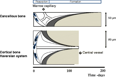
Bone remodeling is a tightly regulated process securing repair of microdamage (targeted remodeling) and replacement of old bone with new bone through sequential osteoclastic resorption and osteoblastic bone formation. The rate of remodeling is regulated by a wide variety of calcitropic hormones (PTH, thyroid hormone, sex steroids etc.). In recent years we have come to appreciate that bone remodeling proceeds in a specialized vascular structure,—the Bone Remodeling Compartment (BRC). The outer lining of this compartment is made up of flattened cells, displaying all the characteristics of lining cells in bone including expression of OPG and RANKL. Reduced bone turnover leads to a decrease in the number of BRCs, while increased turnover causes an increase in the number of BRCs. The secretion of regulatory factors inside a confined space separated from the bone marrow would facilitate local regulation of the remodeling process without interference from growth factors secreted by blood cells in the marrow space. The BRC also creates an environment where cells inside the structure are exposed to denuded bone, which may enable direct cellular interactions with integrins and other matrix factors known to regulate osteoclast/osteoblast activity. However, the denuded bone surface inside the BRC also constitutes an ideal environment for the seeding of bone metastases, known to have high affinity for bone matrix. Circulating osteoclast- and osteoblast precursor cells have been demonstrated in peripheral blood. The dominant pathway regulating osteoclast recruitment is the RANKL/OPG system, while many different factors (RUNX, Osterix) are involved in osteoblast differentiation. Both pathways are modulated by calcitropic hormones.
Dynamic Landscape Analysis of cell fate decisions provides predictive models of neural development from single-cell data
Building a mechanistic understanding of cell fate decisions remains a fundamental goal of developmental biology, with implications for stem cell therapies, regenerative medicine and understanding disease mechanisms. Single-cell transcriptomics provides a detailed picture of the cellular states observed during these decisions, but building dynamic and predictive models from these data remains a challenge. Here, we present dynamic landscape analysis (DLA), an integrative framework that applies dynamical systems theory to identify stable cell states, map transition pathways, and generate a predictive cell fate decision landscape from single-cell data. Applying this framework to vertebrate neural tube development revealed that progenitor specification by Sonic Hedgehog (Shh) can be captured in a landscape with an unexpected topology in which initially divergent lineages converge to the same fate through multiple distinct routes. The model accurately predicted cellular responses and cell fate allocation for unseen dynamic signalling regimes. Cross-species validation using human embryonic organoid data demonstrated conservation of this decision-making architecture. By modelling the dynamic responses that drive cell fate decisions, the DLA framework provides a quantitative and generative framework for extracting mechanistic insights from high-dimensional single-cell data.
Comparing phenotypic manifolds with Kompot: Detecting differential abundance and gene expression at single-cell resolution
Single-cell studies are frequently designed to compare across conditions such as health and disease. However, existing computational approaches typically rely on grouping cells into discrete populations before making comparisons, which can limit resolution for detecting state-dependent changes. Here, we introduce Kompot, a statistical framework for comparative analysis of multi-condition single-cell data. Kompot quantifies both differential abundance, capturing how cells redistribute across the phenotypic space, and differential expression, identifying condition-specific transcriptional changes that may be localized, heterogeneous, or oppositely regulated across states. By modeling cell density and gene expression as continuous functions over a shared cell-state representation, Kompot enables single-cell–resolution inference with principled uncertainty estimates, without requiring predefined clusters or cell types. Applying Kompot to aging murine bone marrow, we identified a continuum of shifts in hematopoietic stem cell and mature cell states, transcriptional remodeling of monocytes independent of compositional changes, and divergent regulation of oxidative stress response genes across cell types. We demonstrate the utility of Kompot in disease settings by identifying cell-state and gene expression changes associated with improved efficacy of combinatorial immunotherapy in melanoma. Additionally, Kompot enables multi-sample comparative analysis by accounting for sample-to-sample heterogeneity. By capturing both global and cell-state–specific effects of perturbation, the Kompot framework is broadly applicable to dissecting condition-specific effects in complex single-cell landscapes. ### Competing Interest Statement The authors have declared no competing interest. National Institutes of Health, R35GM147125, R01CA292932, T32GM136534, S10OD028685 The Mark Foundation for Cancer Research, https://ror.org/00v7th354, Endeavor Award Edward P. Evans Foundation, https://ror.org/03h22gm35, Discovery Research Grant Brotman Baty Institute, https://ror.org/03jxvbk42, Pilot Award

Complex Dynamics of Osteoclast Formation and Death in Long-Term Cultures
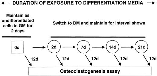
BackgroundOsteoclasts, cells responsible for bone resorption, contribute to the development of degenerative, metabolic and neoplastic bone diseases, which are often characterized by persistent changes in bone microenvironment. We aimed to investigate the dynamics of osteoclast formation and death in cultures that considerably exceeded the length of standard protocol and to design a mathematical model describing osteoclastogenesis.Methodology/Principal FindingsRAW 264.7 monocytic cells fuse to form multinucleated osteoclasts upon treatment with pro-resorptive cytokine RANKL. We have found that in long-term experiments (15–26 days), the dynamics of changes in osteoclast numbers was remarkably complex and qualitatively variable in different experiments. Whereas 19 of 46 experiments exhibited single peak of osteoclast formation, in 27 experiments we observed development of successive waves of osteoclast formation and death. Periodic changes in osteoclast numbers were confirmed in long-term cultures of mouse bone marrow cells treated with M-CSF and RANKL. Because the dynamics of changes in osteoclast numbers was found to be largely independent of monocytes, a two-species model of ordinary differential equations describing the changes in osteoclasts and monocytes was ineffective in recapitulating the oscillations in osteoclast numbers. Following experimental observation that medium collected from mature osteoclasts inhibited osteoclastogenesis in fresh cultures, we introduced a third variable, factor f, to describe osteoclast-derived inhibitor. This model allowed us to simulate the oscillatory changes in osteoclasts, which were coupled to oscillatory changes in the factor f, whereas monocytes changed exponentially. Importantly, to achieve the experimentally observed oscillations with increasing amplitude, we also had to assume that osteoclast presence stimulates osteoclast formation.Conclusions/SignificanceThis study identifies the critical role for osteoclast autocrine regulation in controlling long-term dynamic of osteoclast formation and death and describes the complementary roles for negative and positive feedback mediators in determining the sharp dynamics of activation and inactivation of osteoclasts.
Osteoclasts Provide Coupling Signals to Osteoblast Lineage Cells Through Multiple Mechanisms
Bone remodeling is essential for the repair and replacement of damaged and old bone. The major principle underlying this process is that osteoclast-mediated resorption of a quantum of bone is followed by osteoblast precursor recruitment; these cells differentiate to matrix-producing osteoblasts, which form new bone to replace what was resorbed. Evidence from osteopetrotic syndromes indicate that osteoclasts not only resorb bone, but also provide signals to promote bone formation. Osteoclasts act upon osteoblast lineage cells throughout their differentiation by facilitating growth factor release from resorbed matrix, producing secreted proteins and microvesicles, and expressing membrane-bound factors. These multiple mechanisms mediate the coupling of bone formation to resorption in remodeling. Additional interactions of osteoclasts with osteoblast lineage cells, including interactions with canopy and reversal cells, are required to achieve coordination between bone formation and resorption during bone remodeling.

The bone remodelling cycle
The bone remodelling cycle replaces old and damaged bone and is a highly regulated, lifelong process essential for preserving bone integrity and maintaining mineral homeostasis. During the bone remodelling cycle, osteoclastic resorption is tightly coupled to osteoblastic bone formation. The remodelling cycle occurs within the basic multicellular unit and comprises five co-ordinated steps; activation, resorption, reversal, formation and termination. These steps occur simultaneously but asynchronously at multiple different locations within the skeleton. Study of rare human bone disease and animal models have helped to elucidate the cellular and molecular mechanisms that regulate the bone remodelling cycle. The key signalling pathways controlling osteoclastic bone resorption and osteoblastic bone formation are receptor activator of nuclear factor-κB (RANK)/RANK ligand/osteoprotegerin and canonical Wnt signalling. Cytokines, growth factors and prostaglandins act as paracrine regulators of the cycle, whereas endocrine regulators include parathyroid hormone, vitamin D, calcitonin, growth hormone, glucocorticoids, sex hormones, and thyroid hormone. Disruption of the bone remodelling cycle and any resulting imbalance between bone resorption and formation leads to metabolic bone disease, most commonly osteoporosis. The advances in understanding the cellular and molecular mechanisms underlying bone remodelling have also provided targets for pharmacological interventions which include antiresorptive and anabolic therapies. This review will describe the remodelling process and its regulation, discuss osteoporosis and summarize the commonest pharmacological interventions used in its management.

An Integrated Computational Model of the Bone Microenvironment in Bone-Metastatic Prostate Cancer
Abstract Bone metastasis will impact most men with advanced prostate cancer. The vicious cycle of bone degradation and formation driven by metastatic prostate cells in bone yields factors that drive cancer growth. Mechanistic insights into this vicious cycle have suggested new therapeutic opportunities, but complex temporal and cellular interactions in the bone microenvironment make drug development challenging. We have integrated biologic and computational approaches to generate a hybrid cellular automata model of normal bone matrix homeostasis and the prostate cancer-bone microenvironment. The model accurately reproduces the basic multicellular unit bone coupling process, such that introduction of a single prostate cancer cell yields a vicious cycle similar in cellular composition and pathophysiology to models of prostate-to-bone metastasis. Notably, the model revealed distinct phases of osteolytic and osteogenic activity, a critical role for mesenchymal stromal cells in osteogenesis, and temporal changes in cellular composition. To evaluate the robustness of the model, we assessed the effect of established bisphosphonate and anti-RANKL therapies on bone metastases. At approximately 100% efficacy, bisphosphonates inhibited cancer progression while, in contrast with clinical observations in humans, anti-RANKL therapy fully eradicated metastases. Reducing anti-RANKL yielded clinically similar results, suggesting that better targeting or dosing could improve patient survival. Our work establishes a computational model that can be tailored for rapid assessment of experimental therapies and delivery of precision medicine to patients with prostate cancer with bone metastases. Cancer Res; 74(9); 2391–401. ©2014 AACR.
ResolVI: addressing noise and bias in spatial transcriptomics
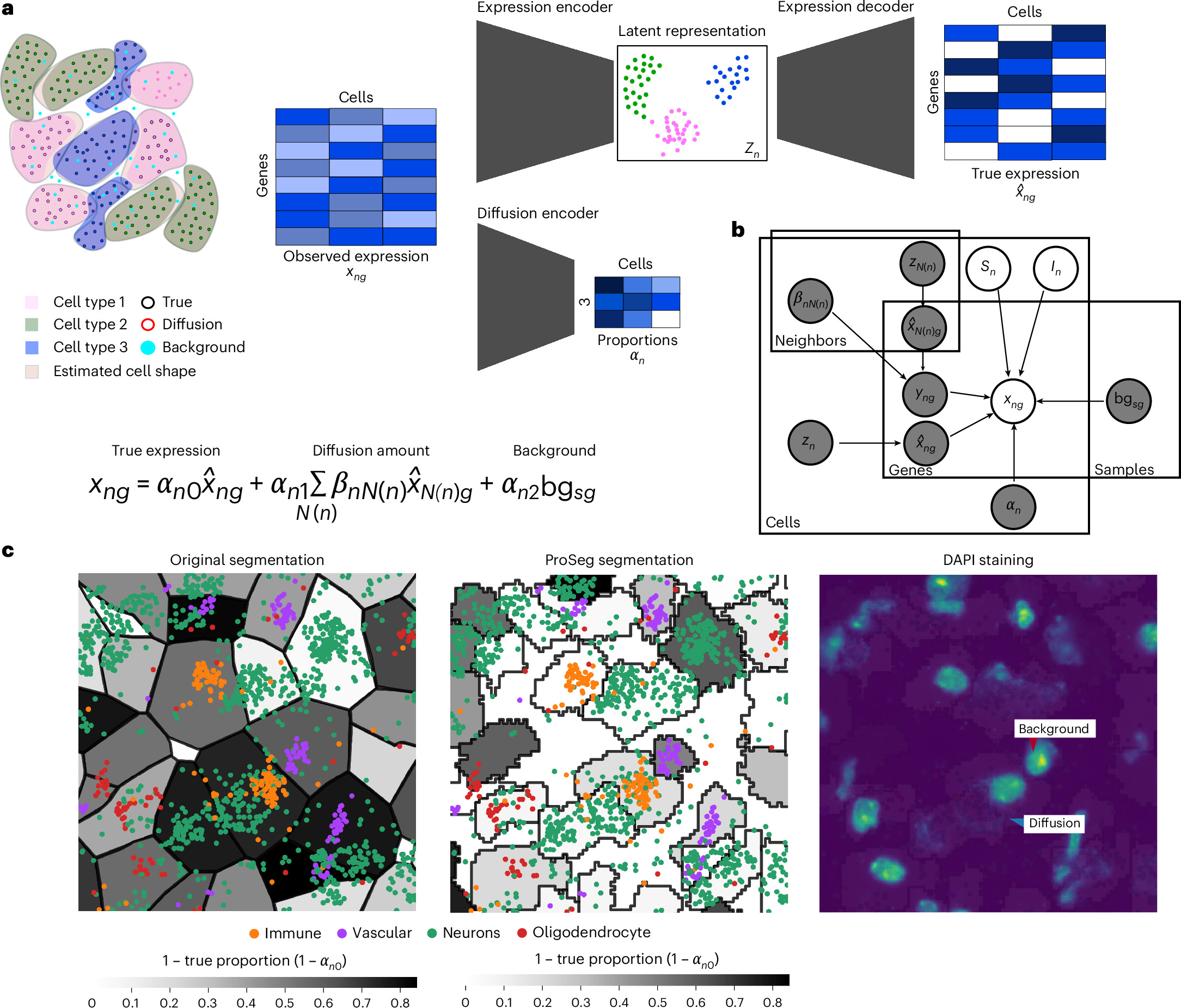
Technologies for estimating RNA expression at high throughput, in intact tissue slices and with high spatial resolution (spatial transcriptomics) shed new light on how cells communicate and tissues function. A fundamental step in analyzing data generated by subcellular resolution spatial transcriptomics technologies is quantification, namely, segmenting the plane into regions, each approximating a cell, and then collating the molecules inside each region to estimate the cellular expression profile. Despite many advances in this area, a persistent problem is that of the incorrect assignment of molecules to cells, which limits many current applications to the level of a priori-defined cell subsets and complicates the discovery of novel cell states. Here we develop resolVI, a model that operates downstream of any segmentation algorithm to generate a probabilistic representation, correcting for the misassignment of molecules, as well as for batch effects and other nuisance factors. We demonstrate that resolVI improves our ability to distinguish between cell states, identify subtle expression changes in space and perform integrated analysis across datasets. ResolVI is available as open source software within scvi-tools.
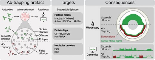
Antibody-trapping presents a widespread pitfall for microscopy and genomics in the nucleus
Abstract. Chromatin has a complex 3D structure and diverse binding proteins that coordinate the genome’s most essential functions. Many microscopy and geno
ABE-ultramax for high-efficiency biallelic adenine base editing in zebrafish
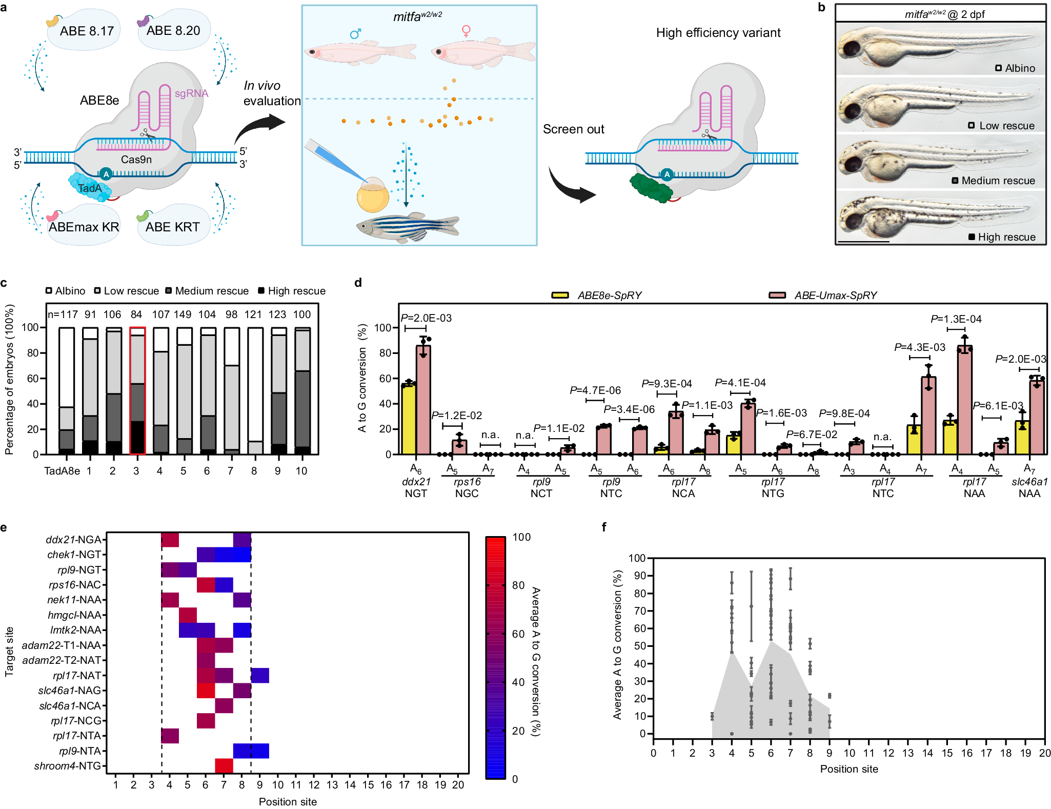
Advancements in CRISPR technology, particularly the development of base editors, revolutionize genetic variant research. When combined with model organisms like zebrafish, base editors significantly accelerate and refine in vivo analysis of genetic variations. However, base editors are restricted by protospacer adjacent motif (PAM) sequences and specific editing windows, hindering their applicability to a broad spectrum of genetic variants. Additionally, base editors can introduce unintended mutations and often exhibit reduced efficiency in living organisms compared to cultured cell lines. Here, we engineer a suite of adenine base editors (ABEs) called ABE-Ultramax (Umax), demonstrating high editing efficiency and low rates of insertions and deletions (indels) in zebrafish. The ABE-Umax suite of editors includes ABEs with shifted, narrowed, or broadened editing windows, reduced bystander mutation frequency, and highly flexible PAM sequence requirements. These advancements have the potential to address previous challenges in disease modeling and advance gene therapy applications.
Molecular Signaling Pathways and Essential Metabolic Elements in Bone Remodeling: An Implication of Therapeutic Targets for Bone Diseases
Bone is one of the dynamic tissues in the human body that undergoes continuous remodelling through subsequent actions of bone cells, osteoclasts, and osteoblasts. Several signal transduction pathways are involved in the transition of mesenchymal stem cells into osteoblasts. These primarily include Runx2, ATF4, Wnt signaling and sympathetic signalling. The differentiation of osteoclasts is controlled by M-CSF, RANKL, and costimulatory signalling. It is well known that bone remodelling is regulated through receptor activator of nuclear factor-kappa B ligand followed by the binding to RANK, which eventually induces the differentiation of osteoclasts. The resorbing osteoclasts secrete TRAP, cathepsin K, MMP-9 and gelatinase to digest the proteinaceous matrix of type I collagen and form a saucer-shaped lacuna along with resorption tunnels in the trabecular bone. Osteoblasts secrete a soluble decoy receptor, osteoprotegerin that prevents the binding of RANK/RANKL and thus moderating osteoclastogenesis. Moreover, bone homeostasis is also regulated by several growth factors, cytokines, calciotropic hormones, parathyroid hormone and sex steroids. The current review presents a correlation of the probable molecular targets underlying the regulation of bone mass and the role of essential metabolic elements in bone remodelling. Targeting these signaling pathways may help design newer therapies for treating bone diseases.

Simulated Interventions to Ameliorate Age-Related Bone Loss Indicate the Importance of Timing
Bone remodeling is the continuous process of bone resorption by osteoclasts and bone formation by osteoblasts, in order to maintain homeostasis. The activity of osteoclasts and osteoblasts is regulated by a network of signaling pathways, including Wnt, parathyroid hormone (PTH), RANKL/OPG and TGF-β, in response to stimuli such as mechanical loading. During aging there is a gradual loss of bone mass due to dysregulation of signaling pathways. This may be due to a decline in physical activity with age and/or changes in hormones and other signaling molecules. In particular, hormones such as PTH have a circadian rhythm which may be disrupted in aging. Due to the complexity of the molecular and cellular networks involved in bone remodeling, several mathematical models have been proposed to aid understanding of the processes involved. However, to date there are no models which explicitly consider the effects of mechanical loading, the circadian rhythm of PTH and the dynamics of signaling molecules on bone remodeling. Therefore, we have constructed a network model of the system using a modular approach which will allow further modifications as required in future research. The model was used to simulate the effects of mechanical loading and also the effects of different interventions such as continuous or intermittent administration of PTH. Our model predicts that the absence of regular mechanical loading and/or an impaired PTH circadian rhythm leads to a gradual decrease in bone mass over time which can be restored by simulated interventions and that the effectiveness of some interventions may depend on their timing.

Osteoprotegerin in Bone Metastases: Mathematical Solution to the Puzzle
Bone is a common site for cancer metastasis. To create space for their growth, cancer cells stimulate bone resorbing osteoclasts. Cytokine RANKL is a key osteoclast activator, while osteoprotegerin (OPG) is a RANKL decoy receptor and an inhibitor of osteoclastogenesis. Consistently, systemic application of OPG decreases metastatic tumor burden in bone. However, OPG produced locally by cancer cells was shown to enhance osteolysis and tumor growth. We propose that OPG produced by cancer cells causes a local reduction in RANKL levels, inducing a steeper RANKL gradient away from the tumor and towards the bone tissue, resulting in faster resorption and tumor expansion. We tested this hypothesis using a mathematical model of nonlinear partial differential equations describing the spatial dynamics of OPG, RANKL, PTHrP, osteoclasts, tumor and bone mass. We demonstrate that at lower expression rates, tumor-derived OPG enhances the chemotactic RANKL gradient and osteolysis, whereas at higher expression rates OPG broadly inhibits RANKL and decreases osteolysis and tumor burden. Moreover, tumor expression of a soluble mediator inducing RANKL in the host tissue, such as PTHrP, is important for correct orientation of the RANKL gradient. A meta-analysis of OPG, RANKL and PTHrP expression in normal prostate, carcinoma and metastatic tissues demonstrated an increase in expression of OPG, but not RANKL, in metastatic prostate cancer, and positive correlation between OPG and PTHrP in metastatic prostate cancer. The proposed mechanism highlights the importance of the spatial distribution of receptors, decoys and ligands, and can be applied to other systems involving regulation of spatially anisotropic processes.
The Expression of Osteoprotegerin and RANK Ligand and the Support of Osteoclast Formation by Stromal-Osteoblast Lineage Cells Is Developmentally Regulated**This work was supported by Grant AG-04875 from the National Institute on Aging.
Abstract. The one or more molecular mechanisms that determine the obligatory sequence of resorption followed by formation during bone remodeling is unclear
Temporal tissue dynamics from a spatial snapshot
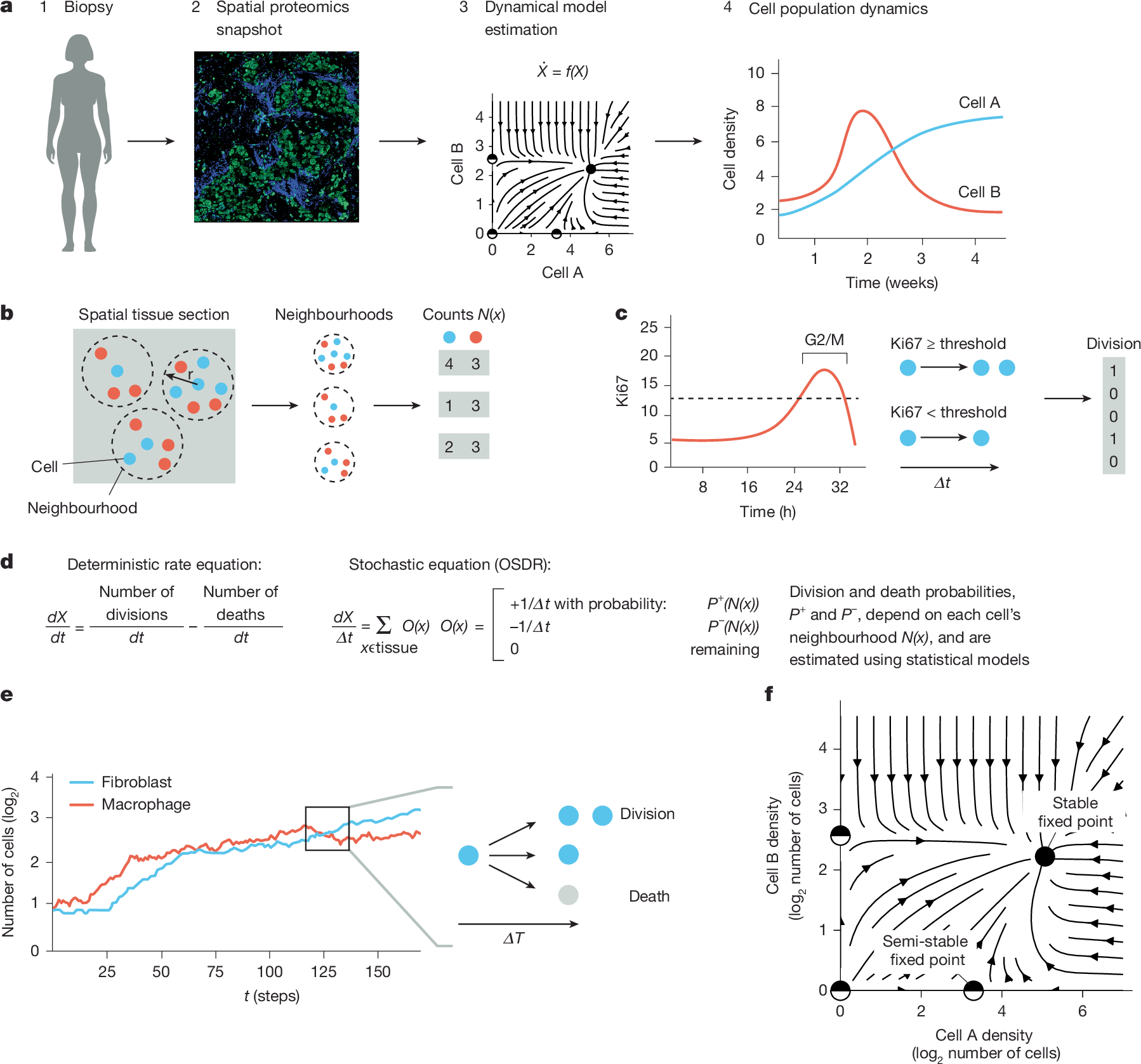
Physiological and pathological processes such as inflammation and cancer emerge from interactions between cells over time1. However, methods to follow cell populations over time within the native context of a human tissue are lacking because a biopsy offers only a single snapshot. Here we present one-shot tissue dynamics reconstruction (OSDR), an approach to estimate a dynamical model of cell populations based on a single tissue sample. OSDR uses spatial proteomics to learn how the composition of cellular neighbourhoods influences division rate, providing a dynamical model of cell population change over time. We apply OSDR to human breast cancer data2–4, and reconstruct two fixed points of fibroblasts and macrophage interactions5,6. These fixed points correspond to hot and cold fibrosis7, in agreement with co-culture experiments that measured these dynamics directly8. We then use OSDR to discover a pulse-generating excitable circuit of T and B cells in the tumour microenvironment, suggesting temporal flares of anticancer immune responses. Finally, we study longitudinal biopsies from a triple-negative breast cancer clinical trial3, in which OSDR predicts the collapse of the tumour cell population in responders but not in non-responders, based on early-treatment biopsies. OSDR can be applied to a wide range of spatial proteomics assays to enable analysis of tissue dynamics based on patient biopsies.