


Oestradiol modulates paracellular permeability and tight junction (TJ) function in endothelia and reproductive tissues, but whether the ovarian hormones and cycle affect the paracellular pathway in the intestinal epithelium remains unclear. Oestrogen receptors (ERs) are expressed in intestinal epithelial cells, and oestradiol regulates epithelium formation. We examined the effects of oestrous cycle stage, oestradiol benzoate (EB), and progesterone (P) on colonic paracellular permeability (CPP) in the female rat, and whether EB affects expression of the TJ proteins in the rat colon and the human colon cell line Caco‐2. In cyclic rats, CPP was determined through lumen‐to‐blood 51 Cr‐labelled EDTA clearance, and in Ussing chambers for dextran permeability. CPP was also examined in ovariectomized (OVX) rats treated with P or EB, with and without the ER antagonist ICI 182,780, or with the selective agonists for ERα (propyl pyrazole triol; PPT) or ERβ (diarylpropionitrile; DPN). In oestrus rats, CPP was reduced ( P < 0.01) relative to dioestrus. In OVX rats, EB dose‐dependently decreased CPP, an effect mimicked by DPN and blocked by ICI 182,780, whereas P had no effect. Oestradiol increased occludin mRNA and protein in the colon ( P < 0.05), but not zona occludens (ZO)‐1. Further, EB and DPN enhanced occludin and junctional adhesion molecule (JAM)‐A expression in Caco‐2 cells without change in ZO‐1, an effect blocked by ICI 182,780. These data show that oestrogen reinforces intestinal epithelial barrier through ERβ‐mediated up‐regulation of the transmembrane proteins occludin and JAM‐A determining paracellular spaces. These findings highlight the importance of the ERβ pathway in the control of colonic paracellular transport and mucosal homeostasis.
The Role of Estrogen in the Control of Rat Osteocyte Apoptosis
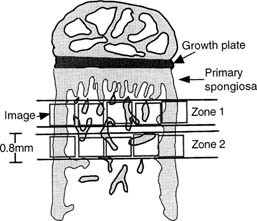
Abstract We have previously shown that estrogen withdrawal by gonadotrophin-releasing hormone analogs (GnRHa) induces osteocyte death via apoptosis in human bone. Although it is likely that the increase in osteocyte death via apoptosis was related to the loss of estrogen, these experiments could not rule out a direct role for the GnRHa. Therefore, in this study, we have used a rat model of ovariectomy (OVX) to determine whether the effect of estrogen withdrawal extends to other species and to clarify the role of estrogen in the maintenance of osteocyte viability. Twelve 9-week-old rats were divided into three treatment groups: sham operated (SHAM) (n = 4), OVX (n = 4), and OVX + estrogen (E2) (25 μg/day) (n = 4). At 3 weeks following the start of treatment, tibial bones were removed. The percentage of osteocytes displaying DNA breaks, using an in situ nick-translation method, was significantly higher in the OVX group compared with the SHAM control in both cortical bone (10.04% vs. 2.31%, respectively; p < 0.0001) and trabecular bone (6.44% vs. 1.58%, respectively; p = 0.003). Addition of estrogen in the OVX animals completely abrogated the increase in osteocyte apoptosis in cortical bone (0.78%) and trabecular bone (1.17%). The percentage of apoptotic osteocytes decreased with increasing distance from the primary/secondary spongiosa interface below the growth plate in the OVX model and the OVX + E2 model. Nuclear morphology and electrophoresis of DNA confirmed the presence of apoptotic cells in the samples. In conclusion, OVX in the rat results in an increase in osteocyte apoptosis as a direct or indirect result of E2 loss. Addition of estrogen in the OVX animals prevents this increase in osteocyte apoptosis. These data confirm an important role for estrogen in the control of osteocyte apoptosis and the maintenance of osteocyte viability. Estrogen deficiency might, through compromising the viability of osteocyte networks, reduce the ability of bone to respond appropriately to loading.
The human ovary exhibits dynamic molecular remodeling in the decades post-menopause
The human ovary is among the first organs to show age-related functional decline, resulting in menopause. Beyond this transition, the postmenopausal ovary is often regarded as quiescent and remains poorly characterized.
Probiotic <i>L. reuteri</i> Treatment Prevents Bone Loss in a Menopausal Ovariectomized Mouse Model
Abstract Estrogen deficiency is a major risk factor for osteoporosis that is associated with bone inflammation and resorption. Half of women over the age of 50 will experience an osteoporosis related fracture in their lifetime, thus novel therapies are needed to combat post‐menopausal bone loss. Recent studies suggest an important role for gut‐bone signaling pathways and the microbiota in regulating bone health. Given that the bacterium Lactobacillus reuteri ATCC PTA 6475 ( L. reuteri ) secretes beneficial immunomodulatory factors, we examined if this candidate probiotic could reduce bone loss associated with estrogen deficiency in an ovariectomized (Ovx) mouse menopausal model. Strikingly, L. reuteri treatment significantly protected Ovx mice from bone loss. Osteoclast bone resorption markers and activators (Trap5 and RANKL) as well as osteoclastogenesis are significantly decreased in L. reuteri ‐treated mice. Consistent with this, L. reuteri suppressed Ovx‐induced increases in bone marrow CD4+ T‐lymphocytes (which promote osteoclastogenesis) and directly suppressed osteoclastogenesis in vitro. We also identified that L. reuteri treatment modifies microbial communities in the Ovx mouse gut. Together, our studies demonstrate that L. reuteri treatment suppresses bone resorption and loss associated with estrogen deficiency. Thus, L. reuteri treatment may be a straightforward and cost‐effective approach to reduce post‐menopausal bone loss. J. Cell. Physiol. 229: 1822–1830, 2014. © 2014 Wiley Periodicals, Inc.

The steroid metabolome in women with premenstrual dysphoric disorder during GnRH agonist-induced ovarian suppression: effects of estradiol and progesterone addback
Clinical evidence suggests that symptoms in premenstrual dysphoric disorder (PMDD) reflect abnormal responsivity to ovarian steroids. This differential steroid sensitivity could be underpinned by abnormal processing of the steroid signal. We used a pharmacometabolomics approach in women with prospectively confirmed PMDD (n=15) and controls without menstrual cycle-related affective symptoms (n=15). All were medication-free with normal menstrual cycle lengths. Notably, women with PMDD were required to show hormone sensitivity in an ovarian suppression protocol. Ovarian suppression was induced for 6 months with gonadotropin-releasing hormone (GnRH)-agonist (Lupron); after 3 months all were randomized to 4 weeks of estradiol (E2) or progesterone (P4). After a 2-week washout, a crossover was performed. Liquid chromatography/tandem mass spectrometry measured 49 steroid metabolites in serum. Values were excluded if >40% were below the limit of detectability (n=21). Analyses were performed with Wilcoxon rank-sum tests using false-discovery rate (q

Epithelial Barrier Function in Gut-Bone Signaling
The intestinal epithelial barrier plays an essential role in maintaining host homeostasis. The barrier regulates nutrient absorption as well as prevents the invasion of pathogenic bacteria in the host. It is composed of epithelial cells, tight junctions, and a mucus layer. Several factors, such as cytokines, diet, and diseases, can affect this barrier. These factors have been shown to increase intestinal permeability, inflammation, and translocation of pathogenic bacteria. In addition, dysregulation of the epithelial barrier can result in inflammatory diseases such as inflammatory bowel disease. Our lab and others have also shown that barrier disruption can have systemic effects including bone loss. In this chapter, we will discuss the current literature to understand the link between intestinal barrier and bone. We will discuss how inflammation, aging, dysbiosis, and metabolic diseases can affect intestinal barrier-bone link. In addition, we will highlight the current suggested mechanism between intestinal barrier and bone.
Lactulose Suppresses Osteoclastogenesis and Ameliorates Estrogen Deficiency-Induced Bone Loss in Mice
<p>Postmenopausal osteoporosis is characterized by excess osteoclastogenesis which leads to net bone loss and brittle fractures. Studies have demonstrated that estrogen deficiency-associated bone loss is microbiota-dependent and could be prevented by probiotics and prebiotics. In this study, we report that orally administered lactulose (20 g/kg, 6 weeks) orally administered significantly inhibited osteoclastogenesis, bone resorption, and prevented ovariectomy (OVX)-induced bone loss in mice. Lactulose increased intestinal <i>Claudin 2, 3 and 15,</i> compared to the OVX group, and lowered pro-osteoclastogenic cytokines levels including tumor necrosis factor-α, interleukin(IL)-6, receptor activator of nuclear factor kappa-Β ligand (RANKL), and IL-17 as well as increased the anti-inflammatory cytokine IL-10 in the intestine, peripheral blood, and bone marrow. Lactulose significantly preserved the number of Foxp3<sup>+</sup> Treg cells in the intestines compared with that in OVX mice. Lactulose altered the composition of intestinal microbiota measured by 16s rDNA sequencing and increased intestinal and serum short-chain fatty acids (SCFAs) levels including acetate, propionate and butyrate which were decreased in OVX mice as measured by gas chromatography. Oral administration of lactulose for 2 weeks significantly lowered the level of bone resorption marker C-telopeptide of type 1 collagen-1 in healthy male young volunteers (aging 20-25 years). In conclusion, lactulose inhibited osteoclastogenesis and bone resorption by altering the intestinal microbiota and increasing SCFAs. Lactulose could serve as an ideal therapeutic agent for postmenopausal osteoporosis.</p>
Critical Role of Estrogens on Bone Homeostasis in Both Male and Female: From Physiology to Medical Implications
Bone is a multi-skilled tissue, protecting major organs, regulating calcium phosphate balance and producing hormones. Its development during childhood determines height and stature as well as resistance against fracture in advanced age. Estrogens are key regulators of bone turnover in both females and males. These hormones play a major role in longitudinal and width growth throughout puberty as well as in the regulation of bone turnover. In women, estrogen deficiency is one of the major causes of postmenopausal osteoporosis. In this review, we will summarize the main clinical and experimental studies reporting the effects of estrogens not only in females but also in males, during different life stages. Effects of estrogens on bone involve either Estrogen Receptor (ER)α or ERβ depending on the type of bone (femur, vertebrae, tibia, mandible), the compartment (trabecular or cortical), cell types involved (osteoclasts, osteoblasts and osteocytes) and sex. Finally, we will discuss new ongoing strategies to increase the benefit/risk ratio of the hormonal treatment of menopause.
Progesterone – Friend or foe?
Estradiol is the “prototypic” sex hormone of women. Yet, women have another sex hormone, which is often disregarded: Progesterone. The goal of this article is to provide a comprehensive review on progesterone, and its metabolite allopregnanolone, emphasizing three key areas: biological properties, main functions, and effects on mood in women. Recent years of intensive research on progesterone and allopregnanolone have paved the way for new treatment of postpartum depression. However, treatment for premenstrual syndrome and premenstrual dysphoric disorder as well as contraception that women can use without risking mental health problems are still needed. As far as progesterone is concerned, we might be dealing with a two-edged sword: while its metabolite allopregnanolone has been proven useful for treatment of PPD, it may trigger negative symptoms in women with PMS and PMDD. Overall, our current knowledge on the beneficial and harmful effects of progesterone is limited and further research is imperative.
Dried Plum’s Polyphenolic Compounds and Carbohydrates Contribute to Its Osteoprotective Effects and Exhibit Prebiotic Activity in Estrogen Deficient C57BL/6 Mice
Evidence of dried plum’s benefits on bone continues to emerge. This study investigated the contribution of the fruit’s polyphenol (PP) and carbohydrate (CHO) components on a bone model of postmenopausal osteoporosis to explore their prebiotic activity. Osteopenic ovariectomized mice were fed diets supplemented with dried plum, a crude extract of dried plum’s polyphenolic compounds, or the PP or CHO fraction of the crude extract. The effects of treatments on the bone phenotype were assessed at 5 and 10 weeks as well as the prebiotic activity of the different components of dried plum. Both the CHO and PP fractions of the extract contributed to the effects on bone with the CHO suppressing bone formation and resorption, and the PP temporally down-regulating formation. The PP and CHO components also altered the gut microbiota and cecal short chain fatty acids. These findings demonstrate that the CHO as well as the PP components of dried plum have potential prebiotic activity, but they have differential roles in mediating the alterations in bone formation and resorption that protect bone in estrogen deficiency.
Estrogens and Androgens in Skeletal Physiology and Pathophysiology
Estrogens and androgens influence the growth and maintenance of the mammalian skeleton and are responsible for its sexual dimorphism. Estrogen deficiency at menopause or loss of both estrogens and androgens in elderly men contribute to the development of osteoporosis, one of the most common and impactful metabolic diseases of old age. In the last 20 years, basic and clinical research advances, genetic insights from humans and rodents, and newer imaging technologies have changed considerably the landscape of our understanding of bone biology as well as the relationship between sex steroids and the physiology and pathophysiology of bone metabolism. Together with the appreciation of the side effects of estrogen-related therapies on breast cancer and cardiovascular diseases, these advances have also drastically altered the treatment of osteoporosis. In this article, we provide a comprehensive review of the molecular and cellular mechanisms of action of estrogens and androgens on bone, their influences on skeletal homeostasis during growth and adulthood, the pathogenetic mechanisms of the adverse effects of their deficiency on the female and male skeleton, as well as the role of natural and synthetic estrogenic or androgenic compounds in the pharmacotherapy of osteoporosis. We highlight latest advances on the crosstalk between hormonal and mechanical signals, the relevance of the antioxidant properties of estrogens and androgens, the difference of their cellular targets in different bone envelopes, the role of estrogen deficiency in male osteoporosis, and the contribution of estrogen or androgen deficiency to the monomorphic effects of aging on skeletal involution.

When Women Initiate Estrogen Therapy Matters | The Menopause Society
New large-scale analysis shows potential long-term benefits of initiating hormones during perimenopause

Progesterone and plasma metabolites in women with and in those without premenstrual dysphoric disorder
BACKGROUND: The molecular mechanisms underpinning the progesterone-triggering mood symptoms in women with premenstrual dysphoric disorder (PMDD) are unknown. Cell metabolism is a potential source of variability. Very little is known about the effect of progesterone sensitivity on the metabolome. In this study, we aimed to characterize the effects of progesterone on the global metabolic profile and explore the differences between women with PMDD and controls. METHODS: Plasma was obtained from 12 women with prospectively confirmed PMDD and 25 controls under two hormone conditions: (1) gonadal suppression induced by leuprolide acetate (3.75 mg IM monthly) and (2) add-back phase with leuprolide and progesterone (200 mg twice daily by vaginal suppository). The global metabolic profile was obtained using liquid and gas chromatography followed by mass spectrometry. Differences between groups and time points were tested using repeated measures analysis of variance. The false discovery rate was calculated to account for multiple testing. RESULTS: Amino acids and their derivatives represented 78% (28/36) of the known compounds that were found in significantly lower plasma concentrations after progesterone administration than during gonadal suppression. The concentration of tyrosine was nominally significantly decreased after progesterone add-back in controls, but not in cases (P = 0.02). CONCLUSION: Plasma levels of some amino acids are decreased in response to progesterone. Albeit preliminary, evidence further suggests that progesterone has a different effect on the metabolic profiles of women with PMDD compared to controls. Further research is needed to replicate our findings in a larger sample and to identify the unknown compounds, especially those differentially expressed.

Mucus barrier, mucins and gut microbiota: the expected slimy partners?
The gastrointestinal tract is often considered as a key organ involved in the digestion of food and providing nutrients to the body for proper maintenance. However, this system is composed of organs that are extremely complex. Among the different parts, the intestine is viewed as an incredible surface of contact with the environment and is colonised by hundreds of trillions of gut microbes. The role of the gut barrier has been studied for decades, but the exact mechanisms involved in the protection of the gut barrier are various and complementary. Among them, the integrity of the mucus barrier is one of the first lines of protection of the gastrointestinal tract. In the past, this ‘slimy’ partner was mostly considered a simple lubricant for facilitating the progression of the food bolus and the stools in the gut. Since then, different researchers have made important progress, and currently, the regulation of this mucus barrier is gaining increasing attention from the scientific community. Among the factors influencing the mucus barrier, the microbiome plays a major role in driving mucus changes. Additionally, our dietary habits (ie, high-fat diet, low-fibre/high-fibre diet, food additives, pre- probiotics) influence the mucus at different levels. Given that the mucus layer has been linked with the appearance of diseases, proper knowledge is highly warranted. Here, we debate different aspects of the mucus layer by focusing on its chemical composition, regulation of synthesis and degradation by the microbiota as well as some characteristics of the mucus layer in both physiological and pathological situations.
Estrogen and the skeleton
Estrogen is the major hormonal regulator of bone metabolism in women and men. Therefore, there is considerable interest in unraveling the pathways by which estrogen exerts its protective effects on bone. Although the major consequence of the loss of estrogen is an increase in bone resorption, estrogen deficiency is associated with a gap between bone resorption and formation, indicating that estrogen is also important for maintaining bone formation at the cellular level. Direct estrogen effects on osteocytes, osteoclasts, and osteoblasts lead to inhibition of bone remodeling, decreased bone resorption, and maintenance of bone formation, respectively.

Prebiotics Enhance Magnesium Absorption and Inulin‐based Fibers Exert Chronic Effects on Calcium Utilization in a Postmenopausal Rodent Model
Abstract: Age‐related changes in calcium metabolism play a role in the development of osteoporosis. A 4‐wk feeding study was conducted in 5‐mo‐old ovariectomized (OVX) Sprague‐Dawley rats to assess the effect of various dietary fibers on mineral metabolism and bone health parameters. There were 6 treatment groups: sham‐Control, OVX‐Control, OVX rats receiving daily estradiol (E 2 ) injections, and OVX rats receiving an AIN‐93M diet supplement with either an inulin‐based fiber (Synergy1® or Fruitafit HD®) or a novel fiber (polydextrose) at 5% wt. of diet. Calcium and magnesium metabolic balances were performed after early (3 d) and late exposure (4 wk) to dietary treatments. Rats receiving polydextrose had significantly higher net calcium absorption efficiency and retention than all control groups and a trend ( P ≤ 0.10) for higher calcium absorption when compared to inulin‐based fibers after early exposure but the advantage did not persist over long‐term exposure. The inulin‐based fibers had positive chronic effects on calcium metabolism that were related to changes in the gut, that is, production of short chain fatty acids and higher cecal wall weights. All fibers improved magnesium absorption and retention in early and late metabolic balances; effects on magnesium metabolism were more pronounced than for calcium. Practical Application: Steady growth in US middle‐aged and elderly populations has led to higher incidences of several chronic diseases including osteoporosis, a bone disease that primarily affects postmenopausal women. Recent research suggests that certain dietary fibers (prebiotics) enhance mineral absorption and may impart bone health benefits. This work examines the impact of prebiotic supplementation on mineral metabolism and bone health using a postmenopausal rat model. Study findings will aid future investigations in ascertaining the factors related to potential bone health benefits of prebiotic which will aid in developing an effective prebiotics food product/supplement that will address the bone health needs of consumers.

Bone Remodeling in Post-menopausal Osteoporosis
Bone mass in the skeleton is dependent on the coordinated activities of bone-forming osteoblasts and bone-resorbing osteoclasts in discrete bone multi-cellular units. Remodeling of bone in these units is important not only for maintaining bone mass, but also to repair microdamage, to prevent accumulation of too much old bone, and for mineral homeostasis. The activities of osteoblasts and osteoclasts are controlled by a variety of hormones and cytokines, as well as by mechanical loading. Most importantly, sex hormones are very crucial for keeping bone mass in balance, and the lack of either estrogen or testosterone leads to decreased bone mass and increased risk for osteoporosis. The prevalence of osteoporotic fractures is increasing dramatically in the Western part of the world and is a major health problem in many countries. In the present review, the cellular and molecular mechanisms controlling bone remodeling and the influence of sex hormones on these processes are summarized. In a separate paper in this issue, the pathogenesis of post-menopausal osteoporosis will be compared with that of inflammation-induced bone remodeling, including the evidence for and against the hypothesis that concomitant post-menopausal osteoporotic disease influences the progression of periodontal disease.
