Protecting the Guts Regulatory T cells (T regs ) in the gut are important sentinels in maintaining the peace between our gut and its trillions of resident bacteria and have been shown to be regulated by specific strains of bacteria in mouse models. Smith et al. (p. 569 , published online 4 July; see the Perspective by Bollrath and Powrie ) asked whether metabolite(s) generated by resident bacterial species may regulate T regs in the gut. Indeed, short-chain fatty acids (SCFAs), bacterial fermentation products of dietary fibers produced by a range of bacteria, restored colonic T reg numbers in mice devoid of a gut microbiota and increased T reg numbers in colonized mice. The effects of SCFAs on T regs were mediated through GPCR43, a receptor for SCFAs, which is expressed on colonic T regs . Mice fed SCFAs were protected against experimentally induced colitis in a manner that was dependent on GPR43-expressing T regs . , Bacterial fermentation products regulate the number and function of regulatory T cells in the mouse colon. [Also see Perspective by Bollrath and Powrie ] , Regulatory T cells (T regs ) that express the transcription factor Foxp3 are critical for regulating intestinal inflammation. Candidate microbe approaches have identified bacterial species and strain-specific molecules that can affect intestinal immune responses, including species that modulate T reg responses. Because neither all humans nor mice harbor the same bacterial strains, we posited that more prevalent factors exist that regulate the number and function of colonic T regs . We determined that short-chain fatty acids, gut microbiota–derived bacterial fermentation products, regulate the size and function of the colonic T reg pool and protect against colitis in a Ffar2 -dependent manner in mice. Our study reveals that a class of abundant microbial metabolites underlies adaptive immune microbiota coadaptation and promotes colonic homeostasis and health.
Regulation of Intestinal Barrier Function by Microbial Metabolites
The human gastrointestinal tract (GI) harbors a diverse population of microbial life that continually shapes host pathophysiological responses. Despite readily available abundant metagenomic data, the functional dynamics of gut microbiota remain to be explored in various health and disease conditions. Microbiota generate a variety of metabolites from dietary products that influence host health and pathophysiological functions. Since gut microbial metabolites are produced in close proximity to gut epithelium, presumably they have significant impact on gut barrier function and immune responses.

Metabolites produced by commensal bacteria promote peripheral regulatory T-cell generation
In mice, provision of butyrate—a short-chain fatty acid produced by commensal microorganisms during starch fermentation—facilitates extrathymic generation and differentiation of Foxp3+ regulatory T cells, demonstrating that metabolic by-products are sensed by cells of the immune system and affect the balance between pro- and anti-inflammatory cells.

Activation of Gpr109a, Receptor for Niacin and the Commensal Metabolite Butyrate, Suppresses Colonic Inflammation and Carcinogenesis
Commensal gut microflora and dietary fiber protect against colonic inflammation and colon cancer through unknown targets. Butyrate, a bacterial product from fermentation of dietary fiber in the colon, has been implicated in this process. GPR109A (encoded by Niacr1) is a receptor for butyrate in the colon. GPR109A is also a receptor for niacin, which is also produced by gut microbiota and suppresses intestinal inflammation. Here we showed that Gpr109a signaling promoted anti-inflammatory properties in colonic macrophages and dendritic cells and enabled them to induce differentiation of Treg cells and IL-10-producing T cells.

Commensal microbe-derived butyrate induces the differentiation of colonic regulatory T cells
The gut microbial metabolite butyrate is shown to induce the differentiation of colonic T regulatory cells in mice and to ameliorate the development of colitis; it also increases histone H3 acetylation at the Foxp3 promoter.

Dietary Fiber and Bacterial SCFA Enhance Oral Tolerance and Protect against Food Allergy through Diverse Cellular Pathways
Tan et al. examine the beneficial roles of dietary fiber in peanut allergy using mice. The authors find that this effect involves reshaping of the gut microbiota as well as increased levels of short-chain fatty acids and activity of their receptors GPR43 and GPR109a. High-fiber feeding also increased tolerogenic CD103+ DCs activity, leading to increased Treg cell differentiation.

The contribution of gut bacterial metabolites in the human immune signaling pathway of non-communicable diseases
The interaction disorder between gut microbiota and its host has been documented in different non-communicable diseases (NCDs) such as metabolic syndrome, neurodegenerative disease, and autoimmune ...

The pleiotropic effects of prebiotic galacto-oligosaccharides on the aging gut
Prebiotic galacto-oligosaccharides (GOS) have an extensively demonstrated beneficial impact on intestinal health. In this study, we determined the impact of GOS diets on hallmarks of gut aging: microbiome dysbiosis, inflammation, and intestinal barrier defects (“leaky gut”). We also evaluated if short-term GOS feeding influenced how the aging gut responded to antibiotic challenges in a mouse model of Clostridioides difficile infection. Finally, we assessed if colonic organoids could reproduce the GOS responder—non-responder phenotypes observed in vivo.

From the gut to the lung: microbiota-associated metabolites as regulators of respiratory immunometabolism
The development and progression of respiratory diseases are influenced by both the local pulmonary microenvironment and the intestinal ecosystem. Research on the gut-lung axis has shown that diverse small-molecule metabolites produced or modified by the gut microbiota can cross the intestinal barrier and enter the systemic circulation, where they may modulate immune-cell response thresholds, functional polarisation, and inflammatory dynamics in the distal lung. The local respiratory microbiota may also contribute to the pulmonary metabolic microenvironment, although its role in metabolite production and immune regulation remains less clearly defined.

Mucus barrier, mucins and gut microbiota: the expected slimy partners?
The gastrointestinal tract is often considered as a key organ involved in the digestion of food and providing nutrients to the body for proper maintenance. However, this system is composed of organs that are extremely complex. Among the different parts, the intestine is viewed as an incredible surface of contact with the environment and is colonised by hundreds of trillions of gut microbes. The role of the gut barrier has been studied for decades, but the exact mechanisms involved in the protection of the gut barrier are various and complementary. Among them, the integrity of the mucus barrier is one of the first lines of protection of the gastrointestinal tract. In the past, this ‘slimy’ partner was mostly considered a simple lubricant for facilitating the progression of the food bolus and the stools in the gut. Since then, different researchers have made important progress, and currently, the regulation of this mucus barrier is gaining increasing attention from the scientific community. Among the factors influencing the mucus barrier, the microbiome plays a major role in driving mucus changes. Additionally, our dietary habits (ie, high-fat diet, low-fibre/high-fibre diet, food additives, pre- probiotics) influence the mucus at different levels. Given that the mucus layer has been linked with the appearance of diseases, proper knowledge is highly warranted. Here, we debate different aspects of the mucus layer by focusing on its chemical composition, regulation of synthesis and degradation by the microbiota as well as some characteristics of the mucus layer in both physiological and pathological situations.
Gut microbiota and bone metabolism
Abstract Osteoporosis is the most common metabolic skeletal disease. It is characterized by the deterioration of the skeletal microarchitecture and bone loss, leading to ostealgia, and even bone fractures. Accumulating evidence has indicated that there is an inextricable relationship between the gut microbiota (GM) and bone homeostasis involving host–microbiota crosstalk. Any perturbation of the GM can play an initiating and reinforcing role in disrupting the bone remodeling balance during the development of osteoporosis. Although the GM is known to influence bone metabolism, the mechanisms associated with these effects remain unclear. Herein, we review the current knowledge of how the GM affects bone metabolism in health and disease, summarize the correlation between pathogen‐associated molecular patterns of GM structural components and bone metabolism, and discuss the potential mechanisms underlying how GM metabolites regulate bone turnover. Deciphering the complicated relationship between the GM and bone health will provide new insights into the prevention and treatment of osteoporosis.

Specific inulin‐type fructan fibers protect against autoimmune diabetes by modulating gut immunity, barrier function, and microbiota homeostasis
Scope Dietary fibers capable of modifying gut barrier and microbiota homeostasis affect the progression of type 1 diabetes (T1D). Here, we aim to compare modulatory effects of inulin‐type fructans (ITFs), natural soluble dietary fibers with different degrees of fermentability from chicory root, on T1D development in nonobese diabetic mice. Methods and results Female nonobese diabetic mice were weaned to long‐ and short‐chain ITFs [ITF(l) and ITF(s), 5%] supplemented diet up to 24 weeks. T1D incidence, pancreatic‐gut immune responses, gut barrier function, and microbiota composition were analyzed. ITF(l) but not ITF(s) supplementation dampened the incidence of T1D. ITF(l) promoted modulatory T‐cell responses, as evidenced by increased CD25 + Foxp3 + CD4 + regulatory T cells, decreased IL17A + CD4 + Th17 cells, and modulated cytokine production profile in the pancreas, spleen, and colon. Furthermore, ITF(l) suppressed NOD like receptor protein 3 caspase‐1‐p20‐IL‐1β inflammasome in the colon. Expression of barrier reinforcing tight junction proteins occludin and claudin‐2, antimicrobial peptides β‐defensin‐1, and cathelicidin‐related antimicrobial peptide as well as short‐chain fatty acid production were enhanced by ITF(l). Next‐generation sequencing analysis revealed that ITF(l) enhanced Firmicutes/Bacteroidetes ratio to an antidiabetogenic balance and enriched modulatory Ruminococcaceae and Lactobacilli . Conclusion Our data demonstrate that ITF(l) but not ITF(s) delays the development of T1D via modulation of gut‐pancreatic immunity, barrier function, and microbiota homeostasis.

The gut microbiota regulates bone mass in mice
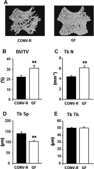
Abstract The gut microbiota modulates host metabolism and development of immune status. Here we show that the gut microbiota is also a major regulator of bone mass in mice. Germ-free (GF) mice exhibit increased bone mass associated with reduced number of osteoclasts per bone surface compared with conventionally raised (CONV-R) mice. Colonization of GF mice with a normal gut microbiota normalizes bone mass. Furthermore, GF mice have decreased frequency of CD4+ T cells and CD11b+/GR 1 osteoclast precursor cells in bone marrow, which could be normalized by colonization. GF mice exhibited reduced expression of inflammatory cytokines in bone and bone marrow compared with CONV-R mice. In summary, the gut microbiota regulates bone mass in mice, and we provide evidence for a mechanism involving altered immune status in bone and thereby affected osteoclast-mediated bone resorption. Further studies are required to evaluate the gut microbiota as a novel therapeutic target for osteoporosis. © 2012 American Society for Bone and Mineral Research.
The mycobiome as integral part of the gut microbiome: crucial role of symbiotic fungi in health and disease
The gut mycobiome significantly affects host health and immunity. However, most studies have focused on symbiotic bacteria in the gut microbiome, whereas less attention has been given to symbiotic fungi. Although fungi constitute only 0.01%-0.1% of the gut microbiome, their larger size and unique immunoregulatory functions make them significant. Factors like diet, antimicrobials use, and age can disrupt the fungal community, leading to dysbiosis. Fungal-bacterial-host immune interactions are critical in maintaining gut homeostasis, with fungi playing a role in mediating immune responses such as Th17 cell activation. This review highlights methods for studying gut fungi, the composition and influencing factors of the gut mycobiome, and its potential in therapeutic interventions for intestinal and hepatic diseases. We aim to provide new insights into the underexplored role of gut fungi in human health.

Epithelial Barrier Function in Gut-Bone Signaling
The intestinal epithelial barrier plays an essential role in maintaining host homeostasis. The barrier regulates nutrient absorption as well as prevents the invasion of pathogenic bacteria in the host. It is composed of epithelial cells, tight junctions, and a mucus layer. Several factors, such as cytokines, diet, and diseases, can affect this barrier. These factors have been shown to increase intestinal permeability, inflammation, and translocation of pathogenic bacteria. In addition, dysregulation of the epithelial barrier can result in inflammatory diseases such as inflammatory bowel disease. Our lab and others have also shown that barrier disruption can have systemic effects including bone loss. In this chapter, we will discuss the current literature to understand the link between intestinal barrier and bone. We will discuss how inflammation, aging, dysbiosis, and metabolic diseases can affect intestinal barrier-bone link. In addition, we will highlight the current suggested mechanism between intestinal barrier and bone.
Galactooligosaccharides Improve Mineral Absorption and Bone Properties in Growing Rats through Gut Fermentation
Galactooligosaccharides (GOS), prebiotic nondigestible oligosaccharides derived from lactose, have the potential for improving mineral balance and bone properties. This study examined the dose–response effect of GOS supplementation on calcium and magnesium absorption, mineral retention, bone properties, and gut microbiota in growing rats. Seventy-five 4-week-old male Sprague–Dawley rats were randomized into one of five treatment groups (n = 15/group) and fed a diet containing 0, 2, 4, 6, or 8% GOS by weight for 8 weeks. Dietary GOS significantly decreased cecal pH and increased cecal wall weight and content weight in a dose-dependent manner (p < 0.0001). Fingerprint patterns of the 16S rRNA gene PCR-DGGE from fecal DNA indicated the variance of bacterial community structure, which was primarily explained by GOS treatments (p = 0.0001). Quantitative PCR of the samples revealed an increase in the relative proportion of bifidobacteria with GOS (p = 0.0001). Net calcium absorption was increased in a dose–response manner (p < 0.01) with GOS supplementation. Dietary GOS also increased (p < 0.02) net magnesium absorption, femur 45Ca uptake, calcium and magnesium retention, and femur and tibia breaking strength. Distal femur total and trabecular volumetric bone mineral density (vBMD) and area and proximal tibia vBMD increased (p < 0.02) with GOS supplementation. Trabecular-rich bones, that is, those that rapidly turn over, were most benefited. Regression modeling showed that GOS benefited calcium and magnesium utilization and vBMD through decreased cecal pH, increased cecal wall and content weight, and increased proportion of bifidobacteria.
Probiotic <i>Lactobacillus reuteri</i> Prevents Postantibiotic Bone Loss by Reducing Intestinal Dysbiosis and Preventing Barrier Disruption
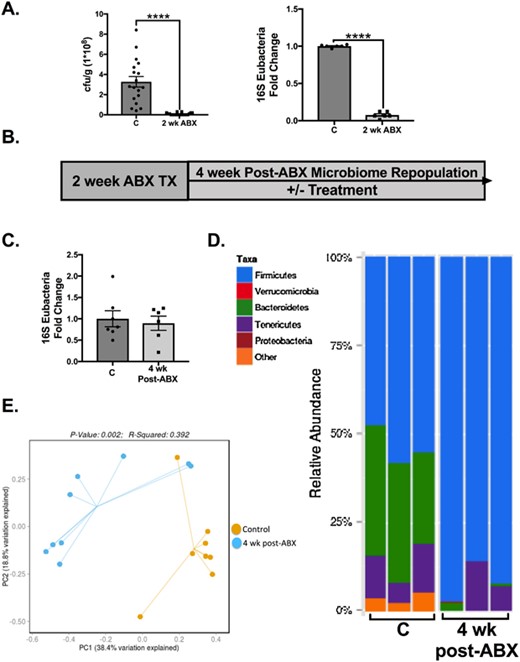
ABSTRACT Antibiotic treatment, commonly prescribed for bacterial infections, depletes and subsequently causes long-term alterations in intestinal microbiota composition. Knowing the importance of the microbiome in the regulation of bone density, we investigated the effect of postantibiotic treatment on gut and bone health. Intestinal microbiome repopulation at 4-weeks postantibiotic treatment resulted in an increase in the Firmicutes:Bacteroidetes ratio, increased intestinal permeability, and notably reduced femoral trabecular bone volume (approximately 30%, p < 0.01). Treatment with a mucus supplement (a high-molecular-weight polymer, MDY-1001 [MDY]) prevented the postantibiotic-induced barrier break as well as bone loss, indicating a mechanistic link between increased intestinal permeability and bone loss. A link between the microbiome composition and bone density was demonstrated by supplementing the mice with probiotic bacteria. Specifically, Lactobacillus reuteri, but not Lactobacillus rhamnosus GG or nonpathogenic Escherichia coli, reduced the postantibiotic elevation of the Firmicutes:Bacteroidetes ratio and prevented femoral and vertebral trabecular bone loss. Consistent with causing bone loss, postantibiotic-induced dysbiosis decreased osteoblast and increased osteoclast activities, changes that were prevented by both L. reuteri and MDY. These data underscore the importance of microbial dysbiosis in the regulation of intestinal permeability and bone health, as well as identify L. reuteri and MDY as novel therapies for preventing these adverse effects. © 2018 American Society for Bone and Mineral Research.