


A new soluble mediator was found in supernatant fluid from cultures of human peripheral blood leukocytes that were stimulated by phytohemagglutinin, or by antigenic material present in human dental plaque deposits. This soluble factor produced bone resorption in organ cultures of fetal rat bones as measured by increased release of calcium-45, and also increased the number of active osteoclasts.
The Effect of Inflammation on Bone
Bone remodeling is the continual process to renew the adult skeleton through the sequential action of osteoblasts and osteoclasts. Nuclear factor RANK, an osteoclast receptor, and its ligand RANKL, expressed on the surface of osteoblasts, result in coordinated control of bone remodeling. Inflammation, a feature of illness and injury, plays a distinct role in skewing this process toward resorption. It does so via the interaction of inflammatory mediators and their related peptides with osteoblasts and osteoclasts, to alter the expression of RANK and RANKL. Such chemical mediators include TNFa, glucocorticoids, histamine, bradykinin, PGE2, systemic RANKL from immune cells, and interleukins 1 and 6. A thorough understanding of bone response to injury and disease, and ability to detect such biomarkers, as well as imaging to identify early structural and mechanical property changes in bone architecture, is important in improving management and outcomes of bone related pathology. While gut health and vitamin and mineral availability appear vitally important, there are very few intervention strategies currently available to favorably alter bone pathology induced by inflammation. Further research is required in this field to advance early detection and treatments.

The amazing osteocyte
Abstract The last decade has provided a virtual explosion of data on the molecular biology and function of osteocytes. Far from being the “passive placeholder in bone,” this cell has been found to have numerous functions, such as acting as an orchestrator of bone remodeling through regulation of both osteoclast and osteoblast activity and also functioning as an endocrine cell. The osteocyte is a source of soluble factors not only to target cells on the bone surface but also to target distant organs, such as kidney, muscle, and other tissues. This cell plays a role in both phosphate metabolism and calcium availability and can remodel its perilacunar matrix. Osteocytes compose 90% to 95% of all bone cells in adult bone and are the longest lived bone cell, up to decades within their mineralized environment. As we age, these cells die, leaving behind empty lacunae that frequently micropetrose. In aged bone such as osteonecrotic bone, empty lacunae are associated with reduced remodeling. Inflammatory factors such as tumor necrosis factor and glucocorticoids used to treat inflammatory disease induce osteocyte cell death, but by different mechanisms with potentially different outcomes. Therefore, healthy, viable osteocytes are necessary for proper functionality of bone and other organs. © 2011 American Society for Bone and Mineral Research.
Biology of Bone Tissue: Structure, Function, and Factors That Influence Bone Cells
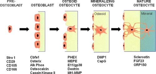
Bone tissue is continuously remodeled through the concerted actions of bone cells, which include bone resorption by osteoclasts and bone formation by osteoblasts, whereas osteocytes act as mechanosensors and orchestrators of the bone remodeling process. This process is under the control of local (e.g., growth factors and cytokines) and systemic (e.g., calcitonin and estrogens) factors that all together contribute for bone homeostasis. An imbalance between bone resorption and formation can result in bone diseases including osteoporosis. Recently, it has been recognized that, during bone remodeling, there are an intricate communication among bone cells. For instance, the coupling from bone resorption to bone formation is achieved by interaction between osteoclasts and osteoblasts. Moreover, osteocytes produce factors that influence osteoblast and osteoclast activities, whereas osteocyte apoptosis is followed by osteoclastic bone resorption. The increasing knowledge about the structure and functions of bone cells contributed to a better understanding of bone biology. It has been suggested that there is a complex communication between bone cells and other organs, indicating the dynamic nature of bone tissue. In this review, we discuss the current data about the structure and functions of bone cells and the factors that influence bone remodeling.

Control of Bone Matrix Properties by Osteocytes
Osteocytes make up 90-95% of the cellular content of bone and form a rich dendritic network with a vastly greater surface area than either osteoblasts or osteoclasts. Osteocytes are well positioned to play a role in bone homeostasis by interacting directly with the matrix; however, the ability for these cells to modify bone matrix remains incompletely understood. With techniques for examining the nano- and microstructure of bone matrix components including hydroxyapatite and type I collagen becoming more widespread, there is great potential to uncover novel roles for the osteocyte in maintaining bone quality. In this review, we begin with an overview of osteocyte biology and the lacunar-canalicular system. Next, we describe recent findings from in vitro models of osteocytes, focusing on the transitions in cellular phenotype as they mature. Finally, we describe historical and current research on matrix alteration by osteocytes in vivo, focusing on the exciting potential for osteocytes to directly form, degrade, and modify the mineral and collagen in their surrounding matrix.

Osteocyte-Related Cytokines Regulate Osteoclast Formation and Bone Resorption
The process of bone remodeling is the result of the regulated balance between bone cell populations, namely bone-forming osteoblasts, bone-resorbing osteoclasts, and the osteocyte, the mechanosensory cell type. Osteoclasts derived from the hematopoietic stem cell lineage are the principal cells involved in bone resorption. In osteolytic diseases such as rheumatoid arthritis, periodontitis, and osteoporosis, the balance is lost and changes in favor of bone resorption. Therefore, it is vital to elucidate the mechanisms of osteoclast formation and bone resorption. It has been reported that osteocytes express Receptor activator of nuclear factor κΒ ligand (RANKL), an essential factor for osteoclast formation. RANKL secreted by osteocytes is the most important factor for physiologically supported osteoclast formation in the developing skeleton and in pathological bone resorption such as experimental periodontal bone loss. TNF-α directly enhances RANKL expression in osteocytes and promotes osteoclast formation. Moreover, TNF-α enhances sclerostin expression in osteocytes, which also increases osteoclast formation. These findings suggest that osteocyte-related cytokines act directly to enhance osteoclast formation and bone resorption. In this review, we outline the most recent knowledge concerning bone resorption-related cytokines and discuss the osteocyte as the master regulator of bone resorption and effector in osteoclast formation.

The Expression of Osteoprotegerin and RANK Ligand and the Support of Osteoclast Formation by Stromal-Osteoblast Lineage Cells Is Developmentally Regulated**This work was supported by Grant AG-04875 from the National Institute on Aging.
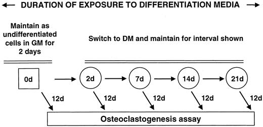
Abstract. The one or more molecular mechanisms that determine the obligatory sequence of resorption followed by formation during bone remodeling is unclear
The Development of Molecular Biology of Osteoporosis
Osteoporosis is one of the major bone disorders that affects both women and men, and causes bone deterioration and bone strength. Bone remodeling maintains bone mass and mineral homeostasis through the balanced action of osteoblasts and osteoclasts, which are responsible for bone formation and bone resorption, respectively. The imbalance in bone remodeling is known to be the main cause of osteoporosis. The imbalance can be the result of the action of various molecules produced by one bone cell that acts on other bone cells and influence cell activity. The understanding of the effect of these molecules on bone can help identify new targets and therapeutics to prevent and treat bone disorders. In this article, we have focused on molecules that are produced by osteoblasts, osteocytes, and osteoclasts and their mechanism of action on these cells. We have also summarized the different pharmacological osteoporosis treatments that target different molecular aspects of these bone cells to minimize osteoporosis.
Osteoclast differentiation and activation
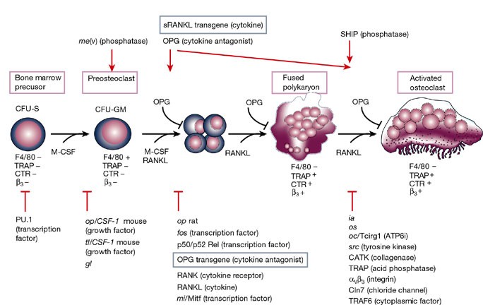
Osteoclasts are specialized cells derived from the monocyte/macrophage haematopoietic lineage that develop and adhere to bone matrix, then secrete acid and lytic enzymes that degrade it in a specialized, extracellular compartment. Discovery of the RANK signalling pathway in the osteoclast has provided insight into the mechanisms of osteoclastogenesis and activation of bone resorption, and how hormonal signals impact bone structure and mass. Further study of this pathway is providing the molecular basis for developing therapeutics to treat osteoporosis and other diseases of bone loss.
Wnt10b Increases Postnatal Bone Formation by Enhancing Osteoblast Differentiation
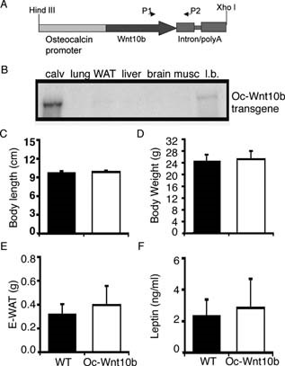
Abstract Overexpression of Wnt10b from the osteocalcin promoter in transgenic mice increases postnatal bone mass. Increases in osteoblast perimeter, mineralizing surface, and bone formation rate without detectable changes in pre-osteoblast proliferation, osteoblast apoptosis, or osteoclast number and activity suggest that, in this animal model, Wnt10b primarily increases bone mass by stimulating osteoblastogenesis. Introduction: Wnt signaling regulates many aspects of development including postnatal accrual of bone. Potential mechanisms for how Wnt signaling increases bone mass include regulation of osteoblast and/or osteoclast number and activity. To help differentiate between these possibilities, we studied mice in which Wnt10b is expressed specifically in osteoblast lineage cells or in mice devoid of Wnt10b. Materials and Methods: Transgenic mice, in which mouse Wnt10b is expressed from the human osteocalcin promoter (Oc-Wnt10b), were generated in C57BL/6 mice. Transgene expression was evaluated by RNase protection assay. Quantitative assessment of bone variables was done by radiography, μCT, and static and dynamic histomorphometry. Mechanisms of bone homeostasis were evaluated with assays for BrdU, TUNEL, and TRACP5b activity, as well as serum levels of C-terminal telopeptide of type I collagen (CTX). The endogenous role of Wnt10b in bone was assessed by dynamic histomorphometry in Wnt10b−/− mice. Results: Oc-Wnt10b mice have increased mandibular bone and impaired eruption of incisors during postnatal development. Analyses of femoral distal metaphyses show significantly higher BMD, bone volume fraction, and trabecular number. Increased bone formation is caused by increases in number of osteoblasts per bone surface, rate of mineral apposition, and percent mineralizing surface. Although number of osteoclasts per bone surface is not altered, Oc-Wnt10b mice have increased total osteoclast activity because of higher bone mass. In Wnt10b−/− mice, changes in mineralizing variables and osteoblast perimeter in femoral distal metaphyses were not observed; however, bone formation rate is reduced because of decreased total bone volume and trabecular number. Conclusions: High bone mass in Oc-Wnt10b mice is primarily caused by increased osteoblastogenesis, with a minor contribution from elevated mineralizing activity of osteoblasts.
Transforming Growth Factor-β1 to the Bone
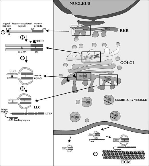
TGF-β1 is a ubiquitous growth factor that is implicated in the control of proliferation, migration, differentiation, and survival of many different cell types. It influences such diverse processes as embryogenesis, angiogenesis, inflammation, and wound healing. In skeletal tissue, TGF-β1 plays a major role in development and maintenance, affecting both cartilage and bone metabolism, the latter being the subject of this review. Because it affects both cells of the osteoblast and osteoclast lineage, TGF-β1 is one of the most important factors in the bone environment, helping to retain the balance between the dynamic processes of bone resorption and bone formation. Many seemingly contradictory reports have been published on the exact functioning of TGF-β1 in the bone milieu. This review provides an overall picture of the bone-specific actions of TGF-β1 and reconciles experimental discrepancies that have been reported for this multifunctional cytokine.
Bone Remodeling in Post-menopausal Osteoporosis
Bone mass in the skeleton is dependent on the coordinated activities of bone-forming osteoblasts and bone-resorbing osteoclasts in discrete bone multi-cellular units. Remodeling of bone in these units is important not only for maintaining bone mass, but also to repair microdamage, to prevent accumulation of too much old bone, and for mineral homeostasis. The activities of osteoblasts and osteoclasts are controlled by a variety of hormones and cytokines, as well as by mechanical loading. Most importantly, sex hormones are very crucial for keeping bone mass in balance, and the lack of either estrogen or testosterone leads to decreased bone mass and increased risk for osteoporosis. The prevalence of osteoporotic fractures is increasing dramatically in the Western part of the world and is a major health problem in many countries. In the present review, the cellular and molecular mechanisms controlling bone remodeling and the influence of sex hormones on these processes are summarized. In a separate paper in this issue, the pathogenesis of post-menopausal osteoporosis will be compared with that of inflammation-induced bone remodeling, including the evidence for and against the hypothesis that concomitant post-menopausal osteoporotic disease influences the progression of periodontal disease.

Physiological Bone Remodeling: Systemic Regulation and Growth Factor Involvement
Bone remodeling is essential for adult bone homeostasis. It comprises two phases: bone formation and resorption. The balance between the two phases is crucial for sustaining bone mass and systemic mineral homeostasis. This review highlights recent work on physiological bone remodeling and discusses our knowledge of how systemic and growth factors regulate this process.

Updates on Osteoimmunology: What's New on the Cross-Talk Between Bone and Immune System
The term osteoimmunology was coined many years ago to describe the research field that deals with the cross-regulation between bone cells and the immune system. As a matter of fact, many factors that are classically considered immune-related, such as interleukins (i.e. IL-6, -11,-17 and -23), tumor necrosis factor (TNF)-α, receptor-activator of nuclear factor kappa B (RANK) and its ligand (RANKL), nuclear factor of activated t-cell, cytoplasmatic-1 (NFATc1) and others have all been found to be crucial in osteoclast and osteoblast biology. Conversely, bone cells, which we used to think would only regulate each other and take care of remodeling bone, actually regulate immune cells, by creating the so-called “endosteal niche”. Both osteoblasts and osteoclasts participate to this niche, either by favoring engraftment, or mobilization of hematopoietic stem cells (HSCs). In this review, we will describe the main milestones at the base of the osteoimmunology and present the key cellular players of the bone-immune system cross-talk, including HSCs, osteoblasts, osteoclasts, bone marrow macrophages, osteomacs, T- and B-lymphocytes, dendritic cells and neutrophils. We will also briefly describe some pathological conditions in which the bone-immune system cross-talk plays a crucial role, with the final aim to portray the state of the art in the mechanisms regulating the bone-immune system interplay, and some of the latest molecular players in the field. This is important to encourage investigation in this field, to identify new targets in the treatment of bone and immune diseases.

The Regulation of Bone Metabolism and Disorders by Wnt Signaling
Wnt, a secreted glycoprotein, has an approximate molecular weight of 40 kDa, and it is a cytokine involved in various biological phenomena including ontogeny, morphogenesis, carcinogenesis, and maintenance of stem cells. The Wnt signaling pathway can be classified into two main pathways: canonical and non-canonical. Of these, the canonical Wnt signaling pathway promotes osteogenesis. Sclerostin produced by osteocytes is an inhibitor of this pathway, thereby inhibiting osteogenesis. Recently, osteoporosis treatment using an anti-sclerostin therapy has been introduced. In this review, the basics of Wnt signaling, its role in bone metabolism and its involvement in skeletal disorders have been covered. Furthermore, the clinical significance and future scopes of Wnt signaling in osteoporosis, osteoarthritis, rheumatoid arthritis and neoplasia are discussed.
Molecular Signaling Pathways and Essential Metabolic Elements in Bone Remodeling: An Implication of Therapeutic Targets for Bone Diseases
Bone is one of the dynamic tissues in the human body that undergoes continuous remodelling through subsequent actions of bone cells, osteoclasts, and osteoblasts. Several signal transduction pathways are involved in the transition of mesenchymal stem cells into osteoblasts. These primarily include Runx2, ATF4, Wnt signaling and sympathetic signalling. The differentiation of osteoclasts is controlled by M-CSF, RANKL, and costimulatory signalling. It is well known that bone remodelling is regulated through receptor activator of nuclear factor-kappa B ligand followed by the binding to RANK, which eventually induces the differentiation of osteoclasts. The resorbing osteoclasts secrete TRAP, cathepsin K, MMP-9 and gelatinase to digest the proteinaceous matrix of type I collagen and form a saucer-shaped lacuna along with resorption tunnels in the trabecular bone. Osteoblasts secrete a soluble decoy receptor, osteoprotegerin that prevents the binding of RANK/RANKL and thus moderating osteoclastogenesis. Moreover, bone homeostasis is also regulated by several growth factors, cytokines, calciotropic hormones, parathyroid hormone and sex steroids. The current review presents a correlation of the probable molecular targets underlying the regulation of bone mass and the role of essential metabolic elements in bone remodelling. Targeting these signaling pathways may help design newer therapies for treating bone diseases.

The osteocyte as a signaling cell
Osteocytes, former osteoblasts encapsulated by mineralized bone matrix, are far from being passive and metabolically inactive bone cells. Instead, osteocytes are multifunctional and dynamic cells capable of integrating hormonal and mechanical signals and transmitting them to effector cells in bone and in distant tissues. Osteocytes are a major source of molecules that regulate bone homeostasis by integrating both mechanical cues and hormonal signals that coordinate the differentiation and function of osteoclasts and osteoblasts. Osteocyte function is altered in both rare and common bone diseases, suggesting that osteocyte dysfunction is directly involved in the pathophysiology of several disorders affecting the skeleton. Advances in osteocyte biology initiated the development of novel therapeutics interfering with osteocyte-secreted molecules. Moreover, osteocytes are targets and key distributors of biological signals mediating the beneficial effects of several bone therapeutics used in the clinic. Here we review the most recent discoveries in osteocyte biology demonstrating that osteocytes regulate bone homeostasis and bone marrow fat via paracrine signaling, influence body composition and energy metabolism via endocrine signaling, and contribute to the damaging effects of diabetes mellitus and hematologic and metastatic cancers in the skeleton.
