

Background Failure of conventional imaging to detect pancreatic ductal adenocarcinoma (PDA) at its visually occult pre-diagnostic stage is a primary barrier to improving its otherwise poor rate of survival. Objective To develop and validate the Radiomics-based Early Detection MODel (REDMOD), an AI framework to identify subvisual radiomic signatures of pre-diagnostic PDA on standard-of-care CT. Designs REDMOD was trained on a multi-institutional cohort (n=969; 156 pre-diagnostic, 813 control) and tested on an independent set (n=493; 63 pre-diagnostic, 430 control), simulating a low prevalence (~1:6) early detection paradigm. The fully automated framework couples AI-driven segmentation with a heterogeneous ensemble architecture trained on a 40-feature radiomic signature derived from Synthetic Minority Over-sampling Technique (SMOTE)-balanced data. A tunable Youden Index-optimised classification threshold enables performance calibration without retraining. Validation included direct comparison with radiologists, longitudinal test–retest analysis and external specificity validation across two independent cohorts (n=539 and n=80). Results On an independent test set (n=493), REDMOD identified occult PDA (AUC 0.82; 73.0% sensitivity) at a median 475-day lead time. This represented nearly twofold higher sensitivity than radiologists (38.9%; p<0.001), which grew to nearly threefold (68.0% vs 23.0%) at >24 months lead time. REDMOD showed strong longitudinal stability (90–92% concordance) and generalisable specificity across multi-institutional (81.3%; n=539) and public (87.5%; n=80) datasets. Mechanistic analyses confirmed predictive power derived principally from multi-scale wavelet-filtered textural features (90% of selected signature), which outperformed unfiltered features (AUC 0.82 vs 0.74; p=0.007) in capturing subvisual architectural disruptions. Conclusions REDMOD is an automated, mechanistically grounded, longitudinally stable, externally validated AI that surpasses radiologists for PDA detection at its visually occult pre-diagnostic stage. These attributes position it for prospective validation in high-risk cohorts, a necessary step towards shifting the paradigm from late-stage symptomatic diagnosis to proactive pre-clinical interception.
AI model detects very early normally ‘invisible’ tissue changes of pancreatic cancer
An AI model (REDMOD) can pick up the very early subtle tissue changes of pancreatic ductal adenocarcinoma, the most common form of pancreatic cancer, which conventional imaging and the human eye find difficult to detect, finds research published online in the journal Gut. As such, it offers the potential to shift an all too common late stage, terminal disease diagnosis to one that is at an early stage (stage 0) and treatable, say the researchers. While REDMOD was more accurate than experienced radiologists, it requires testing in high risk patients, defined as those with unexpected weight loss and newly diagnosed diabetes, before it can be widely used in clinical practice, they add.
AI Spots Pancreatic Cancer Years Before It Shows Up, Study Finds
An artificial intelligence system can spot pancreatic cancer long before it shows up on scans, raising the prospect of catching one of the deadliest tumors early enough to successfully treat, a study found.

GitHub - AIM-KannLab/BrainIAC | Giovanni Giulietti
One of the biggest hurdles in neuroimaging AI has always been the scarcity of labeled data, especially for rare conditions. But a recent study published in Nature Neuroscience introduces a potential solution: BrainIAC (Brain Imaging Adaptive Core). Unlike traditional models that are trained for one specific task, BrainIAC is a "foundation model", a large-scale AI model trained on vast amounts of data that can perform many different tasks and be adapted to specific applications. It was pretrained on a massive dataset of 48965 brain structural MRI sequences (T1w, T2w, FLAIR, T1CE) using self-supervised learning, allowing it to understand the "big picture" of brain anatomy across different ages and conditions. It has been tested across seven diverse and clinically meaningful applications: - brain age prediction - IDH mutation classification - Mild Cognitive Impairment (MCI) classification - diffuse glioma overall survival prediction - MR sequence classification - time-to-stroke prediction - tumor segmentation BrainIAC consistently outperformed traditional supervised learning approaches and other medical-specific models. The real "magic" happens in low-data scenarios. While traditional AI usually needs thousands of examples, BrainIAC can adapt to new tasks using only a few samples. BrainIAC can be used for structural brain MRI analysis through two main methods: - Web Usage (BrainIAC platform): the easiest way to use BrainIAC, providing a user-friendly interface to upload structural brain MRI data and run inference. - Local usage (GitHub repository): for more advanced and customized analysis. It allows complete control over the data preprocessing, model training, and inference pipelines. It requires Python 3.9+ and NVIDIA GPU with CUDA 11.0+. 🔗 BrainIAC paper: https://lnkd.in/dq6Gprgt 🔗 BrainIAC web platform https://lnkd.in/dpreadMp 🔗 BrainIAC GitHub repo: https://lnkd.in/duwSEg4V #MedicalAI #Neuroscience #HealthTech #BrainIAC #Radiology #Innovation #DeepLearning #PrecisionMedicine

GAN-Based Data Augmentation for Prediction Improvement Using Gene Expression Data in Cancer
Within the area of bioinformatics, Deep Learning (DL) models have shown exceptional results in applications in which histological images, scans and tomographies are used. However, when gene expression data is under analysis, the performance is often limited, further hampered by the complexity of these models that require several instances, in the order of thousands, to provide good results. Due to the difficulty and the costs involved in the collection of medical data, the application of Data Augmentation (DA) techniques to alleviate the lack of samples is a topic of great relevance. State-of-the-art models based on Conditional Generative Adversarial Networks (CGAN) and some introduced modifications are used in this work to investigate the effect of DA for prediction of the vital status of patients from RNA-Seq gene expression data. Experimental results on several real-world data sets demonstrate the effectiveness and efficiency of the proposed models. The application of DA methods significantly increase prediction accuracy, leading by 12% with respect to benchmark data sets and 3.15% with respect to data processed with feature selection. Results based on CGAN models outperform in most cases, alternative methods like the SMOTE or noise injection techniques.
The algorithm will see you now - Works in Progress Magazine
Radiology combines digital images, clear benchmarks, and repeatable tasks. But replacing humans with AI is harder than it seems.

Using AI Made Doctors Worse at Spotting Cancer Without Assistance
A new study offers the latest evidence of potential “deskilling” effects on AI users.

Foundation Models in Medical Imaging: A Review and Outlook
Foundation models (FMs) are changing the way medical images are analyzed by learning from large collections of unlabeled data. Instead of relying on manually annotated examples, FMs are...

Towards Expert-level Medical AI for Real-time Video Consultations
Audio-visual interaction is the standard for patient-physician consultations, enabling natural communication and effective assessment of illness through non-verbal cues. While text-based AI has shown promise, it discards essential perceptual dimensions and limits patients who cannot articulate symptoms in writing. Early efforts to extend medical AI to audio-visual interaction have demonstrated feasibility but not reached clinician-level performance. Here, we provide the first demonstration of expert-level AI in real-time clinical video consultations using AMIE (Articulate Medical Intelligence Explorer) in a video configuration. AMIE (Video) is a Gemini-based multi-agent system integrating low-latency dialogue, clinical reasoning, and real-time audio-visual perception. To guide development, we established a taxonomy and automated evaluations for clinical audio-visual cues in telehealth settings. In a randomized Objective Structured Clinical Examination (OSCE) study with 30 primary care physicians (PCPs), 15 patient actors and 100 clinical scenarios, we compared AMIE (Video), its text-only counterpart AMIE (Text), and PCPs consulting via video. Clinical evaluators rated AMIE (Video) on par or better than PCPs in history-taking, diagnosis, management, and physical observation and examination. Patient actors preferred AMIE's approach to assessing and explaining conditions, while PCPs were preferred for rapport and partnership building. In modality ablation, patient actors preferred AMIE (Video)'s interface over text chat for communicative effectiveness, convenience, and feeling understood. Limitations remain in fine anatomical precision, subtle affective nuances, and high-frequency movements. While further research is needed before real-world translation, these results mark an important milestone toward AI systems capable of augmenting care across the sensory complexity of clinical practice.
Hierarchical classification of immune cell transcriptomes at population-scale
Accurate immune cell classification is essential for interpreting single-cell RNA sequencing (scRNA-seq) data. However, progress is constrained by the lack of independent, high-resolution benchmarks, as the routine integration of datasets introduces statistical dependencies that artificially inflate model generalizability. Here, we present the single-cell universal classification omnibus (Suco), a resource of independent, uniform expert annotations, and Compocyte, a modular hierarchical classifier. Together, they establish a framework designed for the scale of human population immunology. This approach substantially outperforms existing classifiers while facilitating expert review of ambiguous annotations. Applying Compocyte across 50 studies, including three newly generated datasets, we classified 15.6 million leukocytes from 3,965 patients. Within this expansive cohort, we identified a new tumor-associated resorptive macrophage phenotype, a non-canonical monocyte subtype in subclinical cytokine release syndrome, and the programmatic erosion of T cell memory stemness across metastatic sites. Suco and Compocyte thus provide a generalizable architecture and benchmark capable of sustaining high-resolution annotation across massive clinical cohorts. ### Competing Interest Statement CMR has consulted regarding oncology drug development with Amgen, AstraZeneca, Daiichi Sankyo, Genentech, Merck, and Novartis, and has received licensing and royalty payments for DLL3-directed therapeutics. T.W. reports stock ownership for Roche, Astra Zeneca, Bayer, Innate Pharma, Kyntra, Illumina, 10x Genomics, and Merck KGaA as well as research funding from Atrandi Biosciences, Vilnius, Lithuania; CanVirex AG, Basel Switzerland; and Institut fuer Klinische Krebsforschung GmbH, Frankfurt, Germany, and travel funding from Roche, Basel, Switzerland. S.Z. reports advisory board membership and honoraria from Amgen, Astellas, AstraZeneca, Bayer, Bristol-Myers Squibb, Daiichi Sankyo, Eisai, EUSA, Gilead, Ipsen, Johnson&Johnson, Lilly, MedSir, Medtoday, Merck, MSD, Novartis, Pfizer, Roche, Sanofi Aventis, StreamedUp, Urotrials, Urotube, Zentiva and resarch funding from Eisai. S.Z. reports clinical trial support from Amgen, AstraZeneca, AVEO, Bayer, Biontech, Bristol-Myers Squibb, Calithera, Exelixis, Gilead, Lilly, MSD, Novartis, Pfizer, Roche, Seagen/Astellas, Urotrials and travels & conference support from Amgen, Astellas, AstraZeneca, Bayer, EISAI, Ipsen, Johnson&Johnson, Merck, MSD, Pfizer. All remaining authors declare no relevant competing interests. Spanish Association Against Cancer, PI049999 Federal Ministry of Research, Technology and Space, 001001KT2322 National Cancer Institute, R35 CA263816 National Cancer Institute, U24 CA213274 National Cancer Institute, P30 CA008748 Research Council of Lithuania, P-MIP-24-93

Mirage: The Illusion of Visual Understanding
Multimodal AI systems have achieved remarkable performance across a broad range of real-world tasks, yet the mechanisms underlying visual–language reasoning remain surprisingly poorly understood. We report three findings that challenge prevailing assumptions about how these systems process and integrate visual information. First, Frontier models readily generate detailed image descriptions and elaborate reasoning traces, including pathology-biased clinical findings, for images never provided; we term this phenomenon mirage reasoning. Second, without any image input, models also attain strikingly high scores across general and medical multimodal benchmarks, bringing into question their utility and design. In the most extreme case, our model achieved the top rank on a standard chest X-ray question-answering benchmark without access to any images. Third, when models were explicitly instructed to guess answers without image access, rather than being implicitly prompted to assume images were present, performance declined markedly. Explicit guessing appears to engage a more conservative response regime, in contrast to the mirage regime in which models behave as though images have been provided. These findings expose fundamental vulnerabilities in how visual–language models reason and are evaluated, pointing to an urgent need for private benchmarks that eliminate textual cues enabling non-visual inference, particularly in medical contexts where miscalibrated AI carries the greatest consequence. We introduce B-Clean as a principled solution for fair, vision-grounded evaluation of multimodal AI systems.
Towards Conversational Medical AI with Eyes, Ears and a Voice
The practice of medicine relies not only upon skillful dialogue but also on the nuanced exchange and interpretation of rich auditory and visual cues between doctors and patients. Building on the low-latency voice and video processing capabilities of Gemini, we introduce AI co-clinician, a first-of-its-kind conversational AI system utilizing continuous streams of audio-visual data from live patient conversations to inform real-time clinical decisions. Its dual-agent architecture balances deep clinical reasoning with the low latency required for natural dialogue. To assess this system, we implemented a video-based interface emulating telemedicine consultations. We crafted 20 standardized outpatient scenarios requiring proactive real-time auditory and visual reasoning and designed "TelePACES" evaluation criteria alongside case-specific rubrics. In a randomized, interface-blinded, crossover simulation study (n = 120 encounters) with 10 internal medicine residents as patient actors, we compared AI co-clinician with primary care physicians (PCPs), GPT-Realtime, and a baseline agent. AI co-clinician approached PCPs in key TelePACES dimensions, including management plans and differential diagnosis, while significantly outperforming GPT-Realtime across all general criteria. While our agent demonstrated parity with PCPs in case-specific triage measures, physicians maintained superior overall performance in case-specific assessments. Although AI co-clinician marks a significant advance in real-time telemedical AI, gaps remain in physical examination and disease-specific reasoning. Our work shows that text-only approaches fail to capture the true challenges of medical consultation and suggests that high-stakes real-time diagnostic AI is most safely advanced in collaborative, triadic models where AI can be a supportive co-clinician for doctors and patients.
Use of Deep Learning for Continuous Prediction of Mortality for All Admissions in Intensive Care Units
<p>The mortality rate in the intensive care unit (ICU) is a key metric of hospital clinical quality. To enhance hospital performance, many methods have been proposed for the stratification of patients’ different risk categories, such as severity scoring systems and machine learning models. However, these methods make capturing time sequence information difficult, posing challenges to the continuous assessment of a patient’s severity during their hospital stay. Therefore, we built a predictive model that can make predictions throughout the patient’s stay and obtain the patient’s risk of death in real time. Our proposed model performed much better than other machine learning methods, including logistic regression, random forest, and XGBoost, in a full set of performance evaluation processes. Thus, the proposed model can support physicians’ decisions by allowing them to pay more attention to high-risk patients and anticipate potential complications to reduce ICU mortality.</p>
Medical Algorithms Are Failing Communities Of Color
Medical algorithms routinely make decisions about patients’ health care, yet they are rife with bias. Health equity must be built into the development and deployment of these health algorithms.
General-purpose large language models outperform specialized clinical AI tools on medical benchmarks
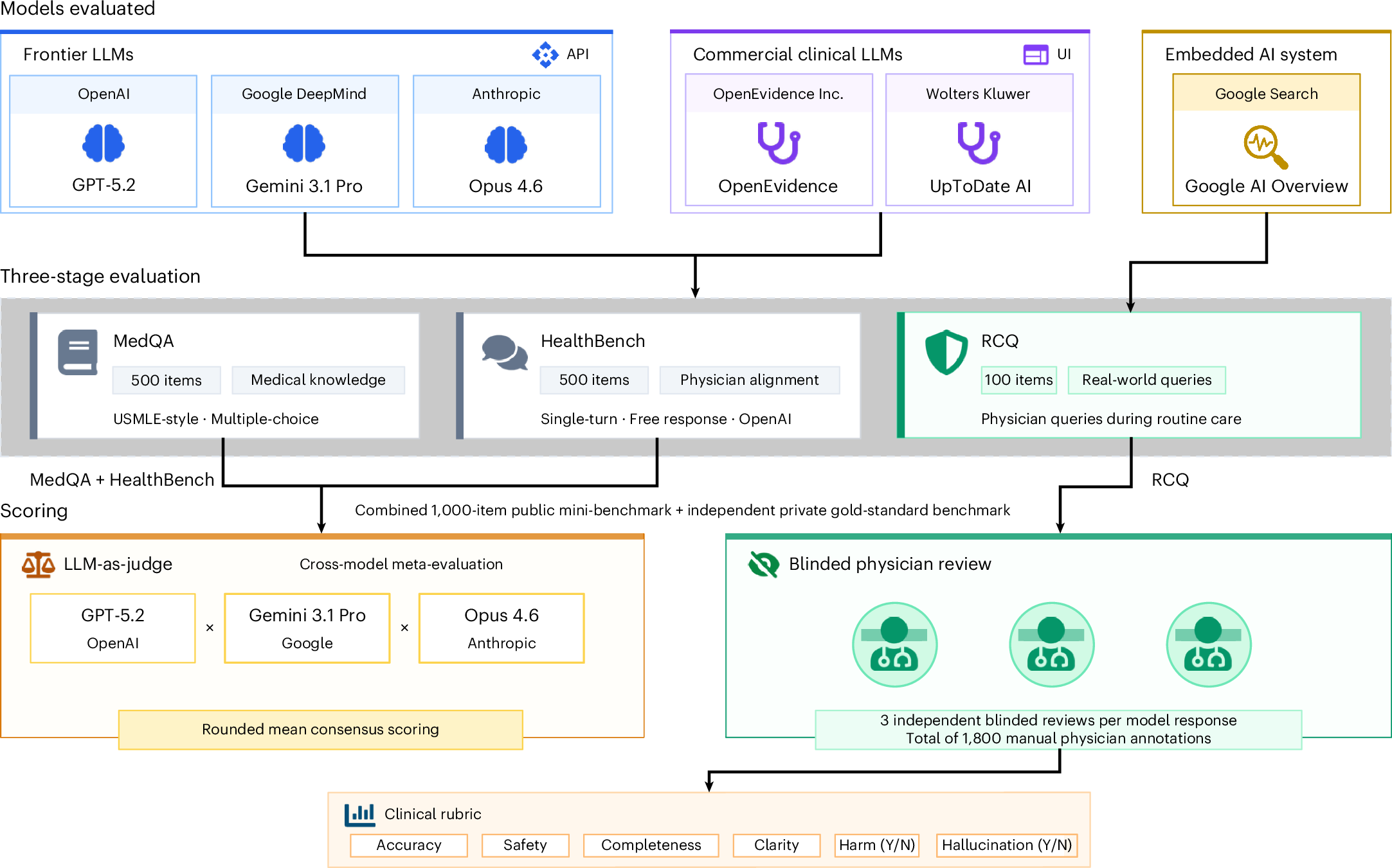
Specialized clinical artificial intelligence (AI) tools are entering medical practice despite scarce independent evaluation. We quantitatively evaluate two clinical AI tools, OpenEvidence and UpToDate Expert AI, built on large language models (LLMs) against three frontier LLMs: GPT-5.2, Gemini 3.1 Pro and Claude Opus 4.6. Our evaluation has three stages: (1) 500 MedQA questions testing medical knowledge, (2) 500 HealthBench items measuring alignment with clinicians and (3) the real clinical queries (RCQ) benchmark, built from 100 de-identified queries from physicians to a general-purpose language model in a live clinical environment. For the RCQ benchmark, 12 US clinicians performed randomized, blinded review of model outputs, producing 1,800 model–question annotations. Frontier LLMs outperformed clinical AI tools in all three evaluations. Clinical AI tools performed comparably to auto-enabled Google Search AI Overview on the RCQ. These findings highlight the need for independent, real-world evaluation of AI tools before they enter clinical settings.
International, cross-sector collaboration commits nearly $2 billion to build foundational data for AI models to predict and treat disease
Biohub, DOE, NIH, and partners will generate open, standardized data to train AI models that predict how cells respond to interventions.
#Pancreaticcancer is usually caught too late to treat. A new study finds an #AI system can pick up early warning signs in routine CT scans more than a year before diagnosis -- outperforming radiologists on scans previously read as normal. Hopefully it can improve survival. shorturl.at/zXUwj

AI Spots Pancreatic Cancer Years Before It Shows Up, Study Finds
www.bloomberg.com