The main determinant of bone Ca accretion is a bimolecular regulatory network on osteoblast (OB) and osteoclast (OC). Even though IGF-1 is known as an important regulator in bone cell cycle, little has been done to model IGF-1 action in bone cell regulation. Thus, the objective is to develop a mathematical model that depicts the regulatory action of IGF-1 onto the OB and OC interaction, and to evaluate adolescent and adult bone Ca accretion in response to differences in IGF-1 levels. As a result, a dynamic model of OB and OC with two main regulatory systems, i.e., Receptor Activator for Nuclear Factor [Formula: see text]B (RANK)-RANK Ligand (RANKL)-osteoprogerin (OPG) system, and TGF-[Formula: see text], was augmented with the IGF-1, and incorporated into Ca kinetic data to predict exchangeable bone Ca. The developed model could predict a change in OB and OC levels in response to perturbations in regulators, producing results consistent with bone physiology and published experimental data. The model also estimated parametric difference in regulators between adults and adolescents, suggesting that RANKL/OPG in adolescents was about 4 times higher than in adults, while adolescent serum PTH and IGF-1 concentrations were 60% and 220% of those of adults, respectively. This study highlighted the influence of IGF-1 on the regulation of bone cells in positively modulating bone Ca, suggesting that IGF-1 may be an effective target for reducing bone loss by promoting mature OB.
Dynamic Biochemical and Cellular Models of Bone Physiology: Integrating Remodeling Processes, Tumor Growth, and Therapy
Bone is an activeCoelho, Rui M. Neto, Joana P. connective tissue composed of different types of cells. The dynamic behavior of bone remodeling processes is typically represented throughValério, Duarte differential equations, which represent the physiological phenomena occurring in this organ. These models takeVinga, Susana into account the tight biochemical regulation between osteoclasts and osteoblasts and have also been enriched with variables and parameters related to bone pathologies and treatment. This chapter reviews some of the more recent models describing bone physiology, focusing on those that include the main cellular processes, along the biochemical control, and also the pharmacokinetic/pharmacodynamic (PK/PD) of the most common treatments for diseases such as cancer. These models are then compared in terms of the simulations obtained and, finally, some highlights on integrating them with the biomechanical component of the system which will be given. These models are expected to provide a valuable insight into this complex system and to support the development of clinical decision systems for bone pathologies.

A novel mathematical model of bone remodelling cycles for trabecular bone at the cellular level
After an initial phase of growth and development, bone undergoes a continuous cycle of repair, renewal and optimisation by a process called remodelling. This paper describes a novel mathematical model of the trabecular bone remodelling cycle. It is essentially formulated to simulate a remodelling event at a fixed position in the bone, integrating bone removal by osteoclasts and formation by osteoblasts. The model is developed to construct the variation in bone thickness at a particular point during the remodelling event, derived from standard bone histomorphometric analyses. The novelties of the approach are the adoption of a predator–prey model to describe the dynamic interaction between osteoclasts and osteoblasts, using a genetic algorithm–based solution; quantitative reconstruction of the bone remodelling cycle; and the introduction of a feedback mechanism in the bone formation activity to co-regulate bone thickness. The application of the model is first demonstrated by using experimental data recorded for normal (healthy) bone remodelling to predict the temporal variation in the number of osteoblasts and osteoclasts. The simulated histomorphometric data and remodelling cycle characteristics compare well with the specified input data. Sensitivity studies then reveal how variations in the model’s parameters affect its output; it is hoped that these parameters can be linked to specific biochemical factors in the future. Two sample pathological conditions, hypothyroidism and primary hyperparathyroidism, are examined to demonstrate how the model could be applied more broadly, and, for the first time, the osteoblast and osteoclast populations are predicted for these conditions. Further data are required to fully validate the model’s predictive capacity, but this work shows it has potential, especially in the modelling of pathological conditions and the optimisation of the treatment of those conditions.
Simulated Interventions to Ameliorate Age-Related Bone Loss Indicate the Importance of Timing
Bone remodeling is the continuous process of bone resorption by osteoclasts and bone formation by osteoblasts, in order to maintain homeostasis. The activity of osteoclasts and osteoblasts is regulated by a network of signaling pathways, including Wnt, parathyroid hormone (PTH), RANKL/OPG and TGF-β, in response to stimuli such as mechanical loading. During aging there is a gradual loss of bone mass due to dysregulation of signaling pathways. This may be due to a decline in physical activity with age and/or changes in hormones and other signaling molecules. In particular, hormones such as PTH have a circadian rhythm which may be disrupted in aging. Due to the complexity of the molecular and cellular networks involved in bone remodeling, several mathematical models have been proposed to aid understanding of the processes involved. However, to date there are no models which explicitly consider the effects of mechanical loading, the circadian rhythm of PTH and the dynamics of signaling molecules on bone remodeling. Therefore, we have constructed a network model of the system using a modular approach which will allow further modifications as required in future research. The model was used to simulate the effects of mechanical loading and also the effects of different interventions such as continuous or intermittent administration of PTH. Our model predicts that the absence of regular mechanical loading and/or an impaired PTH circadian rhythm leads to a gradual decrease in bone mass over time which can be restored by simulated interventions and that the effectiveness of some interventions may depend on their timing.

Molecular Signaling Pathways and Essential Metabolic Elements in Bone Remodeling: An Implication of Therapeutic Targets for Bone Diseases
Bone is one of the dynamic tissues in the human body that undergoes continuous remodelling through subsequent actions of bone cells, osteoclasts, and osteoblasts. Several signal transduction pathways are involved in the transition of mesenchymal stem cells into osteoblasts. These primarily include Runx2, ATF4, Wnt signaling and sympathetic signalling. The differentiation of osteoclasts is controlled by M-CSF, RANKL, and costimulatory signalling. It is well known that bone remodelling is regulated through receptor activator of nuclear factor-kappa B ligand followed by the binding to RANK, which eventually induces the differentiation of osteoclasts. The resorbing osteoclasts secrete TRAP, cathepsin K, MMP-9 and gelatinase to digest the proteinaceous matrix of type I collagen and form a saucer-shaped lacuna along with resorption tunnels in the trabecular bone. Osteoblasts secrete a soluble decoy receptor, osteoprotegerin that prevents the binding of RANK/RANKL and thus moderating osteoclastogenesis. Moreover, bone homeostasis is also regulated by several growth factors, cytokines, calciotropic hormones, parathyroid hormone and sex steroids. The current review presents a correlation of the probable molecular targets underlying the regulation of bone mass and the role of essential metabolic elements in bone remodelling. Targeting these signaling pathways may help design newer therapies for treating bone diseases.

Mathematical Modeling of Spatio-Temporal Dynamics of a Single Bone Multicellular Unit
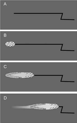
Abstract During bone remodeling, bone-resorbing osteoclasts and bone-forming osteoblasts are organized in bone multicellular units (BMUs), which travel at a rate of 20–40 μm/d for 6–12 mo, maintaining a cylindrical structure. However, the interplay of local BMU geometry with biochemical regulation is poorly understood. We developed a mathematical model of BMU describing changes in time and space of the concentrations of proresorptive cytokine RANKL and its inhibitor osteoprotegerin (OPG), in osteoclast and osteoblast numbers, and in bone mass. We assumed that osteocytes surrounding a microfracture produce RANKL, which attracted osteoclasts. OPG and RANKL were produced by osteoblasts and diffused through bone, RANKL was eliminated by binding to OPG and RANK. Osteoblasts were coupled to osteoclasts through paracrine factors. The evolution of the BMU arising from this model was studied using numerical simulations. Our model recapitulated the spatio-temporal dynamics observed in vivo in a cross-section of bone. In response to a RANKL field, osteoclasts moved as a well-confined cutting cone. The coupling of osteoclasts to osteoblasts allowed for sufficient recruitment of osteoblasts to the resorbed surfaces. The RANKL field was the highest at the microfracture in front of the BMU, whereas the OPG field peaked at the back of the BMU, resulting in the formation of a RANKL/OPG gradient, which strongly affected the rate of BMU progression and its size. Thus, the spatial organization of a BMU provides important constraints on the roles of RANKL and OPG as well as possibly other regulators in determining the outcome of remodeling in the BMU.
A review of mathematical modeling of bone remodeling from a systems biology perspective
Bone remodeling is an essential physiological process in the adult skeleton. Due to the complex nature of this process, many mathematical models of bone remodeling have been developed. Each of these models has unique features, but they have underlying patterns. In this review, the authors highlight the important aspects frequently found in mathematical models for bone remodeling and discuss how and why these aspects are included when considering the physiology of the bone basic multicellular unit, which is the term used for the collection of cells responsible for bone remodeling. The review also emphasizes the view of bone remodeling from a systems biology perspective. Understanding the systemic mechanisms involved in remodeling will help provide information on bone pathology associated with aging, endocrine disorders, cancers, and inflammatory conditions and enhance systems pharmacology. Furthermore, some features of the bone remodeling cycle and interactions with other organ systems that have not yet been modeled mathematically are discussed as promising future directions in the field.
A review of mathematical modeling of bone remodeling from a systems biology perspective
Bone remodeling is an essential, delicately balanced physiological process of coordinated activity of bone cells that remove and deposit new bone tissue in the adult skeleton. Due to the complex nature of this process, many mathematical models of bone remodeling have been developed. Each of these models has unique features, but they have underlying patterns. In this review, the authors highlight the important aspects frequently found in mathematical models for bone remodeling and discuss how and why these aspects are included when considering the physiology of the bone basic multicellular unit, which is the term used for the collection of cells responsible for bone remodeling. The review also emphasizes the view of bone remodeling from a systems biology perspective. Understanding the systemic mechanisms involved in remodeling will help provide information on bone pathology associated with aging, endocrine disorders, cancers, and inflammatory conditions and enhance systems pharmacology. Furthermore, some features of the bone remodeling cycle and interactions with other organ systems that have not yet been modeled mathematically are discussed as promising future directions in the field.

Mechanobiological osteocyte feedback drives mechanostat regulation of bone in a multiscale computational model
Significant progress has been made to identify the cells and signaling molecules involved in the mechanobiological regulation of bone remodeling. It is now well accepted that osteocytes act as mechanosensory cells in bone expressing several signaling molecules such as nitric oxide (NO) and sclerostin (Scl) which are able to control bone remodeling responses. In this paper, we present a comprehensive multiscale computational model of bone remodeling which incorporates biochemical osteocyte feedback. The mechanostat theory is quantitatively incorporated into the model using mechanical feedback to control expression levels of NO and Scl. The catabolic signaling pathway RANK–RANKL–OPG is co-regulated via (continuous) PTH and NO, while the anabolic Wnt signaling pathway is described via competitive binding reactions between Wnt, Scl and the Wnt receptors LRP5/6. Using this novel model of bone remodeling, we investigate the effects of changes in the mechanical loading and hormonal environment on bone balance. Our numerical simulations show that we can calibrate the mechanostat anabolic and catabolic regulatory mechanisms so that they are mutually exclusive. This is consistent with previous models that use a Wolff-type law to regulate bone resorption and formation separately. Furthermore, mechanical feedback provides an effective mechanism to obtain physiological bone loss responses due to mechanical disuse and/or osteoporosis.

Osteocyte-Related Cytokines Regulate Osteoclast Formation and Bone Resorption
The process of bone remodeling is the result of the regulated balance between bone cell populations, namely bone-forming osteoblasts, bone-resorbing osteoclasts, and the osteocyte, the mechanosensory cell type. Osteoclasts derived from the hematopoietic stem cell lineage are the principal cells involved in bone resorption. In osteolytic diseases such as rheumatoid arthritis, periodontitis, and osteoporosis, the balance is lost and changes in favor of bone resorption. Therefore, it is vital to elucidate the mechanisms of osteoclast formation and bone resorption. It has been reported that osteocytes express Receptor activator of nuclear factor κΒ ligand (RANKL), an essential factor for osteoclast formation. RANKL secreted by osteocytes is the most important factor for physiologically supported osteoclast formation in the developing skeleton and in pathological bone resorption such as experimental periodontal bone loss. TNF-α directly enhances RANKL expression in osteocytes and promotes osteoclast formation. Moreover, TNF-α enhances sclerostin expression in osteocytes, which also increases osteoclast formation. These findings suggest that osteocyte-related cytokines act directly to enhance osteoclast formation and bone resorption. In this review, we outline the most recent knowledge concerning bone resorption-related cytokines and discuss the osteocyte as the master regulator of bone resorption and effector in osteoclast formation.

The Role of Osteocytes in Targeted Bone Remodeling: A Mathematical Model
Until recently many studies of bone remodeling at the cellular level have focused on the behavior of mature osteoblasts and osteoclasts, and their respective precursor cells, with the role of osteocytes and bone lining cells left largely unexplored. This is particularly true with respect to the mathematical modeling of bone remodeling. However, there is increasing evidence that osteocytes play important roles in the cycle of targeted bone remodeling, in serving as a significant source of RANKL to support osteoclastogenesis, and in secreting the bone formation inhibitor sclerostin. Moreover, there is also increasing interest in sclerostin, an osteocyte-secreted bone formation inhibitor, and its role in regulating local response to changes in the bone microenvironment. Here we develop a cell population model of bone remodeling that includes the role of osteocytes, sclerostin, and allows for the possibility of RANKL expression by osteocyte cell populations. We have aimed to give a simple, yet still tractable, model that remains faithful to the underlying system based on the known literature. This model extends and complements many of the existing mathematical models for bone remodeling, but can be used to explore aspects of the process of bone remodeling that were previously beyond the scope of prior modeling work. Through numerical simulations we demonstrate that our model can be used to explore theoretically many of the qualitative features of the role of osteocytes in bone biology as presented in recent literature.
Osteoprotegerin in Bone Metastases: Mathematical Solution to the Puzzle
Bone is a common site for cancer metastasis. To create space for their growth, cancer cells stimulate bone resorbing osteoclasts. Cytokine RANKL is a key osteoclast activator, while osteoprotegerin (OPG) is a RANKL decoy receptor and an inhibitor of osteoclastogenesis. Consistently, systemic application of OPG decreases metastatic tumor burden in bone. However, OPG produced locally by cancer cells was shown to enhance osteolysis and tumor growth. We propose that OPG produced by cancer cells causes a local reduction in RANKL levels, inducing a steeper RANKL gradient away from the tumor and towards the bone tissue, resulting in faster resorption and tumor expansion. We tested this hypothesis using a mathematical model of nonlinear partial differential equations describing the spatial dynamics of OPG, RANKL, PTHrP, osteoclasts, tumor and bone mass. We demonstrate that at lower expression rates, tumor-derived OPG enhances the chemotactic RANKL gradient and osteolysis, whereas at higher expression rates OPG broadly inhibits RANKL and decreases osteolysis and tumor burden. Moreover, tumor expression of a soluble mediator inducing RANKL in the host tissue, such as PTHrP, is important for correct orientation of the RANKL gradient. A meta-analysis of OPG, RANKL and PTHrP expression in normal prostate, carcinoma and metastatic tissues demonstrated an increase in expression of OPG, but not RANKL, in metastatic prostate cancer, and positive correlation between OPG and PTHrP in metastatic prostate cancer. The proposed mechanism highlights the importance of the spatial distribution of receptors, decoys and ligands, and can be applied to other systems involving regulation of spatially anisotropic processes.
Analytic approach to explore dynamical osteoporotic bone turnover
The dynamics of the osteoporotic bone turnover is studied in this paper with the aid of stability analysis of the associated mathematical model. Osteoporosis, which is a common bone disorder, is studied in this papper in detail with an emphasis on the relative threshold values. We examine the expository signaling among the bone cells named osteoclast and osteoblast. Main functioning of osteoblasts is bone formation, whereas osteoclasts are bone removal cells.

Biology of Bone Tissue: Structure, Function, and Factors That Influence Bone Cells
Bone tissue is continuously remodeled through the concerted actions of bone cells, which include bone resorption by osteoclasts and bone formation by osteoblasts, whereas osteocytes act as mechanosensors and orchestrators of the bone remodeling process. This process is under the control of local (e.g., growth factors and cytokines) and systemic (e.g., calcitonin and estrogens) factors that all together contribute for bone homeostasis. An imbalance between bone resorption and formation can result in bone diseases including osteoporosis. Recently, it has been recognized that, during bone remodeling, there are an intricate communication among bone cells. For instance, the coupling from bone resorption to bone formation is achieved by interaction between osteoclasts and osteoblasts. Moreover, osteocytes produce factors that influence osteoblast and osteoclast activities, whereas osteocyte apoptosis is followed by osteoclastic bone resorption. The increasing knowledge about the structure and functions of bone cells contributed to a better understanding of bone biology. It has been suggested that there is a complex communication between bone cells and other organs, indicating the dynamic nature of bone tissue. In this review, we discuss the current data about the structure and functions of bone cells and the factors that influence bone remodeling.

The bone remodelling cycle
The bone remodelling cycle replaces old and damaged bone and is a highly regulated, lifelong process essential for preserving bone integrity and maintaining mineral homeostasis. During the bone remodelling cycle, osteoclastic resorption is tightly coupled to osteoblastic bone formation. The remodelling cycle occurs within the basic multicellular unit and comprises five co-ordinated steps; activation, resorption, reversal, formation and termination. These steps occur simultaneously but asynchronously at multiple different locations within the skeleton. Study of rare human bone disease and animal models have helped to elucidate the cellular and molecular mechanisms that regulate the bone remodelling cycle. The key signalling pathways controlling osteoclastic bone resorption and osteoblastic bone formation are receptor activator of nuclear factor-κB (RANK)/RANK ligand/osteoprotegerin and canonical Wnt signalling. Cytokines, growth factors and prostaglandins act as paracrine regulators of the cycle, whereas endocrine regulators include parathyroid hormone, vitamin D, calcitonin, growth hormone, glucocorticoids, sex hormones, and thyroid hormone. Disruption of the bone remodelling cycle and any resulting imbalance between bone resorption and formation leads to metabolic bone disease, most commonly osteoporosis. The advances in understanding the cellular and molecular mechanisms underlying bone remodelling have also provided targets for pharmacological interventions which include antiresorptive and anabolic therapies. This review will describe the remodelling process and its regulation, discuss osteoporosis and summarize the commonest pharmacological interventions used in its management.

Mathematical modelling of the pathogenesis of multiple myeloma‐induced bone disease
SUMMARY Multiple myeloma (MM) is the second most common haematological malignancy and results in destructive bone lesions. The interaction between MM cells and the bone microenvironment plays an important role in the development of the tumour cells and MM‐induced bone disease and forms a ‘vicious cycle’ of tumour development and bone destruction, intensified by suppression of osteoblast activity and promotion of osteoclast activity. In this paper, a mathematical model is proposed to simulate how the interaction between MM cells and the bone microenvironment facilitates the development of the tumour cells and the resultant bone destruction. It includes both the roles of inhibited osteoblast activity and stimulated osteoclast activity. The model is able to mimic the temporal variation of bone cell concentrations and resultant bone volume after the invasion and then removal of the tumour cells and explains why MM‐induced bone lesions rarely heal even after the complete removal of MM cells. The behaviour of the model compares well with published experimental data. The model serves as a first step to understand the development of MM‐induced bone disease and could be applied further to evaluate the current therapies against MM‐induced bone disease and even suggests new potential therapeutic targets. © 2014 The Authors. International Journal for Numerical Methods in Biomedical Engineering published by John Wiley & Sons Ltd

Transforming Growth Factor-β1 to the Bone
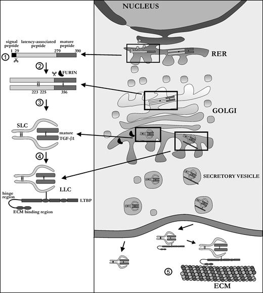
TGF-β1 is a ubiquitous growth factor that is implicated in the control of proliferation, migration, differentiation, and survival of many different cell types. It influences such diverse processes as embryogenesis, angiogenesis, inflammation, and wound healing. In skeletal tissue, TGF-β1 plays a major role in development and maintenance, affecting both cartilage and bone metabolism, the latter being the subject of this review. Because it affects both cells of the osteoblast and osteoclast lineage, TGF-β1 is one of the most important factors in the bone environment, helping to retain the balance between the dynamic processes of bone resorption and bone formation. Many seemingly contradictory reports have been published on the exact functioning of TGF-β1 in the bone milieu. This review provides an overall picture of the bone-specific actions of TGF-β1 and reconciles experimental discrepancies that have been reported for this multifunctional cytokine.