


Bone tissue is continuously remodeled through the concerted actions of bone cells, which include bone resorption by osteoclasts and bone formation by osteoblasts, whereas osteocytes act as mechanosensors and orchestrators of the bone remodeling process. This process is under the control of local (e.g., growth factors and cytokines) and systemic (e.g., calcitonin and estrogens) factors that all together contribute for bone homeostasis. An imbalance between bone resorption and formation can result in bone diseases including osteoporosis. Recently, it has been recognized that, during bone remodeling, there are an intricate communication among bone cells. For instance, the coupling from bone resorption to bone formation is achieved by interaction between osteoclasts and osteoblasts. Moreover, osteocytes produce factors that influence osteoblast and osteoclast activities, whereas osteocyte apoptosis is followed by osteoclastic bone resorption. The increasing knowledge about the structure and functions of bone cells contributed to a better understanding of bone biology. It has been suggested that there is a complex communication between bone cells and other organs, indicating the dynamic nature of bone tissue. In this review, we discuss the current data about the structure and functions of bone cells and the factors that influence bone remodeling.
The Development of Molecular Biology of Osteoporosis
Osteoporosis is one of the major bone disorders that affects both women and men, and causes bone deterioration and bone strength. Bone remodeling maintains bone mass and mineral homeostasis through the balanced action of osteoblasts and osteoclasts, which are responsible for bone formation and bone resorption, respectively. The imbalance in bone remodeling is known to be the main cause of osteoporosis. The imbalance can be the result of the action of various molecules produced by one bone cell that acts on other bone cells and influence cell activity. The understanding of the effect of these molecules on bone can help identify new targets and therapeutics to prevent and treat bone disorders. In this article, we have focused on molecules that are produced by osteoblasts, osteocytes, and osteoclasts and their mechanism of action on these cells. We have also summarized the different pharmacological osteoporosis treatments that target different molecular aspects of these bone cells to minimize osteoporosis.

Osteocyte-Related Cytokines Regulate Osteoclast Formation and Bone Resorption
The process of bone remodeling is the result of the regulated balance between bone cell populations, namely bone-forming osteoblasts, bone-resorbing osteoclasts, and the osteocyte, the mechanosensory cell type. Osteoclasts derived from the hematopoietic stem cell lineage are the principal cells involved in bone resorption. In osteolytic diseases such as rheumatoid arthritis, periodontitis, and osteoporosis, the balance is lost and changes in favor of bone resorption. Therefore, it is vital to elucidate the mechanisms of osteoclast formation and bone resorption. It has been reported that osteocytes express Receptor activator of nuclear factor κΒ ligand (RANKL), an essential factor for osteoclast formation. RANKL secreted by osteocytes is the most important factor for physiologically supported osteoclast formation in the developing skeleton and in pathological bone resorption such as experimental periodontal bone loss. TNF-α directly enhances RANKL expression in osteocytes and promotes osteoclast formation. Moreover, TNF-α enhances sclerostin expression in osteocytes, which also increases osteoclast formation. These findings suggest that osteocyte-related cytokines act directly to enhance osteoclast formation and bone resorption. In this review, we outline the most recent knowledge concerning bone resorption-related cytokines and discuss the osteocyte as the master regulator of bone resorption and effector in osteoclast formation.
The bone remodelling cycle
The bone remodelling cycle replaces old and damaged bone and is a highly regulated, lifelong process essential for preserving bone integrity and maintaining mineral homeostasis. During the bone remodelling cycle, osteoclastic resorption is tightly coupled to osteoblastic bone formation. The remodelling cycle occurs within the basic multicellular unit and comprises five co-ordinated steps; activation, resorption, reversal, formation and termination. These steps occur simultaneously but asynchronously at multiple different locations within the skeleton. Study of rare human bone disease and animal models have helped to elucidate the cellular and molecular mechanisms that regulate the bone remodelling cycle. The key signalling pathways controlling osteoclastic bone resorption and osteoblastic bone formation are receptor activator of nuclear factor-κB (RANK)/RANK ligand/osteoprotegerin and canonical Wnt signalling. Cytokines, growth factors and prostaglandins act as paracrine regulators of the cycle, whereas endocrine regulators include parathyroid hormone, vitamin D, calcitonin, growth hormone, glucocorticoids, sex hormones, and thyroid hormone. Disruption of the bone remodelling cycle and any resulting imbalance between bone resorption and formation leads to metabolic bone disease, most commonly osteoporosis. The advances in understanding the cellular and molecular mechanisms underlying bone remodelling have also provided targets for pharmacological interventions which include antiresorptive and anabolic therapies. This review will describe the remodelling process and its regulation, discuss osteoporosis and summarize the commonest pharmacological interventions used in its management.

Bone Remodeling in Post-menopausal Osteoporosis
Bone mass in the skeleton is dependent on the coordinated activities of bone-forming osteoblasts and bone-resorbing osteoclasts in discrete bone multi-cellular units. Remodeling of bone in these units is important not only for maintaining bone mass, but also to repair microdamage, to prevent accumulation of too much old bone, and for mineral homeostasis. The activities of osteoblasts and osteoclasts are controlled by a variety of hormones and cytokines, as well as by mechanical loading. Most importantly, sex hormones are very crucial for keeping bone mass in balance, and the lack of either estrogen or testosterone leads to decreased bone mass and increased risk for osteoporosis. The prevalence of osteoporotic fractures is increasing dramatically in the Western part of the world and is a major health problem in many countries. In the present review, the cellular and molecular mechanisms controlling bone remodeling and the influence of sex hormones on these processes are summarized. In a separate paper in this issue, the pathogenesis of post-menopausal osteoporosis will be compared with that of inflammation-induced bone remodeling, including the evidence for and against the hypothesis that concomitant post-menopausal osteoporotic disease influences the progression of periodontal disease.
The osteocyte as a signaling cell
Osteocytes, former osteoblasts encapsulated by mineralized bone matrix, are far from being passive and metabolically inactive bone cells. Instead, osteocytes are multifunctional and dynamic cells capable of integrating hormonal and mechanical signals and transmitting them to effector cells in bone and in distant tissues. Osteocytes are a major source of molecules that regulate bone homeostasis by integrating both mechanical cues and hormonal signals that coordinate the differentiation and function of osteoclasts and osteoblasts. Osteocyte function is altered in both rare and common bone diseases, suggesting that osteocyte dysfunction is directly involved in the pathophysiology of several disorders affecting the skeleton. Advances in osteocyte biology initiated the development of novel therapeutics interfering with osteocyte-secreted molecules. Moreover, osteocytes are targets and key distributors of biological signals mediating the beneficial effects of several bone therapeutics used in the clinic. Here we review the most recent discoveries in osteocyte biology demonstrating that osteocytes regulate bone homeostasis and bone marrow fat via paracrine signaling, influence body composition and energy metabolism via endocrine signaling, and contribute to the damaging effects of diabetes mellitus and hematologic and metastatic cancers in the skeleton.

The amazing osteocyte
Abstract The last decade has provided a virtual explosion of data on the molecular biology and function of osteocytes. Far from being the “passive placeholder in bone,” this cell has been found to have numerous functions, such as acting as an orchestrator of bone remodeling through regulation of both osteoclast and osteoblast activity and also functioning as an endocrine cell. The osteocyte is a source of soluble factors not only to target cells on the bone surface but also to target distant organs, such as kidney, muscle, and other tissues. This cell plays a role in both phosphate metabolism and calcium availability and can remodel its perilacunar matrix. Osteocytes compose 90% to 95% of all bone cells in adult bone and are the longest lived bone cell, up to decades within their mineralized environment. As we age, these cells die, leaving behind empty lacunae that frequently micropetrose. In aged bone such as osteonecrotic bone, empty lacunae are associated with reduced remodeling. Inflammatory factors such as tumor necrosis factor and glucocorticoids used to treat inflammatory disease induce osteocyte cell death, but by different mechanisms with potentially different outcomes. Therefore, healthy, viable osteocytes are necessary for proper functionality of bone and other organs. © 2011 American Society for Bone and Mineral Research.
Control of Bone Matrix Properties by Osteocytes
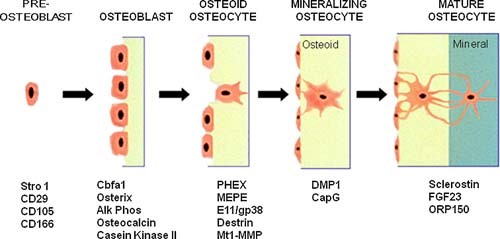
Osteocytes make up 90-95% of the cellular content of bone and form a rich dendritic network with a vastly greater surface area than either osteoblasts or osteoclasts. Osteocytes are well positioned to play a role in bone homeostasis by interacting directly with the matrix; however, the ability for these cells to modify bone matrix remains incompletely understood. With techniques for examining the nano- and microstructure of bone matrix components including hydroxyapatite and type I collagen becoming more widespread, there is great potential to uncover novel roles for the osteocyte in maintaining bone quality. In this review, we begin with an overview of osteocyte biology and the lacunar-canalicular system. Next, we describe recent findings from in vitro models of osteocytes, focusing on the transitions in cellular phenotype as they mature. Finally, we describe historical and current research on matrix alteration by osteocytes in vivo, focusing on the exciting potential for osteocytes to directly form, degrade, and modify the mineral and collagen in their surrounding matrix.

Osteoclast differentiation and activation
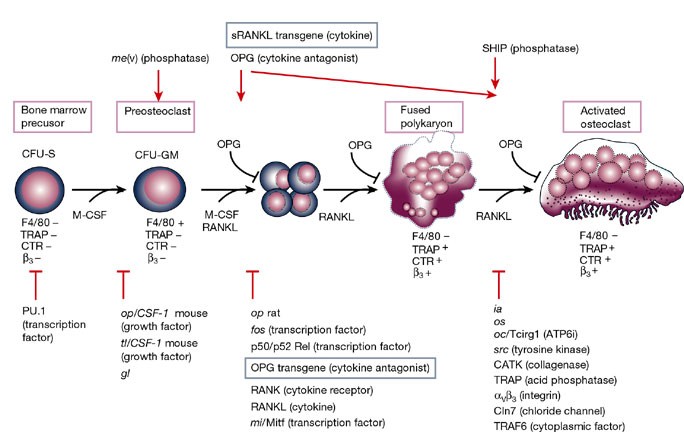
Osteoclasts are specialized cells derived from the monocyte/macrophage haematopoietic lineage that develop and adhere to bone matrix, then secrete acid and lytic enzymes that degrade it in a specialized, extracellular compartment. Discovery of the RANK signalling pathway in the osteoclast has provided insight into the mechanisms of osteoclastogenesis and activation of bone resorption, and how hormonal signals impact bone structure and mass. Further study of this pathway is providing the molecular basis for developing therapeutics to treat osteoporosis and other diseases of bone loss.
Estrogen and the skeleton
Estrogen is the major hormonal regulator of bone metabolism in women and men. Therefore, there is considerable interest in unraveling the pathways by which estrogen exerts its protective effects on bone. Although the major consequence of the loss of estrogen is an increase in bone resorption, estrogen deficiency is associated with a gap between bone resorption and formation, indicating that estrogen is also important for maintaining bone formation at the cellular level. Direct estrogen effects on osteocytes, osteoclasts, and osteoblasts lead to inhibition of bone remodeling, decreased bone resorption, and maintenance of bone formation, respectively.

Cellular mechanisms of bone remodeling
Bone remodeling is a tightly regulated process securing repair of microdamage (targeted remodeling) and replacement of old bone with new bone through sequential osteoclastic resorption and osteoblastic bone formation. The rate of remodeling is regulated by a wide variety of calcitropic hormones (PTH, thyroid hormone, sex steroids etc.). In recent years we have come to appreciate that bone remodeling proceeds in a specialized vascular structure,—the Bone Remodeling Compartment (BRC). The outer lining of this compartment is made up of flattened cells, displaying all the characteristics of lining cells in bone including expression of OPG and RANKL. Reduced bone turnover leads to a decrease in the number of BRCs, while increased turnover causes an increase in the number of BRCs. The secretion of regulatory factors inside a confined space separated from the bone marrow would facilitate local regulation of the remodeling process without interference from growth factors secreted by blood cells in the marrow space. The BRC also creates an environment where cells inside the structure are exposed to denuded bone, which may enable direct cellular interactions with integrins and other matrix factors known to regulate osteoclast/osteoblast activity. However, the denuded bone surface inside the BRC also constitutes an ideal environment for the seeding of bone metastases, known to have high affinity for bone matrix. Circulating osteoclast- and osteoblast precursor cells have been demonstrated in peripheral blood. The dominant pathway regulating osteoclast recruitment is the RANKL/OPG system, while many different factors (RUNX, Osterix) are involved in osteoblast differentiation. Both pathways are modulated by calcitropic hormones.
Application of a mechanism-based disease systems model for osteoporosis to clinical data
A recently proposed mechanism-based disease systems model for osteoporosis (Schmidt et al., J Pharmacokinet Pharmacodyn 38:873–900, 2011) was applied to clinical data from post-menopausal women (n = 767) receiving various doses of the selective estrogen receptor modulator tibolone. Plasma bone-specific alkaline phosphatase activity and urinary N-telopeptide were used as biomarkers reflecting the activity of osteoblasts (bone forming cells) and osteoclasts (bone removing cells), respectively. These data were analyzed in conjunction with data on osteocalcin and on bone mineral density (BMD) (both lumbar spine and total hip), which reflect the activity of both cell types. While the dynamics of bone turnover markers changes rapidly, closely following changes in the activity of bone cells, changes in BMD are slower and have their own dynamics. Application of the mechanism-based disease systems model to the clinical data allowed for an adequate description of the data and yielded parameter estimates that are consistent with physiological values reported in the literature (Lemaire et al., J Theor Biol 229:293–309, 2004). The fitted model enabled characterization of (i) the critical time scales involved in disease progression, (ii) the dynamics of the system during onset and offset of the therapeutic intervention, and (iii) the distinction between responders and low-responders to tibolone treatment.

The Role of Osteocytes in Targeted Bone Remodeling: A Mathematical Model
Until recently many studies of bone remodeling at the cellular level have focused on the behavior of mature osteoblasts and osteoclasts, and their respective precursor cells, with the role of osteocytes and bone lining cells left largely unexplored. This is particularly true with respect to the mathematical modeling of bone remodeling. However, there is increasing evidence that osteocytes play important roles in the cycle of targeted bone remodeling, in serving as a significant source of RANKL to support osteoclastogenesis, and in secreting the bone formation inhibitor sclerostin. Moreover, there is also increasing interest in sclerostin, an osteocyte-secreted bone formation inhibitor, and its role in regulating local response to changes in the bone microenvironment. Here we develop a cell population model of bone remodeling that includes the role of osteocytes, sclerostin, and allows for the possibility of RANKL expression by osteocyte cell populations. We have aimed to give a simple, yet still tractable, model that remains faithful to the underlying system based on the known literature. This model extends and complements many of the existing mathematical models for bone remodeling, but can be used to explore aspects of the process of bone remodeling that were previously beyond the scope of prior modeling work. Through numerical simulations we demonstrate that our model can be used to explore theoretically many of the qualitative features of the role of osteocytes in bone biology as presented in recent literature.
Osteonal and hemi‐osteonal remodeling: The spatial and temporal framework for signal traffic in adult human bone
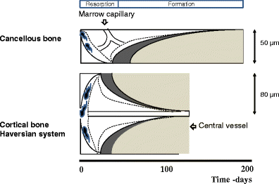
Abstract The bone replacement process in the adult skeleton is known as remodeling. When bone is removed by osteoclasts, new bone is laid down by osteoblasts in the same place, because the load bearing requirement is unchanged. Bone is usually replaced because it is too old to carry out its function, which is mainly mechanical in cortical bone and mainly support for homeostasis and hematopoiesis in cancellous bone. Remodeling always begins on a quiescent bone surface, separated from the marrow by flat lining cells that are one of the two modes of terminal differentiation of osteoblasts. Lining cells are gatekeepers, able to be informed of the need for remodeling, and to either execute or mediate all four components of its activation‐selection and preparation of the site, recruitment of mononuclear preosteoclasts, budding of new capillaries, and attraction of preosteoclasts to the chosen site where they fuse into multinucleated osteoclasts. In cortical bone, osteonal remodeling is carried out by a complex and unique structure, the basic multicellular unit (BMU) that comprises a cutting cone of osteoclasts in front, a closing cone lined by osteoblasts following behind, and connective tissue, blood vessels and nerves filling the cavity. The BMU maintains its size, shape and internal organization for many months as it travels through bone in a controlled direction. Individual osteoclast nuclei are short‐lived, turning over about 8% per d, replaced by new preosteoclasts that originated in the bone marrow and travel in the circulation to the site of resorption. Refilling of bone at each successive cross‐sectional location is accomplished by a team of osteoblasts, probably originating from precursors within the local connective tissue, all assembled within a narrow window of time, at the right location, and in the right orientation to the surface. Each osteoblast team forms bone most rapidly at its onset and slows down progressively. Some of the osteoblasts are buried as osteocytes, some die, and the remainder gradually assume the shape of lining cells. Cancellous bone is more accessible to study than cortical bone, but is geometrically complex. Although remodeling conforms to the same sequence of surface activation, resorption and formation, its three‐dimensional organization is difficult to visualize from two‐dimensional histologic sections. But the average sizes of resorption sites, formation sites, and completed structural units increase progressively, as they do in cortical bone, indicating that the cancellous BMU travels across the surface digging a trench rather than a tunnel, but maintaining its size, shape and individual identity by the continuous recruitment of new cells, just as in cortical bone, a process that can be visualized as hemiosteonal remodeling. The conclusion that all remodeling is carried out by individual BMUs has important implications for bone biology, since many questions about how BMUs operate cannot be answered by studying either intact organisms or isolated cell systems. Many different steps in remodeling and many factors that influence each step have been identified, but very little is known about how the process is regulated in vivo to achieve its biologic purposes; most factors studied to date are likely permissive rather than regulatory in nature. Based on the proposed conceptual model of the BMU, much in vitro experimentation is relevant to the growth, modeling and repair of bone, but not to its remodeling in the adult skeleton. Further progress in the understanding of in vivo physiology will require the characterization of gene expression in individual cells to be related to the spatial and temporal organization of the BMU. This is likely to be possible only for osteonal remodeling in cortical bone in which, because of its geometric simplicity, individual BMUs can consistently be observed in two‐dimensional, longitudinal sections. © 1994 Wiley‐Liss, Inc.

Osteoclasts Provide Coupling Signals to Osteoblast Lineage Cells Through Multiple Mechanisms
Bone remodeling is essential for the repair and replacement of damaged and old bone. The major principle underlying this process is that osteoclast-mediated resorption of a quantum of bone is followed by osteoblast precursor recruitment; these cells differentiate to matrix-producing osteoblasts, which form new bone to replace what was resorbed. Evidence from osteopetrotic syndromes indicate that osteoclasts not only resorb bone, but also provide signals to promote bone formation. Osteoclasts act upon osteoblast lineage cells throughout their differentiation by facilitating growth factor release from resorbed matrix, producing secreted proteins and microvesicles, and expressing membrane-bound factors. These multiple mechanisms mediate the coupling of bone formation to resorption in remodeling. Additional interactions of osteoclasts with osteoblast lineage cells, including interactions with canopy and reversal cells, are required to achieve coordination between bone formation and resorption during bone remodeling.

Molecular Signaling Pathways and Essential Metabolic Elements in Bone Remodeling: An Implication of Therapeutic Targets for Bone Diseases
Bone is one of the dynamic tissues in the human body that undergoes continuous remodelling through subsequent actions of bone cells, osteoclasts, and osteoblasts. Several signal transduction pathways are involved in the transition of mesenchymal stem cells into osteoblasts. These primarily include Runx2, ATF4, Wnt signaling and sympathetic signalling. The differentiation of osteoclasts is controlled by M-CSF, RANKL, and costimulatory signalling. It is well known that bone remodelling is regulated through receptor activator of nuclear factor-kappa B ligand followed by the binding to RANK, which eventually induces the differentiation of osteoclasts. The resorbing osteoclasts secrete TRAP, cathepsin K, MMP-9 and gelatinase to digest the proteinaceous matrix of type I collagen and form a saucer-shaped lacuna along with resorption tunnels in the trabecular bone. Osteoblasts secrete a soluble decoy receptor, osteoprotegerin that prevents the binding of RANK/RANKL and thus moderating osteoclastogenesis. Moreover, bone homeostasis is also regulated by several growth factors, cytokines, calciotropic hormones, parathyroid hormone and sex steroids. The current review presents a correlation of the probable molecular targets underlying the regulation of bone mass and the role of essential metabolic elements in bone remodelling. Targeting these signaling pathways may help design newer therapies for treating bone diseases.

The Effect of Inflammation on Bone
Bone remodeling is the continual process to renew the adult skeleton through the sequential action of osteoblasts and osteoclasts. Nuclear factor RANK, an osteoclast receptor, and its ligand RANKL, expressed on the surface of osteoblasts, result in coordinated control of bone remodeling. Inflammation, a feature of illness and injury, plays a distinct role in skewing this process toward resorption. It does so via the interaction of inflammatory mediators and their related peptides with osteoblasts and osteoclasts, to alter the expression of RANK and RANKL. Such chemical mediators include TNFa, glucocorticoids, histamine, bradykinin, PGE2, systemic RANKL from immune cells, and interleukins 1 and 6. A thorough understanding of bone response to injury and disease, and ability to detect such biomarkers, as well as imaging to identify early structural and mechanical property changes in bone architecture, is important in improving management and outcomes of bone related pathology. While gut health and vitamin and mineral availability appear vitally important, there are very few intervention strategies currently available to favorably alter bone pathology induced by inflammation. Further research is required in this field to advance early detection and treatments.
