


The dynamics of the osteoporotic bone turnover is studied in this paper with the aid of stability analysis of the associated mathematical model. Osteoporosis, which is a common bone disorder, is studied in this papper in detail with an emphasis on the relative threshold values. We examine the expository signaling among the bone cells named osteoclast and osteoblast. Main functioning of osteoblasts is bone formation, whereas osteoclasts are bone removal cells.
Dynamics of bone cell signaling and PTH treatments of osteoporosis
In this paper we analyze and generalize the dynamical system introduced by Lemaire and co-workers [14] as a model of cell signaling in bone remodeling. We show that for large classes of parameter values, including the physically-realistic baseline values, the system has a unique physically relevant equilibrium which is a global attractor. We generalize that model, minimally, to incorporate a mechanism by which parathyroid hormone (PTH) retards osteoblast apoptosis. We show that with this mechanism, which is a simplified version of that proposed by Bellido and co-workers [4], the model exhibits a well-known phenomenon that has puzzled researchers: the system responds catabolically to the continuous administration of PTH andally to appropriately pulsed administration of PTH.
A novel mathematical model of bone remodelling cycles for trabecular bone at the cellular level
After an initial phase of growth and development, bone undergoes a continuous cycle of repair, renewal and optimisation by a process called remodelling. This paper describes a novel mathematical model of the trabecular bone remodelling cycle. It is essentially formulated to simulate a remodelling event at a fixed position in the bone, integrating bone removal by osteoclasts and formation by osteoblasts. The model is developed to construct the variation in bone thickness at a particular point during the remodelling event, derived from standard bone histomorphometric analyses. The novelties of the approach are the adoption of a predator–prey model to describe the dynamic interaction between osteoclasts and osteoblasts, using a genetic algorithm–based solution; quantitative reconstruction of the bone remodelling cycle; and the introduction of a feedback mechanism in the bone formation activity to co-regulate bone thickness. The application of the model is first demonstrated by using experimental data recorded for normal (healthy) bone remodelling to predict the temporal variation in the number of osteoblasts and osteoclasts. The simulated histomorphometric data and remodelling cycle characteristics compare well with the specified input data. Sensitivity studies then reveal how variations in the model’s parameters affect its output; it is hoped that these parameters can be linked to specific biochemical factors in the future. Two sample pathological conditions, hypothyroidism and primary hyperparathyroidism, are examined to demonstrate how the model could be applied more broadly, and, for the first time, the osteoblast and osteoclast populations are predicted for these conditions. Further data are required to fully validate the model’s predictive capacity, but this work shows it has potential, especially in the modelling of pathological conditions and the optimisation of the treatment of those conditions.

Dynamic Biochemical and Cellular Models of Bone Physiology: Integrating Remodeling Processes, Tumor Growth, and Therapy
Bone is an activeCoelho, Rui M. Neto, Joana P. connective tissue composed of different types of cells. The dynamic behavior of bone remodeling processes is typically represented throughValério, Duarte differential equations, which represent the physiological phenomena occurring in this organ. These models takeVinga, Susana into account the tight biochemical regulation between osteoclasts and osteoblasts and have also been enriched with variables and parameters related to bone pathologies and treatment. This chapter reviews some of the more recent models describing bone physiology, focusing on those that include the main cellular processes, along the biochemical control, and also the pharmacokinetic/pharmacodynamic (PK/PD) of the most common treatments for diseases such as cancer. These models are then compared in terms of the simulations obtained and, finally, some highlights on integrating them with the biomechanical component of the system which will be given. These models are expected to provide a valuable insight into this complex system and to support the development of clinical decision systems for bone pathologies.

Biology of Bone Tissue: Structure, Function, and Factors That Influence Bone Cells
Bone tissue is continuously remodeled through the concerted actions of bone cells, which include bone resorption by osteoclasts and bone formation by osteoblasts, whereas osteocytes act as mechanosensors and orchestrators of the bone remodeling process. This process is under the control of local (e.g., growth factors and cytokines) and systemic (e.g., calcitonin and estrogens) factors that all together contribute for bone homeostasis. An imbalance between bone resorption and formation can result in bone diseases including osteoporosis. Recently, it has been recognized that, during bone remodeling, there are an intricate communication among bone cells. For instance, the coupling from bone resorption to bone formation is achieved by interaction between osteoclasts and osteoblasts. Moreover, osteocytes produce factors that influence osteoblast and osteoclast activities, whereas osteocyte apoptosis is followed by osteoclastic bone resorption. The increasing knowledge about the structure and functions of bone cells contributed to a better understanding of bone biology. It has been suggested that there is a complex communication between bone cells and other organs, indicating the dynamic nature of bone tissue. In this review, we discuss the current data about the structure and functions of bone cells and the factors that influence bone remodeling.

A review of recent developments in mathematical modeling of bone remodeling
In this article, we summarize the developments in the mathematical modeling of the mechanics of bone and related biological phenomena. We will devote special attention to the results of the last 10–15 years, although we will cover some relevant classical work to better frame the more recent researches. We will propose a division of the literature based on the main aim of the model (mechanical/biomathematical) and the type of biological phenomena considered (stimulus, growth, cell population dynamics). Finally, we will suggest some possible directions for future investigations.

Bone Dynamics in Metabolic Bone Disease:
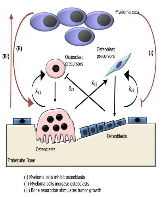
A mathematical model of bone remodeling dynamics for normal bone cell populations and myeloma bone disease
Multiple myeloma is a hematologic malignancy associated with the development of a destructive osteolytic bone disease.
Disease Systems Analysis of Bone Mineral Density and Bone Turnover Markers in Response to Alendronate, Placebo, and Washout in Postmenopausal Women
A previously established mechanism‐based disease systems model for osteoporosis that is based on a mathematically reduced version of a model describing the interactions between osteoclast (bone removing) and osteoblast (bone forming) cells in bone remodeling has been applied to clinical data from women ( n = 1,379) receiving different doses and treatment regimens of alendronate, placebo, and washout. The changes in the biomarkers, plasma bone‐specific alkaline phosphatase activity (BSAP), urinary N‐telopeptide (NTX), lumbar spine bone mineral density (BMD), and total hip BMD, were linked to the underlying mechanistic core of the model. The final model gave an accurate description of all four biomarkers for the different treatments. Simulations were used to visualize the dynamics of the underlying network and the natural disease progression upon alendronate treatment and discontinuation. These results complement the previous applications of this mechanism‐based disease systems model to data from various treatments for osteoporosis.

Mathematical modelling of the pathogenesis of multiple myeloma‐induced bone disease
SUMMARY Multiple myeloma (MM) is the second most common haematological malignancy and results in destructive bone lesions. The interaction between MM cells and the bone microenvironment plays an important role in the development of the tumour cells and MM‐induced bone disease and forms a ‘vicious cycle’ of tumour development and bone destruction, intensified by suppression of osteoblast activity and promotion of osteoclast activity. In this paper, a mathematical model is proposed to simulate how the interaction between MM cells and the bone microenvironment facilitates the development of the tumour cells and the resultant bone destruction. It includes both the roles of inhibited osteoblast activity and stimulated osteoclast activity. The model is able to mimic the temporal variation of bone cell concentrations and resultant bone volume after the invasion and then removal of the tumour cells and explains why MM‐induced bone lesions rarely heal even after the complete removal of MM cells. The behaviour of the model compares well with published experimental data. The model serves as a first step to understand the development of MM‐induced bone disease and could be applied further to evaluate the current therapies against MM‐induced bone disease and even suggests new potential therapeutic targets. © 2014 The Authors. International Journal for Numerical Methods in Biomedical Engineering published by John Wiley & Sons Ltd

Mathematical Modeling of Spatio-Temporal Dynamics of a Single Bone Multicellular Unit
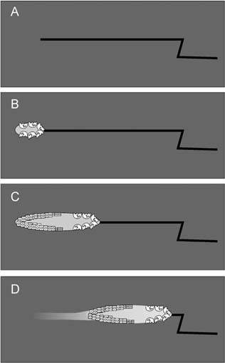
Abstract During bone remodeling, bone-resorbing osteoclasts and bone-forming osteoblasts are organized in bone multicellular units (BMUs), which travel at a rate of 20–40 μm/d for 6–12 mo, maintaining a cylindrical structure. However, the interplay of local BMU geometry with biochemical regulation is poorly understood. We developed a mathematical model of BMU describing changes in time and space of the concentrations of proresorptive cytokine RANKL and its inhibitor osteoprotegerin (OPG), in osteoclast and osteoblast numbers, and in bone mass. We assumed that osteocytes surrounding a microfracture produce RANKL, which attracted osteoclasts. OPG and RANKL were produced by osteoblasts and diffused through bone, RANKL was eliminated by binding to OPG and RANK. Osteoblasts were coupled to osteoclasts through paracrine factors. The evolution of the BMU arising from this model was studied using numerical simulations. Our model recapitulated the spatio-temporal dynamics observed in vivo in a cross-section of bone. In response to a RANKL field, osteoclasts moved as a well-confined cutting cone. The coupling of osteoclasts to osteoblasts allowed for sufficient recruitment of osteoblasts to the resorbed surfaces. The RANKL field was the highest at the microfracture in front of the BMU, whereas the OPG field peaked at the back of the BMU, resulting in the formation of a RANKL/OPG gradient, which strongly affected the rate of BMU progression and its size. Thus, the spatial organization of a BMU provides important constraints on the roles of RANKL and OPG as well as possibly other regulators in determining the outcome of remodeling in the BMU.
Towards a new spatial representation of bone remodeling
Irregular bone remodeling is associated with a number of bone diseases such as osteoporosis and multiple myeloma. Computational and mathematical modeling can aid in therapy and treatment as well as understanding fundamental biology. Different approaches to modeling give insight into different aspects of a phenomena so it is useful to have an arsenal of various computational and mathematical models. Here we develop a mathematical representation of bone remodeling that can effectively describe many aspects of the complicated geometries and spatial behavior observed. There is a sharp interface between bone and marrow regions. Also the surface of bone moves in and out, i.e. in the normal direction, due to remodeling. Based on these observations we employ the use of a level-set function to represent the spatial behavior of remodeling. We elaborate on a temporal model for osteoclast and osteoblast population dynamics to determine the change in bone mass which influences how the interface between bone and marrow changes. We exhibit simulations based on our computational model that show the motion of the interface between bone and marrow as a consequence of bone remodeling. The simulations show that it is possible to capture spatial behavior of bone remodeling in complicated geometries as they occur in vitro and in vivo. By employing the level set approach it is possible to develop computational and mathematical representations of the spatialbehavior of bone remodeling. By including in this formalism further details, such as more complex cytokine interactions and accurate parameter values, it is possible to obtain simulations of phenomena related to bone remodeling with spatial behavior much as in vitro and in vivo. This makes it possible to perform in silica experiments more closely resembling experimental observations.
A review of mathematical modeling of bone remodeling from a systems biology perspective
Bone remodeling is an essential, delicately balanced physiological process of coordinated activity of bone cells that remove and deposit new bone tissue in the adult skeleton. Due to the complex nature of this process, many mathematical models of bone remodeling have been developed. Each of these models has unique features, but they have underlying patterns. In this review, the authors highlight the important aspects frequently found in mathematical models for bone remodeling and discuss how and why these aspects are included when considering the physiology of the bone basic multicellular unit, which is the term used for the collection of cells responsible for bone remodeling. The review also emphasizes the view of bone remodeling from a systems biology perspective. Understanding the systemic mechanisms involved in remodeling will help provide information on bone pathology associated with aging, endocrine disorders, cancers, and inflammatory conditions and enhance systems pharmacology. Furthermore, some features of the bone remodeling cycle and interactions with other organ systems that have not yet been modeled mathematically are discussed as promising future directions in the field.

MODEL-BASED ANALYSIS OF IGF-1 EFFECT ON OSTEOBLAST AND OSTEOCLAST REGULATION IN BONE TURNOVER
The main determinant of bone Ca accretion is a bimolecular regulatory network on osteoblast (OB) and osteoclast (OC). Even though IGF-1 is known as an important regulator in bone cell cycle, little has been done to model IGF-1 action in bone cell regulation. Thus, the objective is to develop a mathematical model that depicts the regulatory action of IGF-1 onto the OB and OC interaction, and to evaluate adolescent and adult bone Ca accretion in response to differences in IGF-1 levels. As a result, a dynamic model of OB and OC with two main regulatory systems, i.e., Receptor Activator for Nuclear Factor [Formula: see text]B (RANK)-RANK Ligand (RANKL)-osteoprogerin (OPG) system, and TGF-[Formula: see text], was augmented with the IGF-1, and incorporated into Ca kinetic data to predict exchangeable bone Ca. The developed model could predict a change in OB and OC levels in response to perturbations in regulators, producing results consistent with bone physiology and published experimental data. The model also estimated parametric difference in regulators between adults and adolescents, suggesting that RANKL/OPG in adolescents was about 4 times higher than in adults, while adolescent serum PTH and IGF-1 concentrations were 60% and 220% of those of adults, respectively. This study highlighted the influence of IGF-1 on the regulation of bone cells in positively modulating bone Ca, suggesting that IGF-1 may be an effective target for reducing bone loss by promoting mature OB.
Simulated Interventions to Ameliorate Age-Related Bone Loss Indicate the Importance of Timing
Bone remodeling is the continuous process of bone resorption by osteoclasts and bone formation by osteoblasts, in order to maintain homeostasis. The activity of osteoclasts and osteoblasts is regulated by a network of signaling pathways, including Wnt, parathyroid hormone (PTH), RANKL/OPG and TGF-β, in response to stimuli such as mechanical loading. During aging there is a gradual loss of bone mass due to dysregulation of signaling pathways. This may be due to a decline in physical activity with age and/or changes in hormones and other signaling molecules. In particular, hormones such as PTH have a circadian rhythm which may be disrupted in aging. Due to the complexity of the molecular and cellular networks involved in bone remodeling, several mathematical models have been proposed to aid understanding of the processes involved. However, to date there are no models which explicitly consider the effects of mechanical loading, the circadian rhythm of PTH and the dynamics of signaling molecules on bone remodeling. Therefore, we have constructed a network model of the system using a modular approach which will allow further modifications as required in future research. The model was used to simulate the effects of mechanical loading and also the effects of different interventions such as continuous or intermittent administration of PTH. Our model predicts that the absence of regular mechanical loading and/or an impaired PTH circadian rhythm leads to a gradual decrease in bone mass over time which can be restored by simulated interventions and that the effectiveness of some interventions may depend on their timing.

Osteoblast Differentiation at a Glance
ABSTRACT Ossification is a tightly regulated process, performed by specialized cells called osteoblasts. Dysregulation of this process may cause inade...

A review of mathematical modeling of bone remodeling from a systems biology perspective
Bone remodeling is an essential physiological process in the adult skeleton. Due to the complex nature of this process, many mathematical models of bone remodeling have been developed. Each of these models has unique features, but they have underlying patterns. In this review, the authors highlight the important aspects frequently found in mathematical models for bone remodeling and discuss how and why these aspects are included when considering the physiology of the bone basic multicellular unit, which is the term used for the collection of cells responsible for bone remodeling. The review also emphasizes the view of bone remodeling from a systems biology perspective. Understanding the systemic mechanisms involved in remodeling will help provide information on bone pathology associated with aging, endocrine disorders, cancers, and inflammatory conditions and enhance systems pharmacology. Furthermore, some features of the bone remodeling cycle and interactions with other organ systems that have not yet been modeled mathematically are discussed as promising future directions in the field.