

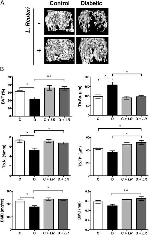
Type 1 diabetes (T1D)–induced osteoporosis is characterized by a predominant suppression of osteoblast number and activity, as well as increased bone marrow adiposity but no change in osteoclast activity. The fundamental mechanisms and alternative anabolic treatments (with few side effects) for T1D bone loss remain undetermined. Recent studies by our laboratory and others indicate that probiotics can benefit bone health. Here, we demonstrate that Lactobacillus reuteri, a probiotic with anti-inflammatory and bone health properties, prevents T1D-induced bone loss and marrow adiposity in mice. We further found that L. reuteri treatment prevented the suppression of Wnt10b in T1D bone. Consistent with a role for attenuated bone Wnt10b expression in T1D osteoporosis, we observed that bone-specific Wnt10b transgenic mice are protected from T1D bone loss. To examine the mechanisms of this protection, we focused on TNF-α, a cytokine up-regulated in T1D that causes suppression of osteoblast Wnt10b expression in vitro. Addition of L. reuteri prevented TNF-α–mediated suppression of Wnt10b and osteoblast maturation markers. Taken together, our findings reveal a mechanism by which T1D causes bone loss and open new avenues for use of probiotics to benefit the bone.
Probiotic <i>L. reuteri</i> Treatment Prevents Bone Loss in a Menopausal Ovariectomized Mouse Model
Abstract Estrogen deficiency is a major risk factor for osteoporosis that is associated with bone inflammation and resorption. Half of women over the age of 50 will experience an osteoporosis related fracture in their lifetime, thus novel therapies are needed to combat post‐menopausal bone loss. Recent studies suggest an important role for gut‐bone signaling pathways and the microbiota in regulating bone health. Given that the bacterium Lactobacillus reuteri ATCC PTA 6475 ( L. reuteri ) secretes beneficial immunomodulatory factors, we examined if this candidate probiotic could reduce bone loss associated with estrogen deficiency in an ovariectomized (Ovx) mouse menopausal model. Strikingly, L. reuteri treatment significantly protected Ovx mice from bone loss. Osteoclast bone resorption markers and activators (Trap5 and RANKL) as well as osteoclastogenesis are significantly decreased in L. reuteri ‐treated mice. Consistent with this, L. reuteri suppressed Ovx‐induced increases in bone marrow CD4+ T‐lymphocytes (which promote osteoclastogenesis) and directly suppressed osteoclastogenesis in vitro. We also identified that L. reuteri treatment modifies microbial communities in the Ovx mouse gut. Together, our studies demonstrate that L. reuteri treatment suppresses bone resorption and loss associated with estrogen deficiency. Thus, L. reuteri treatment may be a straightforward and cost‐effective approach to reduce post‐menopausal bone loss. J. Cell. Physiol. 229: 1822–1830, 2014. © 2014 Wiley Periodicals, Inc.

The Microbial Metabolite Butyrate Stimulates Bone Formation via T Regulatory Cell-Mediated Regulation of WNT10B Expression
Tyagi et al. show that oral supplementation with the widely used probiotic, Lactobacillus rhamnosus GG (LGG), increases bone mass in mice by increasing the serum levels of the short chain fatty acid butyrate. LGG or butyrate increase the frequency of regulatory T (Treg) cells in the intestine and in the bone marrow. Treg cells stimulate CD8+ T cells to secrete the Wnt ligand Wnt10b, which stimulates bone formation by activating Wnt signaling in osteoblasts. Therefore, LGG and butyrate may represent new interventions for the prevention and treatment of osteoporosis.

Probiotics Protect Mice from Ovariectomy-Induced Cortical Bone Loss
The gut microbiota (GM) modulates the hosts metabolism and immune system. Probiotic bacteria are defined as live microorganisms which when administered in adequate amounts confer a health benefit on the host and can alter the composition of the GM. Germ-free mice have increased bone mass associated with reduced bone resorption indicating that the GM also regulates bone mass. Ovariectomy (ovx) results in bone loss associated with altered immune status. The purpose of this study was to determine if probiotic treatment protects mice from ovx-induced bone loss. Mice were treated with either a single Lactobacillus (L) strain, L. paracasei DSM13434 (L. para) or a mixture of three strains, L. paracasei DSM13434, L. plantarum DSM 15312 and DSM 15313 (L. mix) given in the drinking water during 6 weeks, starting two weeks before ovx. Both the L. para and the L. mix treatment protected mice from ovx-induced cortical bone loss and bone resorption. Cortical bone mineral content was higher in both L. para and L. mix treated ovx mice compared to vehicle (veh) treated ovx mice. Serum levels of the resorption marker C-terminal telopeptides and the urinary fractional excretion of calcium were increased by ovx in the veh treated but not in the L. para or the L. mix treated mice. Probiotic treatment reduced the expression of the two inflammatory cytokines, TNFα and IL-1β, and increased the expression of OPG, a potent inhibitor of osteoclastogenesis, in cortical bone of ovx mice. In addition, ovx decreased the frequency of regulatory T cells in bone marrow of veh treated but not probiotic treated mice. In conclusion, treatment with L. para or the L. mix prevents ovx-induced cortical bone loss. Our findings indicate that these probiotic treatments alter the immune status in bone resulting in attenuated bone resorption in ovx mice.
Lactulose Suppresses Osteoclastogenesis and Ameliorates Estrogen Deficiency-Induced Bone Loss in Mice
<p>Postmenopausal osteoporosis is characterized by excess osteoclastogenesis which leads to net bone loss and brittle fractures. Studies have demonstrated that estrogen deficiency-associated bone loss is microbiota-dependent and could be prevented by probiotics and prebiotics. In this study, we report that orally administered lactulose (20 g/kg, 6 weeks) orally administered significantly inhibited osteoclastogenesis, bone resorption, and prevented ovariectomy (OVX)-induced bone loss in mice. Lactulose increased intestinal <i>Claudin 2, 3 and 15,</i> compared to the OVX group, and lowered pro-osteoclastogenic cytokines levels including tumor necrosis factor-α, interleukin(IL)-6, receptor activator of nuclear factor kappa-Β ligand (RANKL), and IL-17 as well as increased the anti-inflammatory cytokine IL-10 in the intestine, peripheral blood, and bone marrow. Lactulose significantly preserved the number of Foxp3<sup>+</sup> Treg cells in the intestines compared with that in OVX mice. Lactulose altered the composition of intestinal microbiota measured by 16s rDNA sequencing and increased intestinal and serum short-chain fatty acids (SCFAs) levels including acetate, propionate and butyrate which were decreased in OVX mice as measured by gas chromatography. Oral administration of lactulose for 2 weeks significantly lowered the level of bone resorption marker C-telopeptide of type 1 collagen-1 in healthy male young volunteers (aging 20-25 years). In conclusion, lactulose inhibited osteoclastogenesis and bone resorption by altering the intestinal microbiota and increasing SCFAs. Lactulose could serve as an ideal therapeutic agent for postmenopausal osteoporosis.</p>
Probiotic <i>Lactobacillus reuteri</i> Prevents Postantibiotic Bone Loss by Reducing Intestinal Dysbiosis and Preventing Barrier Disruption
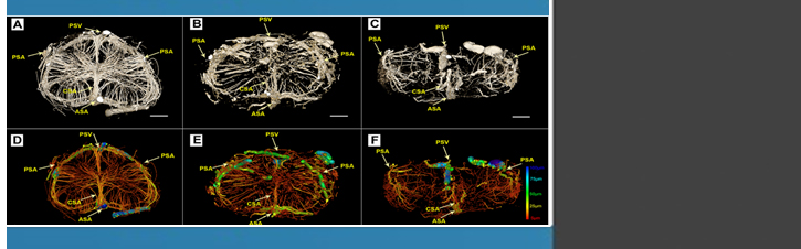
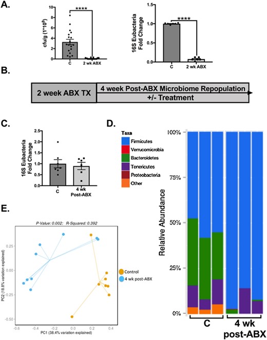
ABSTRACT Antibiotic treatment, commonly prescribed for bacterial infections, depletes and subsequently causes long-term alterations in intestinal microbiota composition. Knowing the importance of the microbiome in the regulation of bone density, we investigated the effect of postantibiotic treatment on gut and bone health. Intestinal microbiome repopulation at 4-weeks postantibiotic treatment resulted in an increase in the Firmicutes:Bacteroidetes ratio, increased intestinal permeability, and notably reduced femoral trabecular bone volume (approximately 30%, p < 0.01). Treatment with a mucus supplement (a high-molecular-weight polymer, MDY-1001 [MDY]) prevented the postantibiotic-induced barrier break as well as bone loss, indicating a mechanistic link between increased intestinal permeability and bone loss. A link between the microbiome composition and bone density was demonstrated by supplementing the mice with probiotic bacteria. Specifically, Lactobacillus reuteri, but not Lactobacillus rhamnosus GG or nonpathogenic Escherichia coli, reduced the postantibiotic elevation of the Firmicutes:Bacteroidetes ratio and prevented femoral and vertebral trabecular bone loss. Consistent with causing bone loss, postantibiotic-induced dysbiosis decreased osteoblast and increased osteoclast activities, changes that were prevented by both L. reuteri and MDY. These data underscore the importance of microbial dysbiosis in the regulation of intestinal permeability and bone health, as well as identify L. reuteri and MDY as novel therapies for preventing these adverse effects. © 2018 American Society for Bone and Mineral Research.
The gut microbiota regulates bone mass in mice
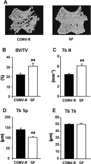
Abstract The gut microbiota modulates host metabolism and development of immune status. Here we show that the gut microbiota is also a major regulator of bone mass in mice. Germ-free (GF) mice exhibit increased bone mass associated with reduced number of osteoclasts per bone surface compared with conventionally raised (CONV-R) mice. Colonization of GF mice with a normal gut microbiota normalizes bone mass. Furthermore, GF mice have decreased frequency of CD4+ T cells and CD11b+/GR 1 osteoclast precursor cells in bone marrow, which could be normalized by colonization. GF mice exhibited reduced expression of inflammatory cytokines in bone and bone marrow compared with CONV-R mice. In summary, the gut microbiota regulates bone mass in mice, and we provide evidence for a mechanism involving altered immune status in bone and thereby affected osteoclast-mediated bone resorption. Further studies are required to evaluate the gut microbiota as a novel therapeutic target for osteoporosis. © 2012 American Society for Bone and Mineral Research.
Gut microbiota and bone metabolism
Abstract Osteoporosis is the most common metabolic skeletal disease. It is characterized by the deterioration of the skeletal microarchitecture and bone loss, leading to ostealgia, and even bone fractures. Accumulating evidence has indicated that there is an inextricable relationship between the gut microbiota (GM) and bone homeostasis involving host–microbiota crosstalk. Any perturbation of the GM can play an initiating and reinforcing role in disrupting the bone remodeling balance during the development of osteoporosis. Although the GM is known to influence bone metabolism, the mechanisms associated with these effects remain unclear. Herein, we review the current knowledge of how the GM affects bone metabolism in health and disease, summarize the correlation between pathogen‐associated molecular patterns of GM structural components and bone metabolism, and discuss the potential mechanisms underlying how GM metabolites regulate bone turnover. Deciphering the complicated relationship between the GM and bone health will provide new insights into the prevention and treatment of osteoporosis.

Sex steroid deficiency–associated bone loss is microbiota dependent and prevented by probiotics
1Division of Endocrinology, Metabolism and Lipids, Department of Medicine, Emory University, Atlanta, Georgia, USA.
Effect of Bacillus subtilis C-3102 on bone mineral density in healthy postmenopausal Japanese women: a randomized, placebo-controlled, double-blind clinical trial
Gut microbiota influence the host immune system and are associated with various diseases. In recent years, postmenopausal bone loss has been suggested to be related to gut microbiota. In the present study, we investigated the treatment effect of the probiotic Bacillus subtilis C-3102 (C-3102) on bone mineral density (BMD) and its influence on gut microbiota in healthy postmenopausal Japanese women. Seventy-six healthy postmenopausal Japanese women were treated with a placebo or C-3102 spore-containing tablets for 24 weeks. When compared with the placebo, C-3102 significantly increased total hip BMD (placebo = 0.83 ± 0.63%, C-3102 = 2.53 ± 0.52%, p=0.043). There was a significant group-by-time interaction effect for urinary type I collagen cross-linked N-telopeptide (uNTx) (p=0.033), a marker of bone resorption. Specifically, the C-3102 group showed significantly lower uNTx when compared with the placebo group at 12 weeks of treatment (p=0.015). In addition, in the C-3102 group, there was a trend towards a decrease in the bone resorption marker tartrate-resistant acid phosphatase isoform 5b (TRACP-5b) when compared with the placebo group at 12 weeks of treatment (p=0.052). The relative abundance of genus Bifidobacterium significantly increased at 12 weeks of treatment compared with the baseline in the C-3102 group. The relative abundance of genus Fusobacterium was significantly decreased in the C-3102 group at 12 and 24 weeks of treatment compared with the baseline. These data suggested that C-3102 improves BMD by inhibiting bone resorption and modulating gut microbiota in healthy postmenopausal women.

Physiological and pathophysiological bone turnover — role of the immune system
Receptor activator of NF-κB ligand (RANKL) and osteoprotegerin (OPG) are key downstream effectors of bone resorption; tumour necrosis factor (TNF) might synergize with RANKL to superinduce osteoclastic bone resorptionB cells, regulated by T cells, are a key source of basal OPG whereas activated T cells and B cells are key sources of TNF and RANKL in inflammatory conditionsShort-term antiresorptive therapies might safely prevent bone loss associated with combination antiretroviral therapyAnergic and parathyroid-hormone-treated T cells secrete the protein Wnt-10b, which promotes bone formationNovel therapeutic strategies targeting the immune system might promote bone formation and decrease bone resorption to manage osteoporotic bone loss and prevent fracture
Mathematical Modeling of the Gut–Bone Axis and Implications of Butyrate Treatment on Osteoimmunology
Butyrate, a short-chain fatty acid produced by the gut microbiota, has pivotal roles in the regulation of the immune system. Recent studies have revealed that butyrate increases the differentiation of peripheral regulatory T cells in the gut–bone axis and promotes osteoblasts’ bone forming activity. However, the mechanism of the therapeutic benefit of butyrate in bone remodeling remains incompletely understood. Here, we develop a multicompartment mathematical model to quantitatively predict the contribution of butyrate on the expansion of regulatory T cells in the gut, blood, and bone compartments. We investigate the interplay between regulatory T cell-derived TGF-β and CD8+ T cell-derived Wnt-10b with changes in gut butyrate concentration. In addition, we connect our model to a detailed model of bone metabolism to study the impacts of butyrate and Wnt-10b on trabecular bone volume. Our results indicate both direct and indirect immune-mediated impacts of butyrate on bone metabolism.

The Effect of Inflammation on Bone
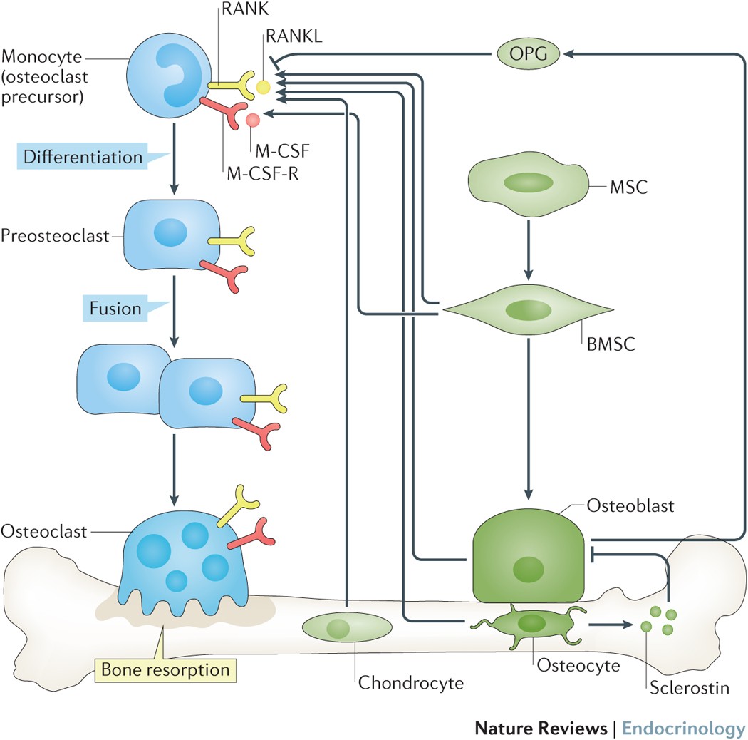
Bone remodeling is the continual process to renew the adult skeleton through the sequential action of osteoblasts and osteoclasts. Nuclear factor RANK, an osteoclast receptor, and its ligand RANKL, expressed on the surface of osteoblasts, result in coordinated control of bone remodeling. Inflammation, a feature of illness and injury, plays a distinct role in skewing this process toward resorption. It does so via the interaction of inflammatory mediators and their related peptides with osteoblasts and osteoclasts, to alter the expression of RANK and RANKL. Such chemical mediators include TNFa, glucocorticoids, histamine, bradykinin, PGE2, systemic RANKL from immune cells, and interleukins 1 and 6. A thorough understanding of bone response to injury and disease, and ability to detect such biomarkers, as well as imaging to identify early structural and mechanical property changes in bone architecture, is important in improving management and outcomes of bone related pathology. While gut health and vitamin and mineral availability appear vitally important, there are very few intervention strategies currently available to favorably alter bone pathology induced by inflammation. Further research is required in this field to advance early detection and treatments.

Montmorency tart cherry protects against age-related bone loss in female C57BL/6 mice and demonstrates some anabolic effects
Age-related bone loss is a consequence of endocrine and immune changes that disrupt bone remodeling. Functional foods (e.g., tart cherries) with antioxidant, anti-inflammatory and prebiotic activity can potentially counter this age-related phenomenon. The aim of this study was to determine if Montmorency tart cherry protects against early age-related bone loss and the culpable alterations in bone metabolism.

The Regulation of Bone Metabolism and Disorders by Wnt Signaling
Wnt, a secreted glycoprotein, has an approximate molecular weight of 40 kDa, and it is a cytokine involved in various biological phenomena including ontogeny, morphogenesis, carcinogenesis, and maintenance of stem cells. The Wnt signaling pathway can be classified into two main pathways: canonical and non-canonical. Of these, the canonical Wnt signaling pathway promotes osteogenesis. Sclerostin produced by osteocytes is an inhibitor of this pathway, thereby inhibiting osteogenesis. Recently, osteoporosis treatment using an anti-sclerostin therapy has been introduced. In this review, the basics of Wnt signaling, its role in bone metabolism and its involvement in skeletal disorders have been covered. Furthermore, the clinical significance and future scopes of Wnt signaling in osteoporosis, osteoarthritis, rheumatoid arthritis and neoplasia are discussed.
The osteocyte as a signaling cell
Osteocytes, former osteoblasts encapsulated by mineralized bone matrix, are far from being passive and metabolically inactive bone cells. Instead, osteocytes are multifunctional and dynamic cells capable of integrating hormonal and mechanical signals and transmitting them to effector cells in bone and in distant tissues. Osteocytes are a major source of molecules that regulate bone homeostasis by integrating both mechanical cues and hormonal signals that coordinate the differentiation and function of osteoclasts and osteoblasts. Osteocyte function is altered in both rare and common bone diseases, suggesting that osteocyte dysfunction is directly involved in the pathophysiology of several disorders affecting the skeleton. Advances in osteocyte biology initiated the development of novel therapeutics interfering with osteocyte-secreted molecules. Moreover, osteocytes are targets and key distributors of biological signals mediating the beneficial effects of several bone therapeutics used in the clinic. Here we review the most recent discoveries in osteocyte biology demonstrating that osteocytes regulate bone homeostasis and bone marrow fat via paracrine signaling, influence body composition and energy metabolism via endocrine signaling, and contribute to the damaging effects of diabetes mellitus and hematologic and metastatic cancers in the skeleton.

Regulation of osteoblastogenesis and bone mass by Wnt10b
Wnts comprise a family of secreted signaling proteins that regulate diverse developmental processes. Activation of Wnt signaling by Wnt10b inhibits differentiation of preadipocytes and blocks adipose tissue development; however, the effect of Wnt10b on other mesenchymal lineages has not been defined. To explore the physiological role of Wnt signaling in bone development, we analyzed FABP4-Wnt10b mice, which express the Wnt10b transgene in marrow. Femurs from FABP4-Wnt10b mice have almost four times as much bone in the distal metaphyses and are mechanically stronger. These mice maintain elevated bone mass at least through 23 months of age. In addition, FABP4-Wnt10b mice are protected from the bone loss characteristic of estrogen deficiency. We used pharmacological and genetic approaches to demonstrate that canonical Wnt signaling stimulates osteoblastogenesis and inhibits adipogenesis of bipotential mesenchymal precursors. Wnt10b shifts cell fate toward the osteoblast lineage by induction of the osteoblastogenic transcription factors Runx2, Dlx5, and osterix and suppression of the adipogenic transcription factors C/EBPα and PPARγ. One mechanism whereby Wnt10b promotes osteoblastogenesis is suppression of PPARγ expression. Finally, Wnt10b-/- mice have decreased trabecular bone and serum osteocalcin, confirming that Wnt10b is an endogenous regulator of bone formation.
