


Background: With increase in the incidence of resistance to antibiotics, probiotics are emerging as a promising adjunctive periodontal therapy. The authors of this study evaluate the influence of probiotic (PROB) supplementation on ligature‐induced periodontitis (LIP) and intestinal morphology in rats. Methods: Thirty‐two rats were randomly divided into four groups: control (C), LIP, PROB, and LIP/PROB. In groups PROB and LIP/PROB, the PROB was administered orally by addition to the drinking water of the animals for 44 days. In groups LIP and LIP/PROB, the mandibular right first molar of the animals received a cotton ligature that was left in the same position for 14 days. All animals were euthanized 44 days after the start of the PROB supplementation. The jaws were resected and histomorphometric analyses were performed. The measurements included evaluation of attachment loss (AL) and alveolar bone level (ABL) on the distal root of the mandibular first molar. Samples of the duodenum, jejunum, and ileum were also dissected from each animal to evaluate the villous height (VH) and crypt depth (CD). The data obtained were subjected to statistical analyses (analysis of variance, Tukey; P <0.05). Results: Mean values of AL and ABL were significantly higher in group LIP compared with group LIP/PROB (AL: 3.05 ± 0.57 mm and 1.78 ± 0.63 mm, respectively; ABL: 4.21 ± 0.42 mm and 3.38 ± 0.17 mm, respectively). In group LIP/PROB, the mean values of VH and CD of the jejunum were significantly higher than the ones from group LIP (VH: 672.1 ± 83.3 µm and 528.0 ± 51.7 µm, respectively; CD: 463.8 ± 100.9 µm and 269.0 ± 48.4 µm, respectively). Conclusions: It can be concluded that PROB supplementation 1) reduces AL and alveolar bone loss in rats with LIP and 2) can protect the small intestine from reactive changes induced by LIP.
Probiotics Protect Mice from Ovariectomy-Induced Cortical Bone Loss
The gut microbiota (GM) modulates the hosts metabolism and immune system. Probiotic bacteria are defined as live microorganisms which when administered in adequate amounts confer a health benefit on the host and can alter the composition of the GM. Germ-free mice have increased bone mass associated with reduced bone resorption indicating that the GM also regulates bone mass. Ovariectomy (ovx) results in bone loss associated with altered immune status. The purpose of this study was to determine if probiotic treatment protects mice from ovx-induced bone loss. Mice were treated with either a single Lactobacillus (L) strain, L. paracasei DSM13434 (L. para) or a mixture of three strains, L. paracasei DSM13434, L. plantarum DSM 15312 and DSM 15313 (L. mix) given in the drinking water during 6 weeks, starting two weeks before ovx. Both the L. para and the L. mix treatment protected mice from ovx-induced cortical bone loss and bone resorption. Cortical bone mineral content was higher in both L. para and L. mix treated ovx mice compared to vehicle (veh) treated ovx mice. Serum levels of the resorption marker C-terminal telopeptides and the urinary fractional excretion of calcium were increased by ovx in the veh treated but not in the L. para or the L. mix treated mice. Probiotic treatment reduced the expression of the two inflammatory cytokines, TNFα and IL-1β, and increased the expression of OPG, a potent inhibitor of osteoclastogenesis, in cortical bone of ovx mice. In addition, ovx decreased the frequency of regulatory T cells in bone marrow of veh treated but not probiotic treated mice. In conclusion, treatment with L. para or the L. mix prevents ovx-induced cortical bone loss. Our findings indicate that these probiotic treatments alter the immune status in bone resulting in attenuated bone resorption in ovx mice.
Probiotic <i>Lactobacillus reuteri</i> Prevents Postantibiotic Bone Loss by Reducing Intestinal Dysbiosis and Preventing Barrier Disruption
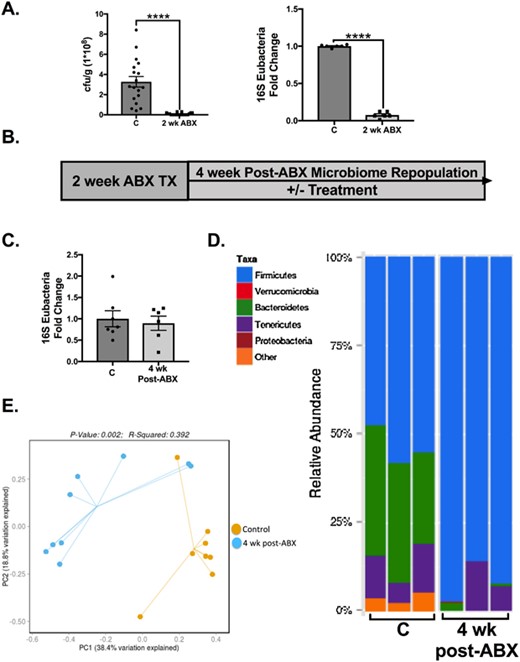
ABSTRACT Antibiotic treatment, commonly prescribed for bacterial infections, depletes and subsequently causes long-term alterations in intestinal microbiota composition. Knowing the importance of the microbiome in the regulation of bone density, we investigated the effect of postantibiotic treatment on gut and bone health. Intestinal microbiome repopulation at 4-weeks postantibiotic treatment resulted in an increase in the Firmicutes:Bacteroidetes ratio, increased intestinal permeability, and notably reduced femoral trabecular bone volume (approximately 30%, p < 0.01). Treatment with a mucus supplement (a high-molecular-weight polymer, MDY-1001 [MDY]) prevented the postantibiotic-induced barrier break as well as bone loss, indicating a mechanistic link between increased intestinal permeability and bone loss. A link between the microbiome composition and bone density was demonstrated by supplementing the mice with probiotic bacteria. Specifically, Lactobacillus reuteri, but not Lactobacillus rhamnosus GG or nonpathogenic Escherichia coli, reduced the postantibiotic elevation of the Firmicutes:Bacteroidetes ratio and prevented femoral and vertebral trabecular bone loss. Consistent with causing bone loss, postantibiotic-induced dysbiosis decreased osteoblast and increased osteoclast activities, changes that were prevented by both L. reuteri and MDY. These data underscore the importance of microbial dysbiosis in the regulation of intestinal permeability and bone health, as well as identify L. reuteri and MDY as novel therapies for preventing these adverse effects. © 2018 American Society for Bone and Mineral Research.
Lactulose Suppresses Osteoclastogenesis and Ameliorates Estrogen Deficiency-Induced Bone Loss in Mice
<p>Postmenopausal osteoporosis is characterized by excess osteoclastogenesis which leads to net bone loss and brittle fractures. Studies have demonstrated that estrogen deficiency-associated bone loss is microbiota-dependent and could be prevented by probiotics and prebiotics. In this study, we report that orally administered lactulose (20 g/kg, 6 weeks) orally administered significantly inhibited osteoclastogenesis, bone resorption, and prevented ovariectomy (OVX)-induced bone loss in mice. Lactulose increased intestinal <i>Claudin 2, 3 and 15,</i> compared to the OVX group, and lowered pro-osteoclastogenic cytokines levels including tumor necrosis factor-α, interleukin(IL)-6, receptor activator of nuclear factor kappa-Β ligand (RANKL), and IL-17 as well as increased the anti-inflammatory cytokine IL-10 in the intestine, peripheral blood, and bone marrow. Lactulose significantly preserved the number of Foxp3<sup>+</sup> Treg cells in the intestines compared with that in OVX mice. Lactulose altered the composition of intestinal microbiota measured by 16s rDNA sequencing and increased intestinal and serum short-chain fatty acids (SCFAs) levels including acetate, propionate and butyrate which were decreased in OVX mice as measured by gas chromatography. Oral administration of lactulose for 2 weeks significantly lowered the level of bone resorption marker C-telopeptide of type 1 collagen-1 in healthy male young volunteers (aging 20-25 years). In conclusion, lactulose inhibited osteoclastogenesis and bone resorption by altering the intestinal microbiota and increasing SCFAs. Lactulose could serve as an ideal therapeutic agent for postmenopausal osteoporosis.</p>
Probiotic <i>L. reuteri</i> Treatment Prevents Bone Loss in a Menopausal Ovariectomized Mouse Model
Abstract Estrogen deficiency is a major risk factor for osteoporosis that is associated with bone inflammation and resorption. Half of women over the age of 50 will experience an osteoporosis related fracture in their lifetime, thus novel therapies are needed to combat post‐menopausal bone loss. Recent studies suggest an important role for gut‐bone signaling pathways and the microbiota in regulating bone health. Given that the bacterium Lactobacillus reuteri ATCC PTA 6475 ( L. reuteri ) secretes beneficial immunomodulatory factors, we examined if this candidate probiotic could reduce bone loss associated with estrogen deficiency in an ovariectomized (Ovx) mouse menopausal model. Strikingly, L. reuteri treatment significantly protected Ovx mice from bone loss. Osteoclast bone resorption markers and activators (Trap5 and RANKL) as well as osteoclastogenesis are significantly decreased in L. reuteri ‐treated mice. Consistent with this, L. reuteri suppressed Ovx‐induced increases in bone marrow CD4+ T‐lymphocytes (which promote osteoclastogenesis) and directly suppressed osteoclastogenesis in vitro. We also identified that L. reuteri treatment modifies microbial communities in the Ovx mouse gut. Together, our studies demonstrate that L. reuteri treatment suppresses bone resorption and loss associated with estrogen deficiency. Thus, L. reuteri treatment may be a straightforward and cost‐effective approach to reduce post‐menopausal bone loss. J. Cell. Physiol. 229: 1822–1830, 2014. © 2014 Wiley Periodicals, Inc.

Galactooligosaccharides Improve Mineral Absorption and Bone Properties in Growing Rats through Gut Fermentation
Galactooligosaccharides (GOS), prebiotic nondigestible oligosaccharides derived from lactose, have the potential for improving mineral balance and bone properties. This study examined the dose–response effect of GOS supplementation on calcium and magnesium absorption, mineral retention, bone properties, and gut microbiota in growing rats. Seventy-five 4-week-old male Sprague–Dawley rats were randomized into one of five treatment groups (n = 15/group) and fed a diet containing 0, 2, 4, 6, or 8% GOS by weight for 8 weeks. Dietary GOS significantly decreased cecal pH and increased cecal wall weight and content weight in a dose-dependent manner (p < 0.0001). Fingerprint patterns of the 16S rRNA gene PCR-DGGE from fecal DNA indicated the variance of bacterial community structure, which was primarily explained by GOS treatments (p = 0.0001). Quantitative PCR of the samples revealed an increase in the relative proportion of bifidobacteria with GOS (p = 0.0001). Net calcium absorption was increased in a dose–response manner (p < 0.01) with GOS supplementation. Dietary GOS also increased (p < 0.02) net magnesium absorption, femur 45Ca uptake, calcium and magnesium retention, and femur and tibia breaking strength. Distal femur total and trabecular volumetric bone mineral density (vBMD) and area and proximal tibia vBMD increased (p < 0.02) with GOS supplementation. Trabecular-rich bones, that is, those that rapidly turn over, were most benefited. Regression modeling showed that GOS benefited calcium and magnesium utilization and vBMD through decreased cecal pH, increased cecal wall and content weight, and increased proportion of bifidobacteria.
Effect of Bacillus subtilis C-3102 on bone mineral density in healthy postmenopausal Japanese women: a randomized, placebo-controlled, double-blind clinical trial
Gut microbiota influence the host immune system and are associated with various diseases. In recent years, postmenopausal bone loss has been suggested to be related to gut microbiota. In the present study, we investigated the treatment effect of the probiotic Bacillus subtilis C-3102 (C-3102) on bone mineral density (BMD) and its influence on gut microbiota in healthy postmenopausal Japanese women. Seventy-six healthy postmenopausal Japanese women were treated with a placebo or C-3102 spore-containing tablets for 24 weeks. When compared with the placebo, C-3102 significantly increased total hip BMD (placebo = 0.83 ± 0.63%, C-3102 = 2.53 ± 0.52%, p=0.043). There was a significant group-by-time interaction effect for urinary type I collagen cross-linked N-telopeptide (uNTx) (p=0.033), a marker of bone resorption. Specifically, the C-3102 group showed significantly lower uNTx when compared with the placebo group at 12 weeks of treatment (p=0.015). In addition, in the C-3102 group, there was a trend towards a decrease in the bone resorption marker tartrate-resistant acid phosphatase isoform 5b (TRACP-5b) when compared with the placebo group at 12 weeks of treatment (p=0.052). The relative abundance of genus Bifidobacterium significantly increased at 12 weeks of treatment compared with the baseline in the C-3102 group. The relative abundance of genus Fusobacterium was significantly decreased in the C-3102 group at 12 and 24 weeks of treatment compared with the baseline. These data suggested that C-3102 improves BMD by inhibiting bone resorption and modulating gut microbiota in healthy postmenopausal women.

Dried Plum’s Polyphenolic Compounds and Carbohydrates Contribute to Its Osteoprotective Effects and Exhibit Prebiotic Activity in Estrogen Deficient C57BL/6 Mice
Evidence of dried plum’s benefits on bone continues to emerge. This study investigated the contribution of the fruit’s polyphenol (PP) and carbohydrate (CHO) components on a bone model of postmenopausal osteoporosis to explore their prebiotic activity. Osteopenic ovariectomized mice were fed diets supplemented with dried plum, a crude extract of dried plum’s polyphenolic compounds, or the PP or CHO fraction of the crude extract. The effects of treatments on the bone phenotype were assessed at 5 and 10 weeks as well as the prebiotic activity of the different components of dried plum. Both the CHO and PP fractions of the extract contributed to the effects on bone with the CHO suppressing bone formation and resorption, and the PP temporally down-regulating formation. The PP and CHO components also altered the gut microbiota and cecal short chain fatty acids. These findings demonstrate that the CHO as well as the PP components of dried plum have potential prebiotic activity, but they have differential roles in mediating the alterations in bone formation and resorption that protect bone in estrogen deficiency.

Loss of Bone and Wnt10b Expression in Male Type 1 Diabetic Mice Is Blocked by the Probiotic Lactobacillus reuteri
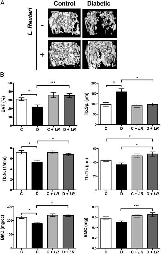
Type 1 diabetes (T1D)–induced osteoporosis is characterized by a predominant suppression of osteoblast number and activity, as well as increased bone marrow adiposity but no change in osteoclast activity. The fundamental mechanisms and alternative anabolic treatments (with few side effects) for T1D bone loss remain undetermined. Recent studies by our laboratory and others indicate that probiotics can benefit bone health. Here, we demonstrate that Lactobacillus reuteri, a probiotic with anti-inflammatory and bone health properties, prevents T1D-induced bone loss and marrow adiposity in mice. We further found that L. reuteri treatment prevented the suppression of Wnt10b in T1D bone. Consistent with a role for attenuated bone Wnt10b expression in T1D osteoporosis, we observed that bone-specific Wnt10b transgenic mice are protected from T1D bone loss. To examine the mechanisms of this protection, we focused on TNF-α, a cytokine up-regulated in T1D that causes suppression of osteoblast Wnt10b expression in vitro. Addition of L. reuteri prevented TNF-α–mediated suppression of Wnt10b and osteoblast maturation markers. Taken together, our findings reveal a mechanism by which T1D causes bone loss and open new avenues for use of probiotics to benefit the bone.
Sex steroid deficiency–associated bone loss is microbiota dependent and prevented by probiotics
1Division of Endocrinology, Metabolism and Lipids, Department of Medicine, Emory University, Atlanta, Georgia, USA.
The Microbial Metabolite Butyrate Stimulates Bone Formation via T Regulatory Cell-Mediated Regulation of WNT10B Expression
Tyagi et al. show that oral supplementation with the widely used probiotic, Lactobacillus rhamnosus GG (LGG), increases bone mass in mice by increasing the serum levels of the short chain fatty acid butyrate. LGG or butyrate increase the frequency of regulatory T (Treg) cells in the intestine and in the bone marrow. Treg cells stimulate CD8+ T cells to secrete the Wnt ligand Wnt10b, which stimulates bone formation by activating Wnt signaling in osteoblasts. Therefore, LGG and butyrate may represent new interventions for the prevention and treatment of osteoporosis.

The gut microbiota regulates bone mass in mice
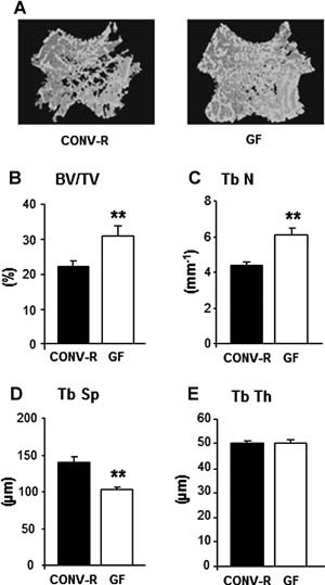
Abstract The gut microbiota modulates host metabolism and development of immune status. Here we show that the gut microbiota is also a major regulator of bone mass in mice. Germ-free (GF) mice exhibit increased bone mass associated with reduced number of osteoclasts per bone surface compared with conventionally raised (CONV-R) mice. Colonization of GF mice with a normal gut microbiota normalizes bone mass. Furthermore, GF mice have decreased frequency of CD4+ T cells and CD11b+/GR 1 osteoclast precursor cells in bone marrow, which could be normalized by colonization. GF mice exhibited reduced expression of inflammatory cytokines in bone and bone marrow compared with CONV-R mice. In summary, the gut microbiota regulates bone mass in mice, and we provide evidence for a mechanism involving altered immune status in bone and thereby affected osteoclast-mediated bone resorption. Further studies are required to evaluate the gut microbiota as a novel therapeutic target for osteoporosis. © 2012 American Society for Bone and Mineral Research.
Prebiotics Enhance Magnesium Absorption and Inulin‐based Fibers Exert Chronic Effects on Calcium Utilization in a Postmenopausal Rodent Model
Abstract: Age‐related changes in calcium metabolism play a role in the development of osteoporosis. A 4‐wk feeding study was conducted in 5‐mo‐old ovariectomized (OVX) Sprague‐Dawley rats to assess the effect of various dietary fibers on mineral metabolism and bone health parameters. There were 6 treatment groups: sham‐Control, OVX‐Control, OVX rats receiving daily estradiol (E 2 ) injections, and OVX rats receiving an AIN‐93M diet supplement with either an inulin‐based fiber (Synergy1® or Fruitafit HD®) or a novel fiber (polydextrose) at 5% wt. of diet. Calcium and magnesium metabolic balances were performed after early (3 d) and late exposure (4 wk) to dietary treatments. Rats receiving polydextrose had significantly higher net calcium absorption efficiency and retention than all control groups and a trend ( P ≤ 0.10) for higher calcium absorption when compared to inulin‐based fibers after early exposure but the advantage did not persist over long‐term exposure. The inulin‐based fibers had positive chronic effects on calcium metabolism that were related to changes in the gut, that is, production of short chain fatty acids and higher cecal wall weights. All fibers improved magnesium absorption and retention in early and late metabolic balances; effects on magnesium metabolism were more pronounced than for calcium. Practical Application: Steady growth in US middle‐aged and elderly populations has led to higher incidences of several chronic diseases including osteoporosis, a bone disease that primarily affects postmenopausal women. Recent research suggests that certain dietary fibers (prebiotics) enhance mineral absorption and may impart bone health benefits. This work examines the impact of prebiotic supplementation on mineral metabolism and bone health using a postmenopausal rat model. Study findings will aid future investigations in ascertaining the factors related to potential bone health benefits of prebiotic which will aid in developing an effective prebiotics food product/supplement that will address the bone health needs of consumers.

Epithelial Barrier Function in Gut-Bone Signaling
The intestinal epithelial barrier plays an essential role in maintaining host homeostasis. The barrier regulates nutrient absorption as well as prevents the invasion of pathogenic bacteria in the host. It is composed of epithelial cells, tight junctions, and a mucus layer. Several factors, such as cytokines, diet, and diseases, can affect this barrier. These factors have been shown to increase intestinal permeability, inflammation, and translocation of pathogenic bacteria. In addition, dysregulation of the epithelial barrier can result in inflammatory diseases such as inflammatory bowel disease. Our lab and others have also shown that barrier disruption can have systemic effects including bone loss. In this chapter, we will discuss the current literature to understand the link between intestinal barrier and bone. We will discuss how inflammation, aging, dysbiosis, and metabolic diseases can affect intestinal barrier-bone link. In addition, we will highlight the current suggested mechanism between intestinal barrier and bone.
Effect of Galactooligosaccharides on Calcium Absorption and Preventing Bone Loss in Ovariectomized Rats
Abstract. The effects of galactooligosaccharides (GOS), a mixture of galactosyl oligosaccharides formed from lactose by the transgalactosyl reaction of β-D
The Role of Estrogen in the Control of Rat Osteocyte Apoptosis
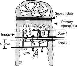
Abstract We have previously shown that estrogen withdrawal by gonadotrophin-releasing hormone analogs (GnRHa) induces osteocyte death via apoptosis in human bone. Although it is likely that the increase in osteocyte death via apoptosis was related to the loss of estrogen, these experiments could not rule out a direct role for the GnRHa. Therefore, in this study, we have used a rat model of ovariectomy (OVX) to determine whether the effect of estrogen withdrawal extends to other species and to clarify the role of estrogen in the maintenance of osteocyte viability. Twelve 9-week-old rats were divided into three treatment groups: sham operated (SHAM) (n = 4), OVX (n = 4), and OVX + estrogen (E2) (25 μg/day) (n = 4). At 3 weeks following the start of treatment, tibial bones were removed. The percentage of osteocytes displaying DNA breaks, using an in situ nick-translation method, was significantly higher in the OVX group compared with the SHAM control in both cortical bone (10.04% vs. 2.31%, respectively; p < 0.0001) and trabecular bone (6.44% vs. 1.58%, respectively; p = 0.003). Addition of estrogen in the OVX animals completely abrogated the increase in osteocyte apoptosis in cortical bone (0.78%) and trabecular bone (1.17%). The percentage of apoptotic osteocytes decreased with increasing distance from the primary/secondary spongiosa interface below the growth plate in the OVX model and the OVX + E2 model. Nuclear morphology and electrophoresis of DNA confirmed the presence of apoptotic cells in the samples. In conclusion, OVX in the rat results in an increase in osteocyte apoptosis as a direct or indirect result of E2 loss. Addition of estrogen in the OVX animals prevents this increase in osteocyte apoptosis. These data confirm an important role for estrogen in the control of osteocyte apoptosis and the maintenance of osteocyte viability. Estrogen deficiency might, through compromising the viability of osteocyte networks, reduce the ability of bone to respond appropriately to loading.
The pleiotropic effects of prebiotic galacto-oligosaccharides on the aging gut
Prebiotic galacto-oligosaccharides (GOS) have an extensively demonstrated beneficial impact on intestinal health. In this study, we determined the impact of GOS diets on hallmarks of gut aging: microbiome dysbiosis, inflammation, and intestinal barrier defects (“leaky gut”). We also evaluated if short-term GOS feeding influenced how the aging gut responded to antibiotic challenges in a mouse model of Clostridioides difficile infection. Finally, we assessed if colonic organoids could reproduce the GOS responder—non-responder phenotypes observed in vivo.
