


Bone remodeling is the continuous process of bone resorption by osteoclasts and bone formation by osteoblasts, in order to maintain homeostasis. The activity of osteoclasts and osteoblasts is regulated by a network of signaling pathways, including Wnt, parathyroid hormone (PTH), RANKL/OPG and TGF-β, in response to stimuli such as mechanical loading. During aging there is a gradual loss of bone mass due to dysregulation of signaling pathways. This may be due to a decline in physical activity with age and/or changes in hormones and other signaling molecules. In particular, hormones such as PTH have a circadian rhythm which may be disrupted in aging. Due to the complexity of the molecular and cellular networks involved in bone remodeling, several mathematical models have been proposed to aid understanding of the processes involved. However, to date there are no models which explicitly consider the effects of mechanical loading, the circadian rhythm of PTH and the dynamics of signaling molecules on bone remodeling. Therefore, we have constructed a network model of the system using a modular approach which will allow further modifications as required in future research. The model was used to simulate the effects of mechanical loading and also the effects of different interventions such as continuous or intermittent administration of PTH. Our model predicts that the absence of regular mechanical loading and/or an impaired PTH circadian rhythm leads to a gradual decrease in bone mass over time which can be restored by simulated interventions and that the effectiveness of some interventions may depend on their timing.
Regulation of Bone Remodeling by Parathyroid Hormone
Parathyroid hormone (PTH) exerts profound effects on skeletal homeostasis through multiple cellular and molecular mechanisms. Continuous hyperparathyroidism causes net loss of bone mass, despite accelerating bone formation by osteoblasts. Intermittent treatment with PTH analogs represents the only Food and Drug Administration (FDA)-approved bone anabolic osteoporosis treatment strategy. Functional PTH receptors are present on cells of the osteoblast lineage, ranging from early skeletal stem cells to matrix-embedded osteocytes. In addition, bone remodeling by osteoclasts liberates latent growth factors present within bone matrix. Here, we will provide an overview of the multiple cellular and molecular mechanisms through which PTH influences bone homeostasis. Notably, net skeletal effects of continuous versus intermittent can differ significantly. Where possible, we will highlight mechanisms through which continuous hyperparathyroidism leads to bone loss, and through which intermittent hyperparathyroidism boosts bone mass. Given the therapeutic usage of intermittent PTH (iPTH) treatment for osteoporosis, particular attention will be paid toward mechanisms underlying the bone anabolic effects of once daily PTH administration.
The bone remodelling cycle
The bone remodelling cycle replaces old and damaged bone and is a highly regulated, lifelong process essential for preserving bone integrity and maintaining mineral homeostasis. During the bone remodelling cycle, osteoclastic resorption is tightly coupled to osteoblastic bone formation. The remodelling cycle occurs within the basic multicellular unit and comprises five co-ordinated steps; activation, resorption, reversal, formation and termination. These steps occur simultaneously but asynchronously at multiple different locations within the skeleton. Study of rare human bone disease and animal models have helped to elucidate the cellular and molecular mechanisms that regulate the bone remodelling cycle. The key signalling pathways controlling osteoclastic bone resorption and osteoblastic bone formation are receptor activator of nuclear factor-κB (RANK)/RANK ligand/osteoprotegerin and canonical Wnt signalling. Cytokines, growth factors and prostaglandins act as paracrine regulators of the cycle, whereas endocrine regulators include parathyroid hormone, vitamin D, calcitonin, growth hormone, glucocorticoids, sex hormones, and thyroid hormone. Disruption of the bone remodelling cycle and any resulting imbalance between bone resorption and formation leads to metabolic bone disease, most commonly osteoporosis. The advances in understanding the cellular and molecular mechanisms underlying bone remodelling have also provided targets for pharmacological interventions which include antiresorptive and anabolic therapies. This review will describe the remodelling process and its regulation, discuss osteoporosis and summarize the commonest pharmacological interventions used in its management.

Mathematical Model of Bone Remodeling Captures the Antiresorptive and Anabolic Actions of Various Therapies
A better understanding of the molecular pathways regulating the bone remodeling process should help in the development of new antiresorptive regulators and anabolic regulators, that is, regulators of bone resorption and of bone formation. Understanding the mechanisms by which parathyroid hormone (PTH) influences bone formation and how it switches from anabolic to catabolic action is important for treating osteoporosis (Poole and Reeve in Curr Opin Pharmacol 5:612–617, 2005). In this paper we describe a mathematical model of bone remodeling that incorporates, extends, and integrates several models of particular aspects of this biochemical system (Cabal et al. in J Bone Miner Res 28(8):1830–1836, 2013; Lemaire et al. in J Theor Biol 229:293–309, 2004; Peterson and Riggs in Bone 46:49–63, 2010; Raposo et al. in J Clin Endocrinol Metab 87(9):4330–4340, 2002; Ross et al. in J Disc Cont Dyn Sys Series B 17(6):2185–2200, 2012). We plan to use this model as a bone homeostasis platform to develop anabolic and antiresorptive compounds. The model will allow us to test hypotheses about the dynamics of compounds and to test the potential benefits of combination therapies. At the core of the model is the idealized account of osteoclast and osteoblast signaling given by Lemaire et al. (J Theor Biol 229:293–309, 2004). We have relaxed some of their assumptions about the roles of osteoprotegerin, transforming growth factor $$\upbeta $$, and receptor activator of nuclear factor $$\upkappa $$B ligand; we have devised more detailed models of the interactions of these species. We have incorporated a model of the effect of calcium sensing receptor antagonists on remodeling (Cabal et al. in J Bone Miner Res 28(8):1830–1836, 2013). We have also incorporated a basic model of the effects of vitamin D on calcium homeostasis. We have included a simple model of the mechanism proposed by Bellido et al. (2003), Ross et al. (J Disc Cont Dyn Sys Series B 17(6):2185–2200, 2012), of the influence of PTH on osteoblast apoptosis, a mechanism that accounts for the anabolic response to pulsatile PTH administration. Finally, we have devised a simple model of the administration and effects of bisphosphonates. The biomarkers in the model are procollagen type 1 amino-terminal propeptide and C-terminal telopeptide. Bone mineral density is the model’s principal endpoint.

Parathyroid hormone temporal effects on bone formation and resorption
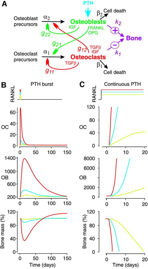
Parathyroid hormone (PTH) paradoxically causes net bone loss (resorption) when administered in a continuous fashion, and net bone formation (deposition) when administered intermittently. Currently no pharmacological formulations are available to promote bone formation, as needed for the treatment of osteoporosis. The paradoxical behavior of PTH confuses endocrinologists, thus, a model bone resorption or deposition dependent on the timing of PTH administration would de-mystify this behavior and provide the basis for logical drug formulation. We developed a mathematical model that accounts for net bone loss with continuous PTH administration and net bone formation with intermittent PTH administration, based on the differential effects of PTH on the osteoblastic and osteoclastic populations of cells.

Dynamics of bone cell signaling and PTH treatments of osteoporosis
In this paper we analyze and generalize the dynamical system introduced by Lemaire and co-workers [14] as a model of cell signaling in bone remodeling. We show that for large classes of parameter values, including the physically-realistic baseline values, the system has a unique physically relevant equilibrium which is a global attractor. We generalize that model, minimally, to incorporate a mechanism by which parathyroid hormone (PTH) retards osteoblast apoptosis. We show that with this mechanism, which is a simplified version of that proposed by Bellido and co-workers [4], the model exhibits a well-known phenomenon that has puzzled researchers: the system responds catabolically to the continuous administration of PTH andally to appropriately pulsed administration of PTH.
Mechanobiological osteocyte feedback drives mechanostat regulation of bone in a multiscale computational model
Significant progress has been made to identify the cells and signaling molecules involved in the mechanobiological regulation of bone remodeling. It is now well accepted that osteocytes act as mechanosensory cells in bone expressing several signaling molecules such as nitric oxide (NO) and sclerostin (Scl) which are able to control bone remodeling responses. In this paper, we present a comprehensive multiscale computational model of bone remodeling which incorporates biochemical osteocyte feedback. The mechanostat theory is quantitatively incorporated into the model using mechanical feedback to control expression levels of NO and Scl. The catabolic signaling pathway RANK–RANKL–OPG is co-regulated via (continuous) PTH and NO, while the anabolic Wnt signaling pathway is described via competitive binding reactions between Wnt, Scl and the Wnt receptors LRP5/6. Using this novel model of bone remodeling, we investigate the effects of changes in the mechanical loading and hormonal environment on bone balance. Our numerical simulations show that we can calibrate the mechanostat anabolic and catabolic regulatory mechanisms so that they are mutually exclusive. This is consistent with previous models that use a Wolff-type law to regulate bone resorption and formation separately. Furthermore, mechanical feedback provides an effective mechanism to obtain physiological bone loss responses due to mechanical disuse and/or osteoporosis.

A review of mathematical modeling of bone remodeling from a systems biology perspective
Bone remodeling is an essential physiological process in the adult skeleton. Due to the complex nature of this process, many mathematical models of bone remodeling have been developed. Each of these models has unique features, but they have underlying patterns. In this review, the authors highlight the important aspects frequently found in mathematical models for bone remodeling and discuss how and why these aspects are included when considering the physiology of the bone basic multicellular unit, which is the term used for the collection of cells responsible for bone remodeling. The review also emphasizes the view of bone remodeling from a systems biology perspective. Understanding the systemic mechanisms involved in remodeling will help provide information on bone pathology associated with aging, endocrine disorders, cancers, and inflammatory conditions and enhance systems pharmacology. Furthermore, some features of the bone remodeling cycle and interactions with other organ systems that have not yet been modeled mathematically are discussed as promising future directions in the field.
A review of mathematical modeling of bone remodeling from a systems biology perspective
Bone remodeling is an essential, delicately balanced physiological process of coordinated activity of bone cells that remove and deposit new bone tissue in the adult skeleton. Due to the complex nature of this process, many mathematical models of bone remodeling have been developed. Each of these models has unique features, but they have underlying patterns. In this review, the authors highlight the important aspects frequently found in mathematical models for bone remodeling and discuss how and why these aspects are included when considering the physiology of the bone basic multicellular unit, which is the term used for the collection of cells responsible for bone remodeling. The review also emphasizes the view of bone remodeling from a systems biology perspective. Understanding the systemic mechanisms involved in remodeling will help provide information on bone pathology associated with aging, endocrine disorders, cancers, and inflammatory conditions and enhance systems pharmacology. Furthermore, some features of the bone remodeling cycle and interactions with other organ systems that have not yet been modeled mathematically are discussed as promising future directions in the field.

Mathematical Model of Paracrine Interactions between Osteoclasts and Osteoblasts Predicts Anabolic Action of Parathyroid Hormone on Bone
Abstract. To restore falling plasma calcium levels, PTH promotes calcium liberation from bone. PTH targets bone-forming cells, osteoblasts, to increase exp
Molecular Signaling Pathways and Essential Metabolic Elements in Bone Remodeling: An Implication of Therapeutic Targets for Bone Diseases
Bone is one of the dynamic tissues in the human body that undergoes continuous remodelling through subsequent actions of bone cells, osteoclasts, and osteoblasts. Several signal transduction pathways are involved in the transition of mesenchymal stem cells into osteoblasts. These primarily include Runx2, ATF4, Wnt signaling and sympathetic signalling. The differentiation of osteoclasts is controlled by M-CSF, RANKL, and costimulatory signalling. It is well known that bone remodelling is regulated through receptor activator of nuclear factor-kappa B ligand followed by the binding to RANK, which eventually induces the differentiation of osteoclasts. The resorbing osteoclasts secrete TRAP, cathepsin K, MMP-9 and gelatinase to digest the proteinaceous matrix of type I collagen and form a saucer-shaped lacuna along with resorption tunnels in the trabecular bone. Osteoblasts secrete a soluble decoy receptor, osteoprotegerin that prevents the binding of RANK/RANKL and thus moderating osteoclastogenesis. Moreover, bone homeostasis is also regulated by several growth factors, cytokines, calciotropic hormones, parathyroid hormone and sex steroids. The current review presents a correlation of the probable molecular targets underlying the regulation of bone mass and the role of essential metabolic elements in bone remodelling. Targeting these signaling pathways may help design newer therapies for treating bone diseases.

A novel mathematical model of bone remodelling cycles for trabecular bone at the cellular level
After an initial phase of growth and development, bone undergoes a continuous cycle of repair, renewal and optimisation by a process called remodelling. This paper describes a novel mathematical model of the trabecular bone remodelling cycle. It is essentially formulated to simulate a remodelling event at a fixed position in the bone, integrating bone removal by osteoclasts and formation by osteoblasts. The model is developed to construct the variation in bone thickness at a particular point during the remodelling event, derived from standard bone histomorphometric analyses. The novelties of the approach are the adoption of a predator–prey model to describe the dynamic interaction between osteoclasts and osteoblasts, using a genetic algorithm–based solution; quantitative reconstruction of the bone remodelling cycle; and the introduction of a feedback mechanism in the bone formation activity to co-regulate bone thickness. The application of the model is first demonstrated by using experimental data recorded for normal (healthy) bone remodelling to predict the temporal variation in the number of osteoblasts and osteoclasts. The simulated histomorphometric data and remodelling cycle characteristics compare well with the specified input data. Sensitivity studies then reveal how variations in the model’s parameters affect its output; it is hoped that these parameters can be linked to specific biochemical factors in the future. Two sample pathological conditions, hypothyroidism and primary hyperparathyroidism, are examined to demonstrate how the model could be applied more broadly, and, for the first time, the osteoblast and osteoclast populations are predicted for these conditions. Further data are required to fully validate the model’s predictive capacity, but this work shows it has potential, especially in the modelling of pathological conditions and the optimisation of the treatment of those conditions.
The Role of Osteocytes in Targeted Bone Remodeling: A Mathematical Model
Until recently many studies of bone remodeling at the cellular level have focused on the behavior of mature osteoblasts and osteoclasts, and their respective precursor cells, with the role of osteocytes and bone lining cells left largely unexplored. This is particularly true with respect to the mathematical modeling of bone remodeling. However, there is increasing evidence that osteocytes play important roles in the cycle of targeted bone remodeling, in serving as a significant source of RANKL to support osteoclastogenesis, and in secreting the bone formation inhibitor sclerostin. Moreover, there is also increasing interest in sclerostin, an osteocyte-secreted bone formation inhibitor, and its role in regulating local response to changes in the bone microenvironment. Here we develop a cell population model of bone remodeling that includes the role of osteocytes, sclerostin, and allows for the possibility of RANKL expression by osteocyte cell populations. We have aimed to give a simple, yet still tractable, model that remains faithful to the underlying system based on the known literature. This model extends and complements many of the existing mathematical models for bone remodeling, but can be used to explore aspects of the process of bone remodeling that were previously beyond the scope of prior modeling work. Through numerical simulations we demonstrate that our model can be used to explore theoretically many of the qualitative features of the role of osteocytes in bone biology as presented in recent literature.
A mechano-chemo-biological model for bone remodeling with a new mechano-chemo-transduction approach
Bone remodeling is a fundamental biological process that develops in bone tissue along its whole lifetime. It refers to a continuous bone transformation with new bone formation and old bone resorption that changes the internal microstructure and composition of the tissue. The main objectives of bone remodeling are: repair of the internal microcracks; adaptation of the macroscopic stiffness and strength to the actual changing mechanical demands; and control of the calcium homeostasis. Understanding this process and predicting its evolution is critical to reduce the effects of long-term disuse as happens during periods of reduced mobility. It is also important in the design of bone implants to avoid long-term stress shielding. Many mathematical models have been proposed from the earliest purely phenomenological to the latest that include biological knowledge. However, there still exists a lack of connection between the mechanical driving force and the biochemical and cell processes it triggers. Here, and following previous works that model independently the mechanobiological and biochemical processes in bone remodeling, we present a more complete model, useful for both cortical and trabecular bone, that uses a new mechanotransduction approach based on the effect of strains onto the bonding–unbonding rate of RANK/RANKL/OPG receptor–ligand reactions. We compare the results of this model with previous ones, showing a good agreement in similar conditions. We also apply it to realistic situations such as a femoral bone after implantation of a hip prosthesis, getting similar results to the clinical ones in the final bone density distribution. Finally, we extend this approach to the anisotropic case, getting not only the mean density, but also the directional homogenization of the microstructure. This biochemical approach permits, not only to predict the bone evolution under changes in the mechanical loads, but also, to consider anabolic and catabolic drugs to control bone density, such as those used in osteoporosis.

The Cellular Dynamics of Bone Remodeling: A Mathematical Model
The mechanical properties of vertebrate bone are largely determined by a process which involves the complex interplay of three different cell types. This process is called bone remodeling and occurs asynchronously at multiple sites in the mature skeleton. The cells involved are bone resorbing osteoclasts, bone matrix producing osteoblasts, and mechanosensing osteocytes. These cells communicate with each other by means of autocrine and paracrine signaling factors and operate in complex entities, the so-called bone multicellular units (BMUs). To investigate the BMU dynamics in silico, we develop a novel mathematical model resulting in a system of nonlinear partial differential equations (PDEs) with time delays. The model describes the osteoblast and osteoclast populations together with the dynamics of the key messenger molecule RANKL and its decoy receptor OPG. Scaling theory is used to address parameter sensitivity and predict the emergence of pathological remodeling regimes. The model is studied numerically in one and two space dimensions using finite difference schemes in space and explicit delay equation solvers in time. The computational results are in agreement with in vivo observations and provide new insights into the role of the RANKL/OPG pathway in the spatial regulation of bone remodeling.

TRANCE Is Necessary and Sufficient for Osteoblast-mediated Activation of Bone Resorption in Osteoclasts
TRANCE (tumor necrosis factor–related activation-induced cytokine) is a recently described member of the tumor necrosis factor superfamily that stimulates dendritic cell survival and has also been found to induce osteoclastic differentiation from hemopoietic precursors. However, its effects on mature osteoclasts have not been defined. It has long been recognized that stimulation of osteoclasts by agents such as parathyroid hormone (PTH) occurs through a hormonal interaction with osteoblastic cells, which are thereby induced to activate osteoclasts. To determine whether TRANCE accounts for this activity, we tested its effects on mature osteoclasts. TRANCE rapidly induced a dramatic change in osteoclast motility and spreading and inhibited apoptosis. In populations of osteoclasts that were unresponsive to PTH, TRANCE caused activation of bone resorption equivalent to that induced by PTH in the presence of osteoblastic cells. Moreover, osteoblast-mediated stimulation of bone resorption was abrogated by soluble TRANCE receptor and by the soluble decoy receptor osteoprotegerin (OPG), and stimulation of isolated osteoclasts by TRANCE was neutralized by OPG. Thus, TRANCE expression by osteoblasts appears to be both necessary and sufficient for hormone-mediated activation of mature osteoclasts, and TRANCE-R is likely to be a receptor for signal transduction for activation of the osteoclast and its survival.

Biology of Bone Tissue: Structure, Function, and Factors That Influence Bone Cells
Bone tissue is continuously remodeled through the concerted actions of bone cells, which include bone resorption by osteoclasts and bone formation by osteoblasts, whereas osteocytes act as mechanosensors and orchestrators of the bone remodeling process. This process is under the control of local (e.g., growth factors and cytokines) and systemic (e.g., calcitonin and estrogens) factors that all together contribute for bone homeostasis. An imbalance between bone resorption and formation can result in bone diseases including osteoporosis. Recently, it has been recognized that, during bone remodeling, there are an intricate communication among bone cells. For instance, the coupling from bone resorption to bone formation is achieved by interaction between osteoclasts and osteoblasts. Moreover, osteocytes produce factors that influence osteoblast and osteoclast activities, whereas osteocyte apoptosis is followed by osteoclastic bone resorption. The increasing knowledge about the structure and functions of bone cells contributed to a better understanding of bone biology. It has been suggested that there is a complex communication between bone cells and other organs, indicating the dynamic nature of bone tissue. In this review, we discuss the current data about the structure and functions of bone cells and the factors that influence bone remodeling.
