


Discovery and characterization of the cytokine receptor-cytokine-decoy receptor triad formed by RANKL-RANK-OPG have led not only to immense advances in understanding the biology of bone homeostasis, but have also crystalized appreciation of the critical regulatory relationship that exists between bone and immunity, resulting in the emergence of the burgeoning field of osteoimmunology. RANKL-RANK-OPG are members of the tumor necrosis factor (TNF) and TNF receptor superfamilies, and share signaling characteristics common to many members of each. Developmentally regulated and cell-type specific expression patterns of each of these factors have revealed key regulatory functions for RANKL-RANK-OPG in bone homeostasis, organogenesis, immune tolerance and cancer. Successful efforts at designing and developing therapeutic agents targeting RANKL-RANK-OPG have been undertaken for osteoporosis, and additional efforts are underway for other conditions. In this review, we will summarize the basic biology of the RANKL-RANK-OPG system, relate its cell-type specific functions to system-wide mechanisms of development and homeostasis, and highlight emerging areas of interest for this cytokine group.
Osteoimmunology: The Conceptual Framework Unifying the Immune and Skeletal Systems
The immune and skeletal systems share a variety of molecules, including cytokines, chemokines, hormones, receptors, and transcription factors. Bone cells interact with immune cells under physiological and pathological conditions. Osteoimmunology was created as a new interdisciplinary field in large part to highlight the shared molecules and reciprocal interactions between the two systems in both heath and disease. Receptor activator of NF-κB ligand (RANKL) plays an essential role not only in the development of immune organs and bones, but also in autoimmune diseases affecting bone, thus effectively comprising the molecule that links the two systems. Here we review the function, gene regulation, and signal transduction of osteoimmune molecules, including RANKL, in the context of osteoclastogenesis as well as multiple other regulatory functions. Osteoimmunology has become indispensable for understanding the pathogenesis of a number of diseases such as rheumatoid arthritis (RA). We review the various osteoimmune pathologies, including the bone destruction in RA, in which pathogenic helper T cell subsets [such as IL-17-expressing helper T (Th17) cells] induce bone erosion through aberrant RANKL expression. We also focus on cellular interactions and the identification of the communication factors in the bone marrow, discussing the contribution of bone cells to the maintenance and regulation of hematopoietic stem and progenitors cells. Thus the time has come for a basic reappraisal of the framework for understanding both the immune and bone systems. The concept of a unified osteoimmune system will be absolutely indispensable for basic and translational approaches to diseases related to bone and/or the immune system.

Osteoporosis in Rheumatoid Arthritis: Dangerous Liaisons
Osteoporosis has been classically considered a comorbidity of rheumatoid arthritis (RA). However, recent advances in the pathogenesis of osteoporosis in RA have shown a close interplay between cells of the immune system and those involved in bone remodeling, introducing new actors into the classic route in which osteoclast activation is related to the RANK/RANKL/OPG pathway. In fact, the inflammatory state in early stages of RA, mediated by interleukin (IL)-1, IL-6, IL-8 and tumour necrosis factor (TNF)-α has the ability to activate and differentiate osteoclasts not only through their relationship with RANKL, but also through the Wnt/DKK1/sclerostin pathway, leading to bone loss. The role of synovial fibroblasts and activated T lymphocytes in the expression of the RANKL system and its connection to bone destruction is also depicted. In addition, autoantibodies such as rheumatoid factor and anti-citrullinated protein antibodies are other pathogenic mechanisms for the development of bone erosions and systemic osteoporosis in RA, even before the onset of arthritis. The aim of this review is to unravel the relationship between different factors involved in the development of osteoporosis in RA patients, both the classic factors and the most novel, based on the relationship of autoantibodies with bone remodeling. Furthermore, we propose that bone mineral density measured by different techniques may be helpful as a biomarker of severity in early arthritis patients.

Physiological and pathophysiological bone turnover — role of the immune system
Receptor activator of NF-κB ligand (RANKL) and osteoprotegerin (OPG) are key downstream effectors of bone resorption; tumour necrosis factor (TNF) might synergize with RANKL to superinduce osteoclastic bone resorptionB cells, regulated by T cells, are a key source of basal OPG whereas activated T cells and B cells are key sources of TNF and RANKL in inflammatory conditionsShort-term antiresorptive therapies might safely prevent bone loss associated with combination antiretroviral therapyAnergic and parathyroid-hormone-treated T cells secrete the protein Wnt-10b, which promotes bone formationNovel therapeutic strategies targeting the immune system might promote bone formation and decrease bone resorption to manage osteoporotic bone loss and prevent fracture
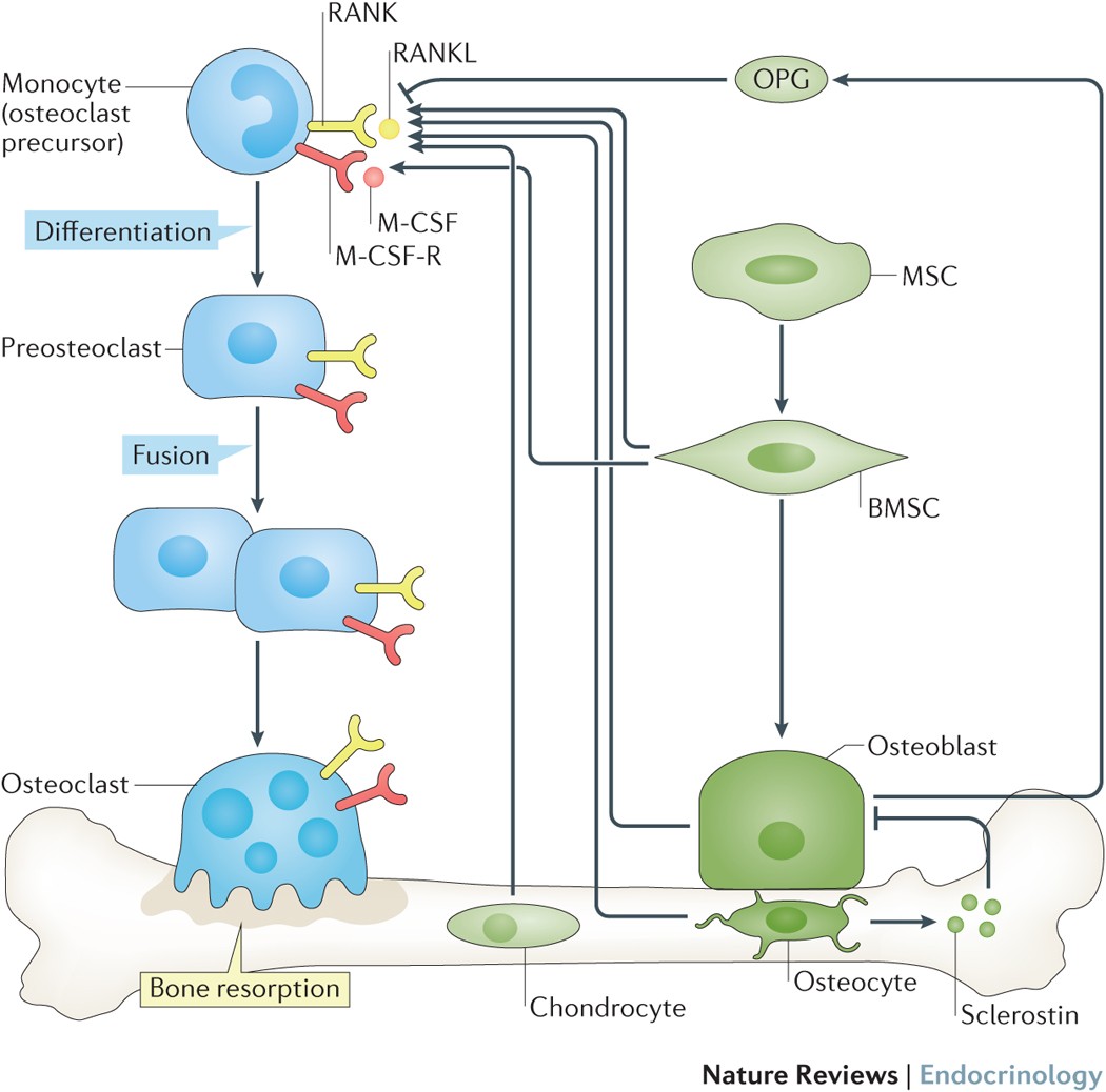
Osteocyte-Related Cytokines Regulate Osteoclast Formation and Bone Resorption
The process of bone remodeling is the result of the regulated balance between bone cell populations, namely bone-forming osteoblasts, bone-resorbing osteoclasts, and the osteocyte, the mechanosensory cell type. Osteoclasts derived from the hematopoietic stem cell lineage are the principal cells involved in bone resorption. In osteolytic diseases such as rheumatoid arthritis, periodontitis, and osteoporosis, the balance is lost and changes in favor of bone resorption. Therefore, it is vital to elucidate the mechanisms of osteoclast formation and bone resorption. It has been reported that osteocytes express Receptor activator of nuclear factor κΒ ligand (RANKL), an essential factor for osteoclast formation. RANKL secreted by osteocytes is the most important factor for physiologically supported osteoclast formation in the developing skeleton and in pathological bone resorption such as experimental periodontal bone loss. TNF-α directly enhances RANKL expression in osteocytes and promotes osteoclast formation. Moreover, TNF-α enhances sclerostin expression in osteocytes, which also increases osteoclast formation. These findings suggest that osteocyte-related cytokines act directly to enhance osteoclast formation and bone resorption. In this review, we outline the most recent knowledge concerning bone resorption-related cytokines and discuss the osteocyte as the master regulator of bone resorption and effector in osteoclast formation.

Osteoimmunology: shared mechanisms and crosstalk between the immune and bone systems
Osteoimmunology is an interdisciplinary field, that covers the shared mechanisms and interactions between bone cells and immune cellsReceptor activator of nuclear factor-κB (NF-κB) ligand (RANKL) is an osteoclast-differentiation factor that links the activated immune system and bone loss. In addition, abnormal bone homeostasis has been observed in various mice deficient in immunomodulatory molecules.Osteoclast differentiation is dependent on the transcription factor nuclear factor of activated T cells, cytoplasmic 1 (NFATc1), which is induced and activated by RANKL and its co-stimulatory (immunoglobulin-like) receptors.Interleukin-17 (IL-17)-producing T helper cells (TH17 cells) are the key T-cell subset that links T-cell activation and bone destruction in autoimmune arthritis.Bone cells are involved in the maintenance and mobilization of haematopoietic stem cells.Osteoimmunology is becoming increasingly important for understanding the pathogenesis of, and developing new therapeutic strategies for, diseases that affect both systems.
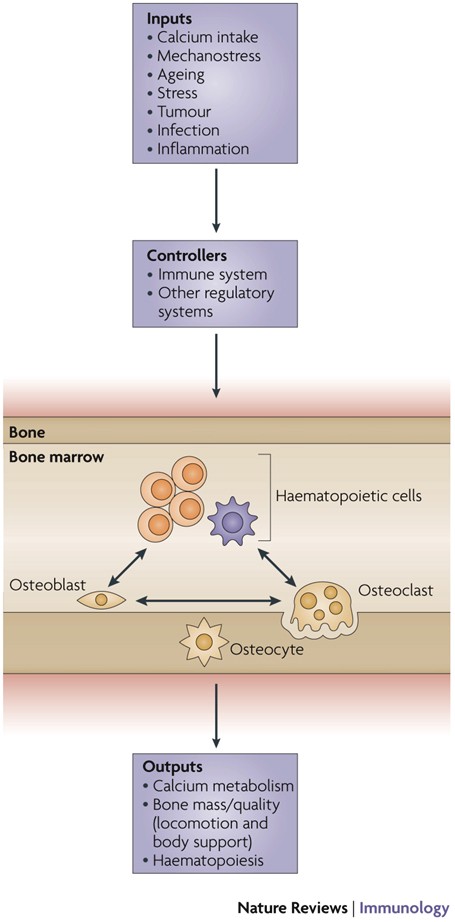
Updates on Osteoimmunology: What's New on the Cross-Talk Between Bone and Immune System
The term osteoimmunology was coined many years ago to describe the research field that deals with the cross-regulation between bone cells and the immune system. As a matter of fact, many factors that are classically considered immune-related, such as interleukins (i.e. IL-6, -11,-17 and -23), tumor necrosis factor (TNF)-α, receptor-activator of nuclear factor kappa B (RANK) and its ligand (RANKL), nuclear factor of activated t-cell, cytoplasmatic-1 (NFATc1) and others have all been found to be crucial in osteoclast and osteoblast biology. Conversely, bone cells, which we used to think would only regulate each other and take care of remodeling bone, actually regulate immune cells, by creating the so-called “endosteal niche”. Both osteoblasts and osteoclasts participate to this niche, either by favoring engraftment, or mobilization of hematopoietic stem cells (HSCs). In this review, we will describe the main milestones at the base of the osteoimmunology and present the key cellular players of the bone-immune system cross-talk, including HSCs, osteoblasts, osteoclasts, bone marrow macrophages, osteomacs, T- and B-lymphocytes, dendritic cells and neutrophils. We will also briefly describe some pathological conditions in which the bone-immune system cross-talk plays a crucial role, with the final aim to portray the state of the art in the mechanisms regulating the bone-immune system interplay, and some of the latest molecular players in the field. This is important to encourage investigation in this field, to identify new targets in the treatment of bone and immune diseases.

Osteoprotegerin in Bone Metastases: Mathematical Solution to the Puzzle
Bone is a common site for cancer metastasis. To create space for their growth, cancer cells stimulate bone resorbing osteoclasts. Cytokine RANKL is a key osteoclast activator, while osteoprotegerin (OPG) is a RANKL decoy receptor and an inhibitor of osteoclastogenesis. Consistently, systemic application of OPG decreases metastatic tumor burden in bone. However, OPG produced locally by cancer cells was shown to enhance osteolysis and tumor growth. We propose that OPG produced by cancer cells causes a local reduction in RANKL levels, inducing a steeper RANKL gradient away from the tumor and towards the bone tissue, resulting in faster resorption and tumor expansion. We tested this hypothesis using a mathematical model of nonlinear partial differential equations describing the spatial dynamics of OPG, RANKL, PTHrP, osteoclasts, tumor and bone mass. We demonstrate that at lower expression rates, tumor-derived OPG enhances the chemotactic RANKL gradient and osteolysis, whereas at higher expression rates OPG broadly inhibits RANKL and decreases osteolysis and tumor burden. Moreover, tumor expression of a soluble mediator inducing RANKL in the host tissue, such as PTHrP, is important for correct orientation of the RANKL gradient. A meta-analysis of OPG, RANKL and PTHrP expression in normal prostate, carcinoma and metastatic tissues demonstrated an increase in expression of OPG, but not RANKL, in metastatic prostate cancer, and positive correlation between OPG and PTHrP in metastatic prostate cancer. The proposed mechanism highlights the importance of the spatial distribution of receptors, decoys and ligands, and can be applied to other systems involving regulation of spatially anisotropic processes.
The Effect of Inflammation on Bone
Bone remodeling is the continual process to renew the adult skeleton through the sequential action of osteoblasts and osteoclasts. Nuclear factor RANK, an osteoclast receptor, and its ligand RANKL, expressed on the surface of osteoblasts, result in coordinated control of bone remodeling. Inflammation, a feature of illness and injury, plays a distinct role in skewing this process toward resorption. It does so via the interaction of inflammatory mediators and their related peptides with osteoblasts and osteoclasts, to alter the expression of RANK and RANKL. Such chemical mediators include TNFa, glucocorticoids, histamine, bradykinin, PGE2, systemic RANKL from immune cells, and interleukins 1 and 6. A thorough understanding of bone response to injury and disease, and ability to detect such biomarkers, as well as imaging to identify early structural and mechanical property changes in bone architecture, is important in improving management and outcomes of bone related pathology. While gut health and vitamin and mineral availability appear vitally important, there are very few intervention strategies currently available to favorably alter bone pathology induced by inflammation. Further research is required in this field to advance early detection and treatments.

Osteoclast differentiation and activation
Osteoclasts are specialized cells derived from the monocyte/macrophage haematopoietic lineage that develop and adhere to bone matrix, then secrete acid and lytic enzymes that degrade it in a specialized, extracellular compartment. Discovery of the RANK signalling pathway in the osteoclast has provided insight into the mechanisms of osteoclastogenesis and activation of bone resorption, and how hormonal signals impact bone structure and mass. Further study of this pathway is providing the molecular basis for developing therapeutics to treat osteoporosis and other diseases of bone loss.
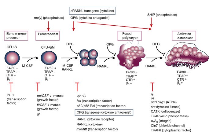
Immunoporosis: Role of Innate Immune Cells in Osteoporosis
Osteoporosis or porous bone disorder is the result of an imbalance in an otherwise highly balanced physiological process known as ‘bone remodeling’. The immune system is intricately involved in bone physiology as well as pathologies. Inflammatory diseases are often correlated with osteoporosis. Inflammatory mediators such as reactive oxygen species (ROS), and pro-inflammatory cytokines and chemokines directly or indirectly act on the bone cells and play a role in the pathogenesis of osteoporosis. Recently, Srivastava et al. (Srivastava RK, Dar HY, Mishra PK. Immunoporosis: Immunology of Osteoporosis-Role of T Cells. Frontiers in immunology. 2018;9:657) have coined the term “immunoporosis” to emphasize the role of immune cells in the pathology of osteoporosis. Accumulated pieces of evidence suggest both innate and adaptive immune cells contribute to osteoporosis. However, innate cells are the major effectors of inflammation. They sense various triggers to inflammation such as pathogen-associated molecular patterns (PAMPS), damage-associated molecular patterns (DAMPS), cellular stress, etc., thus producing pro-inflammatory mediators that play a critical role in the pathogenesis of osteoporosis. In this review, we have discussed the role of the innate immune cells in great detail and divided these cells into different sections in a systemic manner. In the beginning, we talked about cells of the myeloid lineage, including macrophages, monocytes, and dendritic cells. This group of cells explicitly influences the skeletal system by the action of production of pro-inflammatory cytokines and can transdifferentiate into osteoclast. Other cells of the myeloid lineage, such as neutrophils, eosinophils, and mast cells, largely impact osteoporosis via the production of pro-inflammatory cytokines. Further, we talked about the cells of the lymphoid lineage, including natural killer cells and innate lymphoid cells, which share innate-like properties and play a role in osteoporosis. In addition to various innate immune cells, we also discussed the impact of classical pro-inflammatory cytokines on osteoporosis. We also highlighted the studies regarding the impact of physiological and metabolic changes in the body, which results in chronic inflammatory conditions such as ageing, ultimately triggering osteoporosis.

Modelling osteomyelitis
This work focuses on the computational modelling of osteomyelitis, a bone pathology caused by bacteria infection (mostly Staphylococcus aureus). The infection alters the RANK/RANKL/OPG signalling dynamics that regulates osteoblasts and osteoclasts behaviour in bone remodelling, i.e. the resorption and mineralization activity. The infection rapidly leads to severe bone loss, necrosis of the affected portion, and it may even spread to other parts of the body. On the other hand, osteoporosis is not a bacterial infection but similarly is a defective bone pathology arising due to imbalances in the RANK/RANKL/OPG molecular pathway, and due to the progressive weakening of bone structure.
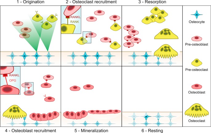
Molecular Signaling Pathways and Essential Metabolic Elements in Bone Remodeling: An Implication of Therapeutic Targets for Bone Diseases
Bone is one of the dynamic tissues in the human body that undergoes continuous remodelling through subsequent actions of bone cells, osteoclasts, and osteoblasts. Several signal transduction pathways are involved in the transition of mesenchymal stem cells into osteoblasts. These primarily include Runx2, ATF4, Wnt signaling and sympathetic signalling. The differentiation of osteoclasts is controlled by M-CSF, RANKL, and costimulatory signalling. It is well known that bone remodelling is regulated through receptor activator of nuclear factor-kappa B ligand followed by the binding to RANK, which eventually induces the differentiation of osteoclasts. The resorbing osteoclasts secrete TRAP, cathepsin K, MMP-9 and gelatinase to digest the proteinaceous matrix of type I collagen and form a saucer-shaped lacuna along with resorption tunnels in the trabecular bone. Osteoblasts secrete a soluble decoy receptor, osteoprotegerin that prevents the binding of RANK/RANKL and thus moderating osteoclastogenesis. Moreover, bone homeostasis is also regulated by several growth factors, cytokines, calciotropic hormones, parathyroid hormone and sex steroids. The current review presents a correlation of the probable molecular targets underlying the regulation of bone mass and the role of essential metabolic elements in bone remodelling. Targeting these signaling pathways may help design newer therapies for treating bone diseases.

Bone and the Immune System
Osteoporosis increases fracture risk, a cause of crippling morbidity and mortality. The immunoskeletal interface (ISI) is a centralization of cell and cytokine effectors shared between skeletal and immune systems. Consequently, the immune system mediates powerful effects on bone turnover. Physiologically, B cells secrete osteoprotegerin (OPG), a potent anti-osteoclastogenic factor that preserves bone mass. However, activated T cells and B cells secrete pro-osteoclastogenic factors including receptor activator of Nuclear factor-kappaB (NF-kB) ligand (RANKL), Interleukin (IL)-17A, and tumor necrosis factor (TNF)-α promoting bone loss in inflammatory states such as rheumatoid arthritis. Recently, ISI disruption has been linked to osteoporosis in human immunodeficiency virus (HIV) infection/acquired immunodeficiency syndrome (AIDS), where elevated B cell RANKL and diminished OPG drive bone resorption. HIV-antiretroviral therapy paradoxically intensifies bone loss during disease reversal, as immune reconstitution produces osteoclastogenic cytokines. Interestingly, in estrogen deficiency, activated T cells secrete RANKL, TNF, and IL-17A that amplify bone resorption and contribute to postmenopausal osteoporosis. T cell–produced TNF and IL-17A further contribute to bone loss in hyperparathyroidism, while T cell production of the anabolic Wingless integration site (Wnt) ligand, Wnt10b, promotes bone formation in response to anabolic parathyroid hormone and the immunomodulatory costimulation inhibitor cytotoxic T lymphocyte–associated protein-4-IgG (abatacept). These findings provide a window into the workings of the ISI and suggest novel targets for future therapeutic interventions to reduce fracture risk.

Osteoimmunology: The Nexus between bone and immune system
Osteoimmunology is an interdisciplinary research field which combines the existing fields of osteology (bone biology) and immunology under one umbrella. The observation that contributed enormously to the emergence of osteoimmunology as an independent field of investigation was the enhanced bone loss in various inflammatory bone diseases such as rheumatoid arthritis, osteoporosis and periodontitis. T helper cells (Th1, Th2, Treg and Th17) along with various other immune cells (B cells, DC, macrophages etc.) are actively involved in bone homeostasis. The present review thus provides an overview of the nexus between these two prominent systems (Bone and Immune system) of an organism, which reside in a common niche (bone marrow) and thus cross-communicate to modulate their respective development. Investigations in the field of osteoimmunology thus promise the advent of new era in the field with novel therapeutics for bone loss in various inflammatory conditions. A molecular insight into the field of osteoimmunology can lead to novel approaches for the prevention and treatment of diverse inflammatory conditions such as osteoporosis, rheumatoid arthritis and osteoarthritis.
OSTEOIMMUNOLOGY: Interplay Between the Immune System and Bone Metabolism
Studies of bone and the immune system have converged in recent years under the banner of osteoimmunology. The immune system is spawned in the bone marrow reservoir, and investigators now recognize that important niches also exist there for memory lymphocytes. At the same time, various factors produced during immune responses are capable of profoundly affecting regulation of bone. Mechanisms have evolved to prevent excessive interference by the immune system with bone homeostasis, yet pathologic bone loss is a common sequela associated with autoimmunity and cancer. There are also developmental links, or parallels, between bone and the immune system. Cells that regulate bone turnover share a common precursor with inflammatory immune cells and may restrict themselves anatomically, in part by utilizing a signaling network analogous to lymphocyte costimulation. Efforts are currently under way to further characterize how these two organ systems overlap and to develop therapeutic strategies that benefit from this understanding.

Gut, inflammation and osteoporosis: basic and clinical concepts
Chronic inflammatory disorders such as inflammatory bowel diseases (IBD) affect bone metabolism and are frequently associated with the presence of osteoporosis. Bone loss is regulated by various mediators of the immune system such as the pro-inflammatory cytokines tumour necrosis factor-alpha (TNF-α), interleukin-1beta (IL-1β), IL-6, or interferon-gamma. TNF-α, a master cytokine in human IBD, causes bone erosions in experimental models and these effects are exerted by osteoclasts. Other TNF-related cytokines such as receptor activator of nuclear factor kappa B (RANK), its ligand, RANKL, and osteoprotegerin are important mediators in inflammatory processes in the gut and are critically involved in the pathophysiology of bone loss. The awareness and early diagnosis of osteoporosis in states of chronic inflammation, together with applied therapies such as bisphosphonates, may be beneficial in inflammation-associated osteoporosis. Although several mechanisms may contribute to osteoporosis in patients with IBD and coeliac disease, inflammation as an important factor has so far been neglected. As key inflammatory mediators in IBD such as TNF-α are involved in the disease process both in gut and bone, we hypothesise that neutralisation of TNF-α could prove an efficient strategy in the treatment of inflammation-related osteoporosis in the future.