


The majority of T cells present in the bone marrow (BM) represent an activated/memory phenotype and most of these, if not all, are circulating T cells. Their lodging in the bone marrow keeps them activated, turning the bone marrow microenvironment into a “memory reservoir”. This article will focus on how T cell activation in the marrow results in both direct and indirect effect on the hematopoiesis. The hematopoietic stem cell niche will be presented, with its main components and organization, along with the role-played by T-lymphocytes in basal and pathologic conditions and their effect on the bone remodeling process. Also discussed herein will be how “normal” bone mass peak is achieved only in the presence of an intact adaptive immune system, with T and B cells playing critical roles in this process. Our main hypothesis is that the partnership between T cells and cells of the bone marrow microenvironment orchestrates numerous processes regulating immunity, hematopoiesis and bone remodeling.
OSTEOIMMUNOLOGY: Interplay Between the Immune System and Bone Metabolism
Studies of bone and the immune system have converged in recent years under the banner of osteoimmunology. The immune system is spawned in the bone marrow reservoir, and investigators now recognize that important niches also exist there for memory lymphocytes. At the same time, various factors produced during immune responses are capable of profoundly affecting regulation of bone. Mechanisms have evolved to prevent excessive interference by the immune system with bone homeostasis, yet pathologic bone loss is a common sequela associated with autoimmunity and cancer. There are also developmental links, or parallels, between bone and the immune system. Cells that regulate bone turnover share a common precursor with inflammatory immune cells and may restrict themselves anatomically, in part by utilizing a signaling network analogous to lymphocyte costimulation. Efforts are currently under way to further characterize how these two organ systems overlap and to develop therapeutic strategies that benefit from this understanding.

Updates on Osteoimmunology: What's New on the Cross-Talk Between Bone and Immune System
The term osteoimmunology was coined many years ago to describe the research field that deals with the cross-regulation between bone cells and the immune system. As a matter of fact, many factors that are classically considered immune-related, such as interleukins (i.e. IL-6, -11,-17 and -23), tumor necrosis factor (TNF)-α, receptor-activator of nuclear factor kappa B (RANK) and its ligand (RANKL), nuclear factor of activated t-cell, cytoplasmatic-1 (NFATc1) and others have all been found to be crucial in osteoclast and osteoblast biology. Conversely, bone cells, which we used to think would only regulate each other and take care of remodeling bone, actually regulate immune cells, by creating the so-called “endosteal niche”. Both osteoblasts and osteoclasts participate to this niche, either by favoring engraftment, or mobilization of hematopoietic stem cells (HSCs). In this review, we will describe the main milestones at the base of the osteoimmunology and present the key cellular players of the bone-immune system cross-talk, including HSCs, osteoblasts, osteoclasts, bone marrow macrophages, osteomacs, T- and B-lymphocytes, dendritic cells and neutrophils. We will also briefly describe some pathological conditions in which the bone-immune system cross-talk plays a crucial role, with the final aim to portray the state of the art in the mechanisms regulating the bone-immune system interplay, and some of the latest molecular players in the field. This is important to encourage investigation in this field, to identify new targets in the treatment of bone and immune diseases.

Immunoporosis: Immunology of Osteoporosis—Role of T Cells
The role of immune system in various bone pathologies such as osteoporosis, osteoarthritis and rheumatoid arthritis is now well established. This had led to the emergence of a modern field of systems biology called as osteoimmunology, an integrated research between fields of immunology and bone biology under one umbrella. Osteoporosis is one of the most common inflammatory bone-loss conditions with more than 200 million individuals affected worldwide. T helper cells along with various other immune cells are major players involved in bone homeostasis. In the present review, we specifically discuss the role of various defined T lymphocyte subsets (Th cells comprising Th1, Th2, Th9, Th17, Th22, regulatory T cells, follicular helper T cells, natural killer T cells, γδ T cells and CD8+ T cells) in the pathophysiology of osteoporosis. The study of the specific role of immune system in osteoporosis has now been proposed by our group as “Immunoporosis: The immunology of osteoporosis” with special emphasis on the role of various subsets of T lymphocytes. The establishment of this new field had been the need of the hour due to the emergence of novel roles of various T cell lymphocytes in accelerated bone loss observed during osteoporosis. Activated T cells either directly or indirectly through the secretion of various cytokines and factors modulate bone health and thereby regulate bone remodelling. Various studies have summarized the role of inflammation in pathogenesis of osteoporosis, but very few reports had delineated the precise role of various T cell subsets in the pathobiology of osteoporosis. The present review thus for the first time clearly highlights and summarizes the role of various T lymphocytes in the development and pathophysiology of osteoporosis, giving birth to a new field of biology termed as “Immunoporosis”. This novel field will thus provide an overview of the nexus between the cellular components of both bone and immune systems, responsible for the observed bone loss in osteoporosis. A molecular insight into the upcoming and novel field of immunoporosis would thus lead to development of innovative approaches for the prevention and treatment of osteoporosis.

Bone versus immune system
A molecule on activated T cells triggers bone loss by switching on bone-resorbing cells. Fortunately, it seems that this mechanism is kept in check by another molecule, secreted by the T cells.
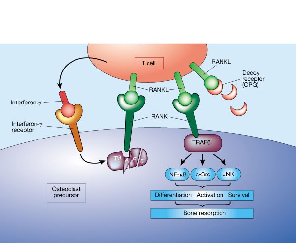
Osteoimmunology: The Nexus between bone and immune system
Osteoimmunology is an interdisciplinary research field which combines the existing fields of osteology (bone biology) and immunology under one umbrella. The observation that contributed enormously to the emergence of osteoimmunology as an independent field of investigation was the enhanced bone loss in various inflammatory bone diseases such as rheumatoid arthritis, osteoporosis and periodontitis. T helper cells (Th1, Th2, Treg and Th17) along with various other immune cells (B cells, DC, macrophages etc.) are actively involved in bone homeostasis. The present review thus provides an overview of the nexus between these two prominent systems (Bone and Immune system) of an organism, which reside in a common niche (bone marrow) and thus cross-communicate to modulate their respective development. Investigations in the field of osteoimmunology thus promise the advent of new era in the field with novel therapeutics for bone loss in various inflammatory conditions. A molecular insight into the field of osteoimmunology can lead to novel approaches for the prevention and treatment of diverse inflammatory conditions such as osteoporosis, rheumatoid arthritis and osteoarthritis.
Osteoimmunology: shared mechanisms and crosstalk between the immune and bone systems
Osteoimmunology is an interdisciplinary field, that covers the shared mechanisms and interactions between bone cells and immune cellsReceptor activator of nuclear factor-κB (NF-κB) ligand (RANKL) is an osteoclast-differentiation factor that links the activated immune system and bone loss. In addition, abnormal bone homeostasis has been observed in various mice deficient in immunomodulatory molecules.Osteoclast differentiation is dependent on the transcription factor nuclear factor of activated T cells, cytoplasmic 1 (NFATc1), which is induced and activated by RANKL and its co-stimulatory (immunoglobulin-like) receptors.Interleukin-17 (IL-17)-producing T helper cells (TH17 cells) are the key T-cell subset that links T-cell activation and bone destruction in autoimmune arthritis.Bone cells are involved in the maintenance and mobilization of haematopoietic stem cells.Osteoimmunology is becoming increasingly important for understanding the pathogenesis of, and developing new therapeutic strategies for, diseases that affect both systems.
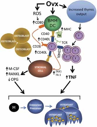
Role of T cells in ovariectomy induced bone loss—revisited
More than a decade has passed since the publication of the first report implicating T cells in the bone loss induced by ovariectomy (ovx).1 Since then ther
Mathematical Modeling of the Gut–Bone Axis and Implications of Butyrate Treatment on Osteoimmunology
Butyrate, a short-chain fatty acid produced by the gut microbiota, has pivotal roles in the regulation of the immune system. Recent studies have revealed that butyrate increases the differentiation of peripheral regulatory T cells in the gut–bone axis and promotes osteoblasts’ bone forming activity. However, the mechanism of the therapeutic benefit of butyrate in bone remodeling remains incompletely understood. Here, we develop a multicompartment mathematical model to quantitatively predict the contribution of butyrate on the expansion of regulatory T cells in the gut, blood, and bone compartments. We investigate the interplay between regulatory T cell-derived TGF-β and CD8+ T cell-derived Wnt-10b with changes in gut butyrate concentration. In addition, we connect our model to a detailed model of bone metabolism to study the impacts of butyrate and Wnt-10b on trabecular bone volume. Our results indicate both direct and indirect immune-mediated impacts of butyrate on bone metabolism.

<p>The Rising Era of “Immunoporosis”: Role of Immune System in the Pathophysiology of Osteoporosis</p>
We exhaustively revisit the characteristics, mechanism of action, and function of both innate and adaptive immune cells .

Cellular mechanisms of bone remodeling
Bone remodeling is a tightly regulated process securing repair of microdamage (targeted remodeling) and replacement of old bone with new bone through sequential osteoclastic resorption and osteoblastic bone formation. The rate of remodeling is regulated by a wide variety of calcitropic hormones (PTH, thyroid hormone, sex steroids etc.). In recent years we have come to appreciate that bone remodeling proceeds in a specialized vascular structure,—the Bone Remodeling Compartment (BRC). The outer lining of this compartment is made up of flattened cells, displaying all the characteristics of lining cells in bone including expression of OPG and RANKL. Reduced bone turnover leads to a decrease in the number of BRCs, while increased turnover causes an increase in the number of BRCs. The secretion of regulatory factors inside a confined space separated from the bone marrow would facilitate local regulation of the remodeling process without interference from growth factors secreted by blood cells in the marrow space. The BRC also creates an environment where cells inside the structure are exposed to denuded bone, which may enable direct cellular interactions with integrins and other matrix factors known to regulate osteoclast/osteoblast activity. However, the denuded bone surface inside the BRC also constitutes an ideal environment for the seeding of bone metastases, known to have high affinity for bone matrix. Circulating osteoclast- and osteoblast precursor cells have been demonstrated in peripheral blood. The dominant pathway regulating osteoclast recruitment is the RANKL/OPG system, while many different factors (RUNX, Osterix) are involved in osteoblast differentiation. Both pathways are modulated by calcitropic hormones.
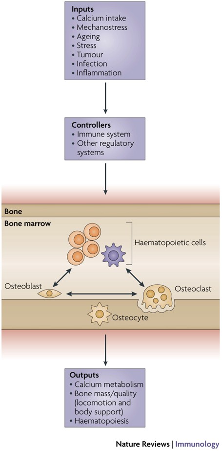
Physiological and pathophysiological bone turnover — role of the immune system
Receptor activator of NF-κB ligand (RANKL) and osteoprotegerin (OPG) are key downstream effectors of bone resorption; tumour necrosis factor (TNF) might synergize with RANKL to superinduce osteoclastic bone resorptionB cells, regulated by T cells, are a key source of basal OPG whereas activated T cells and B cells are key sources of TNF and RANKL in inflammatory conditionsShort-term antiresorptive therapies might safely prevent bone loss associated with combination antiretroviral therapyAnergic and parathyroid-hormone-treated T cells secrete the protein Wnt-10b, which promotes bone formationNovel therapeutic strategies targeting the immune system might promote bone formation and decrease bone resorption to manage osteoporotic bone loss and prevent fracture
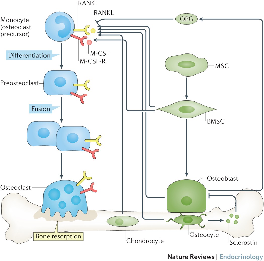
Osteoimmunology: A Current Update of the Interplay Between Bone and the Immune System
Immunology, already a discipline in its own right, has become a major part of many different medical fields. However, its relationship to orthopedics and trauma surgery has unfortunately, and perhaps unjustly, been developing rather slowly. Discover-ies in recent years have emphasized the immense breadth of communication and connection between both systems and, importantly, the highly promising therapeu-tic opportunities.Recent discoveries of factors originally assigned to the immune system have now also been shown to have a significant impact on bone health and disease, which has greatly changed how we approach treatment of bone pathologies. In case of bone fracture, immune cells, especially macrophages, are present through-out the whole healing process, assure defense against pathogens and discharge a complex variety of effectors to regulate bone modelling. In rheumatoid arthritis and osteoporosis, the immune system contributes to the formation of the pathological and chronic conditions. Fascinatingly, prosthesis failure is not at all solely a me-chanical problem of improper strain but works in conjunction with an active contri-bution of the immune system as a reaction to irritant debris from material wear.Unravelling conjoined mechanisms of the immune and osseous systems heralds therapeutic possibilities for ailments of both. Contemplation of the bone as merely an unchanging support pillar is outdated and obsolete. Instead it is manda-tory that this highly diverse network be incorporated in our understanding of the immune system and hematopoiesis.

Osteoimmunology: The Conceptual Framework Unifying the Immune and Skeletal Systems
The immune and skeletal systems share a variety of molecules, including cytokines, chemokines, hormones, receptors, and transcription factors. Bone cells interact with immune cells under physiological and pathological conditions. Osteoimmunology was created as a new interdisciplinary field in large part to highlight the shared molecules and reciprocal interactions between the two systems in both heath and disease. Receptor activator of NF-κB ligand (RANKL) plays an essential role not only in the development of immune organs and bones, but also in autoimmune diseases affecting bone, thus effectively comprising the molecule that links the two systems. Here we review the function, gene regulation, and signal transduction of osteoimmune molecules, including RANKL, in the context of osteoclastogenesis as well as multiple other regulatory functions. Osteoimmunology has become indispensable for understanding the pathogenesis of a number of diseases such as rheumatoid arthritis (RA). We review the various osteoimmune pathologies, including the bone destruction in RA, in which pathogenic helper T cell subsets [such as IL-17-expressing helper T (Th17) cells] induce bone erosion through aberrant RANKL expression. We also focus on cellular interactions and the identification of the communication factors in the bone marrow, discussing the contribution of bone cells to the maintenance and regulation of hematopoietic stem and progenitors cells. Thus the time has come for a basic reappraisal of the framework for understanding both the immune and bone systems. The concept of a unified osteoimmune system will be absolutely indispensable for basic and translational approaches to diseases related to bone and/or the immune system.

Chronic Infection Depletes Hematopoietic Stem Cells through Stress-Induced Terminal Differentiation
Matatall et al. show that chronic infection causes pancytopenia and hematopoietic stem cell (HSC) depletion in mice. HSCs are lost through impaired self-renewal and increased terminal differentiation, which can be triggered by induction of the interferon gamma-responsive transcription factor BATF2. This study elucidates mechanisms underlying bone marrow failure during chronic infections.

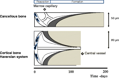
Osteonal and hemi‐osteonal remodeling: The spatial and temporal framework for signal traffic in adult human bone
Abstract The bone replacement process in the adult skeleton is known as remodeling. When bone is removed by osteoclasts, new bone is laid down by osteoblasts in the same place, because the load bearing requirement is unchanged. Bone is usually replaced because it is too old to carry out its function, which is mainly mechanical in cortical bone and mainly support for homeostasis and hematopoiesis in cancellous bone. Remodeling always begins on a quiescent bone surface, separated from the marrow by flat lining cells that are one of the two modes of terminal differentiation of osteoblasts. Lining cells are gatekeepers, able to be informed of the need for remodeling, and to either execute or mediate all four components of its activation‐selection and preparation of the site, recruitment of mononuclear preosteoclasts, budding of new capillaries, and attraction of preosteoclasts to the chosen site where they fuse into multinucleated osteoclasts. In cortical bone, osteonal remodeling is carried out by a complex and unique structure, the basic multicellular unit (BMU) that comprises a cutting cone of osteoclasts in front, a closing cone lined by osteoblasts following behind, and connective tissue, blood vessels and nerves filling the cavity. The BMU maintains its size, shape and internal organization for many months as it travels through bone in a controlled direction. Individual osteoclast nuclei are short‐lived, turning over about 8% per d, replaced by new preosteoclasts that originated in the bone marrow and travel in the circulation to the site of resorption. Refilling of bone at each successive cross‐sectional location is accomplished by a team of osteoblasts, probably originating from precursors within the local connective tissue, all assembled within a narrow window of time, at the right location, and in the right orientation to the surface. Each osteoblast team forms bone most rapidly at its onset and slows down progressively. Some of the osteoblasts are buried as osteocytes, some die, and the remainder gradually assume the shape of lining cells. Cancellous bone is more accessible to study than cortical bone, but is geometrically complex. Although remodeling conforms to the same sequence of surface activation, resorption and formation, its three‐dimensional organization is difficult to visualize from two‐dimensional histologic sections. But the average sizes of resorption sites, formation sites, and completed structural units increase progressively, as they do in cortical bone, indicating that the cancellous BMU travels across the surface digging a trench rather than a tunnel, but maintaining its size, shape and individual identity by the continuous recruitment of new cells, just as in cortical bone, a process that can be visualized as hemiosteonal remodeling. The conclusion that all remodeling is carried out by individual BMUs has important implications for bone biology, since many questions about how BMUs operate cannot be answered by studying either intact organisms or isolated cell systems. Many different steps in remodeling and many factors that influence each step have been identified, but very little is known about how the process is regulated in vivo to achieve its biologic purposes; most factors studied to date are likely permissive rather than regulatory in nature. Based on the proposed conceptual model of the BMU, much in vitro experimentation is relevant to the growth, modeling and repair of bone, but not to its remodeling in the adult skeleton. Further progress in the understanding of in vivo physiology will require the characterization of gene expression in individual cells to be related to the spatial and temporal organization of the BMU. This is likely to be possible only for osteonal remodeling in cortical bone in which, because of its geometric simplicity, individual BMUs can consistently be observed in two‐dimensional, longitudinal sections. © 1994 Wiley‐Liss, Inc.

Immunoporosis: Role of Innate Immune Cells in Osteoporosis
Osteoporosis or porous bone disorder is the result of an imbalance in an otherwise highly balanced physiological process known as ‘bone remodeling’. The immune system is intricately involved in bone physiology as well as pathologies. Inflammatory diseases are often correlated with osteoporosis. Inflammatory mediators such as reactive oxygen species (ROS), and pro-inflammatory cytokines and chemokines directly or indirectly act on the bone cells and play a role in the pathogenesis of osteoporosis. Recently, Srivastava et al. (Srivastava RK, Dar HY, Mishra PK. Immunoporosis: Immunology of Osteoporosis-Role of T Cells. Frontiers in immunology. 2018;9:657) have coined the term “immunoporosis” to emphasize the role of immune cells in the pathology of osteoporosis. Accumulated pieces of evidence suggest both innate and adaptive immune cells contribute to osteoporosis. However, innate cells are the major effectors of inflammation. They sense various triggers to inflammation such as pathogen-associated molecular patterns (PAMPS), damage-associated molecular patterns (DAMPS), cellular stress, etc., thus producing pro-inflammatory mediators that play a critical role in the pathogenesis of osteoporosis. In this review, we have discussed the role of the innate immune cells in great detail and divided these cells into different sections in a systemic manner. In the beginning, we talked about cells of the myeloid lineage, including macrophages, monocytes, and dendritic cells. This group of cells explicitly influences the skeletal system by the action of production of pro-inflammatory cytokines and can transdifferentiate into osteoclast. Other cells of the myeloid lineage, such as neutrophils, eosinophils, and mast cells, largely impact osteoporosis via the production of pro-inflammatory cytokines. Further, we talked about the cells of the lymphoid lineage, including natural killer cells and innate lymphoid cells, which share innate-like properties and play a role in osteoporosis. In addition to various innate immune cells, we also discussed the impact of classical pro-inflammatory cytokines on osteoporosis. We also highlighted the studies regarding the impact of physiological and metabolic changes in the body, which results in chronic inflammatory conditions such as ageing, ultimately triggering osteoporosis.
