


Osteoporosis has been defined as the silent disease of the 21st century, becoming a public health risk due to its severity, chronicity and progression and affecting mainly postmenopausal women and older adults. Osteoporosis is characterized by an imbalance between bone resorption and bone production. It is diagnosed through different methods such as bone densitometry and dual X-rays. The treatment of this pathology focuses on different aspects. On the one hand, pharmacological treatments are characterized by the use of anti-resorptive drugs, as well as emerging regenerative medicine treatments such as cell therapies and the use of bioactive hydrogels. On the other hand, non-pharmacological treatments are associated with lifestyle habits that should be incorporated, such as physical activity, diet and the cessation of harmful habits such as a high consumption of alcohol or smoking. This review seeks to provide an overview of the theoretical basis in relation to bone biology, the existing methods for diagnosis and the treatments of osteoporosis, including the development of new strategies.
The Development of Molecular Biology of Osteoporosis
Osteoporosis is one of the major bone disorders that affects both women and men, and causes bone deterioration and bone strength. Bone remodeling maintains bone mass and mineral homeostasis through the balanced action of osteoblasts and osteoclasts, which are responsible for bone formation and bone resorption, respectively. The imbalance in bone remodeling is known to be the main cause of osteoporosis. The imbalance can be the result of the action of various molecules produced by one bone cell that acts on other bone cells and influence cell activity. The understanding of the effect of these molecules on bone can help identify new targets and therapeutics to prevent and treat bone disorders. In this article, we have focused on molecules that are produced by osteoblasts, osteocytes, and osteoclasts and their mechanism of action on these cells. We have also summarized the different pharmacological osteoporosis treatments that target different molecular aspects of these bone cells to minimize osteoporosis.
Postmenopausal osteoporosis
Osteoporosis is a metabolic bone disorder that is characterized by low bone mass and micro-architectural deterioration of bone tissue. Fractures of the proximal femur, the vertebrae and the distal radius are the most frequent osteoporotic fractures, although most fractures in the elderly are probably at least partly related to bone fragility. The incidence of fractures varies greatly by country, but on average up to 50% of women >50 years of age are at risk of fractures. Fractures severely affect the quality of life of an individual and are becoming a major public health problem owing to the ageing population. Postmenopausal osteoporosis, resulting from oestrogen deficiency, is the most common type of osteoporosis. Oestrogen deficiency results in an increase in bone turnover owing to effects on all types of bone cells. The imbalance in bone formation and resorption has effects on trabecular bone (loss of connectivity) and cortical bone (cortical thinning and porosity). Osteoporosis is diagnosed using bone density measurements of the lumbar spine and proximal femur. Preventive strategies to improve bone health include diet, exercise and abstaining from smoking. Fractures may be prevented by reducing falls in high-risk populations. Several drugs are licensed to reduce fracture risk by slowing down bone resorption (such as bisphosphonates and denosumab) or by stimulating bone formation (such as teriparatide). Improved understanding of the cellular basis for osteoporosis has resulted in new drugs targeted to key pathways, which are under development.

Bone Remodeling in Post-menopausal Osteoporosis
Bone mass in the skeleton is dependent on the coordinated activities of bone-forming osteoblasts and bone-resorbing osteoclasts in discrete bone multi-cellular units. Remodeling of bone in these units is important not only for maintaining bone mass, but also to repair microdamage, to prevent accumulation of too much old bone, and for mineral homeostasis. The activities of osteoblasts and osteoclasts are controlled by a variety of hormones and cytokines, as well as by mechanical loading. Most importantly, sex hormones are very crucial for keeping bone mass in balance, and the lack of either estrogen or testosterone leads to decreased bone mass and increased risk for osteoporosis. The prevalence of osteoporotic fractures is increasing dramatically in the Western part of the world and is a major health problem in many countries. In the present review, the cellular and molecular mechanisms controlling bone remodeling and the influence of sex hormones on these processes are summarized. In a separate paper in this issue, the pathogenesis of post-menopausal osteoporosis will be compared with that of inflammation-induced bone remodeling, including the evidence for and against the hypothesis that concomitant post-menopausal osteoporotic disease influences the progression of periodontal disease.

Biology of Bone Tissue: Structure, Function, and Factors That Influence Bone Cells
Bone tissue is continuously remodeled through the concerted actions of bone cells, which include bone resorption by osteoclasts and bone formation by osteoblasts, whereas osteocytes act as mechanosensors and orchestrators of the bone remodeling process. This process is under the control of local (e.g., growth factors and cytokines) and systemic (e.g., calcitonin and estrogens) factors that all together contribute for bone homeostasis. An imbalance between bone resorption and formation can result in bone diseases including osteoporosis. Recently, it has been recognized that, during bone remodeling, there are an intricate communication among bone cells. For instance, the coupling from bone resorption to bone formation is achieved by interaction between osteoclasts and osteoblasts. Moreover, osteocytes produce factors that influence osteoblast and osteoclast activities, whereas osteocyte apoptosis is followed by osteoclastic bone resorption. The increasing knowledge about the structure and functions of bone cells contributed to a better understanding of bone biology. It has been suggested that there is a complex communication between bone cells and other organs, indicating the dynamic nature of bone tissue. In this review, we discuss the current data about the structure and functions of bone cells and the factors that influence bone remodeling.

U.S. Montmorency Tart Cherry Juice Decreases Bone Resorption in Women Aged 65–80 Years
Pre-clinical studies have demonstrated that tart cherries, rich in hydroxycinnamic acids and anthocyanins, protect against age-related and inflammation-induced bone loss. This study examined how daily consumption of Montmorency tart cherry juice (TC) alters biomarkers of bone metabolism in older women. Healthy women, aged 65–80 years (n = 27), were randomly assigned to consume ~240 mL (8 fl. oz.) of juice once (TC1X) or twice (TC2X) per day for 90 d. Dual-energy x-ray absorptiometry (DXA) scans were performed to determine bone density at baseline, and pre- and post-treatment serum biomarkers of bone formation and resorption, vitamin D, inflammation, and oxidative stress were assessed. Irrespective of osteoporosis risk, the bone resorption marker, tartrate resistant acid phosphatase type 5b, was significantly reduced with the TC2X dose compared to baseline, but not with the TC1X dose. In terms of indicators of bone formation and turnover, neither serum bone-specific alkaline phosphatase nor osteocalcin were altered. No changes in thiobarbituric acid reactive substances or high sensitivity C-reactive protein were observed in response to either TC1X or TC2X. We conclude that short-term supplementation with the higher dose of tart cherry juice decreased bone resorption from baseline without altering bone formation and turnover biomarkers in this cohort.

Estrogen therapy for osteoporosis in the modern era
Menopause predisposes women to osteoporosis due to declining estrogen levels. This results in a decrease in bone mineral density (BMD) and an increase in fractures. Osteoporotic fractures lead to substantial morbidity and mortality, and are considered one of the largest public health priorities by the World Health Organization (WHO). It is therefore essential for menopausal women to receive appropriate guidance for the prevention and management of osteoporosis. The Women’s Health Initiative (WHI) randomized controlled trial first proved hormonal therapy (HT) reduces the incidence of all osteoporosis-related fractures in postmenopausal women. However, the study concluded that the adverse effects outweighed the potential benefits on bone, leading to a significant decrease in HT use for menopausal symptoms. Additionally, HT was not used as first-line therapy for osteoporosis and fractures. Subsequent studies have challenged these initial conclusions and have shown significant efficacy of HT in various doses, durations, regimens, and routes of administration. These studies support that HT improves BMD and reduces fracture risk in women with and without osteoporosis. Furthermore, the studies suggest that low-dose and transdermal HT are less likely associated with the adverse effects of breast cancer, endometrial hyperplasia, coronary artery disease (CAD), and venous thromboembolism (VTE) previously observed in standard-dose oral HT regimens. Given the need for estrogen in menopausal women and evidence supporting the cost effectiveness, safety, and efficacy of HT, we propose that HT should be considered for the primary prevention and treatment of osteoporosis in appropriate candidates. HT should be individualized and the once “lowest dose for shortest period of time” concept should no longer be used. This review will focus on the prior and current studies for various HT formulations used for the prevention and treatment of osteoporosis, exploring the safety profile of low-dose and transdermal HT that have been shown to be safer than oral standard-dose HT.
Disease Systems Analysis of Bone Mineral Density and Bone Turnover Markers in Response to Alendronate, Placebo, and Washout in Postmenopausal Women
A previously established mechanism‐based disease systems model for osteoporosis that is based on a mathematically reduced version of a model describing the interactions between osteoclast (bone removing) and osteoblast (bone forming) cells in bone remodeling has been applied to clinical data from women ( n = 1,379) receiving different doses and treatment regimens of alendronate, placebo, and washout. The changes in the biomarkers, plasma bone‐specific alkaline phosphatase activity (BSAP), urinary N‐telopeptide (NTX), lumbar spine bone mineral density (BMD), and total hip BMD, were linked to the underlying mechanistic core of the model. The final model gave an accurate description of all four biomarkers for the different treatments. Simulations were used to visualize the dynamics of the underlying network and the natural disease progression upon alendronate treatment and discontinuation. These results complement the previous applications of this mechanism‐based disease systems model to data from various treatments for osteoporosis.

Application of a mechanism-based disease systems model for osteoporosis to clinical data
A recently proposed mechanism-based disease systems model for osteoporosis (Schmidt et al., J Pharmacokinet Pharmacodyn 38:873–900, 2011) was applied to clinical data from post-menopausal women (n = 767) receiving various doses of the selective estrogen receptor modulator tibolone. Plasma bone-specific alkaline phosphatase activity and urinary N-telopeptide were used as biomarkers reflecting the activity of osteoblasts (bone forming cells) and osteoclasts (bone removing cells), respectively. These data were analyzed in conjunction with data on osteocalcin and on bone mineral density (BMD) (both lumbar spine and total hip), which reflect the activity of both cell types. While the dynamics of bone turnover markers changes rapidly, closely following changes in the activity of bone cells, changes in BMD are slower and have their own dynamics. Application of the mechanism-based disease systems model to the clinical data allowed for an adequate description of the data and yielded parameter estimates that are consistent with physiological values reported in the literature (Lemaire et al., J Theor Biol 229:293–309, 2004). The fitted model enabled characterization of (i) the critical time scales involved in disease progression, (ii) the dynamics of the system during onset and offset of the therapeutic intervention, and (iii) the distinction between responders and low-responders to tibolone treatment.

Integrated model for denosumab and ibandronate pharmacodynamics in postmenopausal women
ABSTRACT This study aims to characterize the pharmacodynamic properties of denosumab, a RANK ligand inhibitor, and ibandronate, a bisphosphonate, using an integrated bone homeostasis model in postmenopausal women. Mean temporal profiles of denosumab, serum and urine N‐telopeptide (sNTX, uNTX), lumbar spine bone mineral density (BMD) following denosumab administration, and urine C‐telopeptide (uCTX) and lumbar spine BMD upon ibandronate administration were extracted from the literature. A mechanistic model was developed that integrates denosumab pharmacokinetics with binding to RANK ligand and ibandronate inhibition of osteoclast precursor differentiation to active osteoclasts (AOC). Biomarker concentrations were linked to the AOC pool. The BMD was characterized by a turnover model with stimulation of bone formation and degradation by AOB (active osteoblasts) and AOC pools. The estimated basal sNTX, uNTX and uCTX concentrations were 7.24 n m , 14.4 nmol/mmolCr and 31 µg/mmolCr. The BMD degradation rate was 0.00161 day ‐1 with stimulation constants associated with AOB and AOC of 1214 and 790 p m ‐1 . The plasma ibandronate concentration producing 50% of maximum inhibition of osteoclast differentiation was 522 ng/l. The integrated model, which incorporates multiple pathways of therapeutic intervention, quantitatively describes changes in clinical biomarkers of bone turnover and BMD after denosumab and ibandronate exposures in postmenopausal women. Copyright © 2011 John Wiley & Sons, Ltd.

Bone metastases
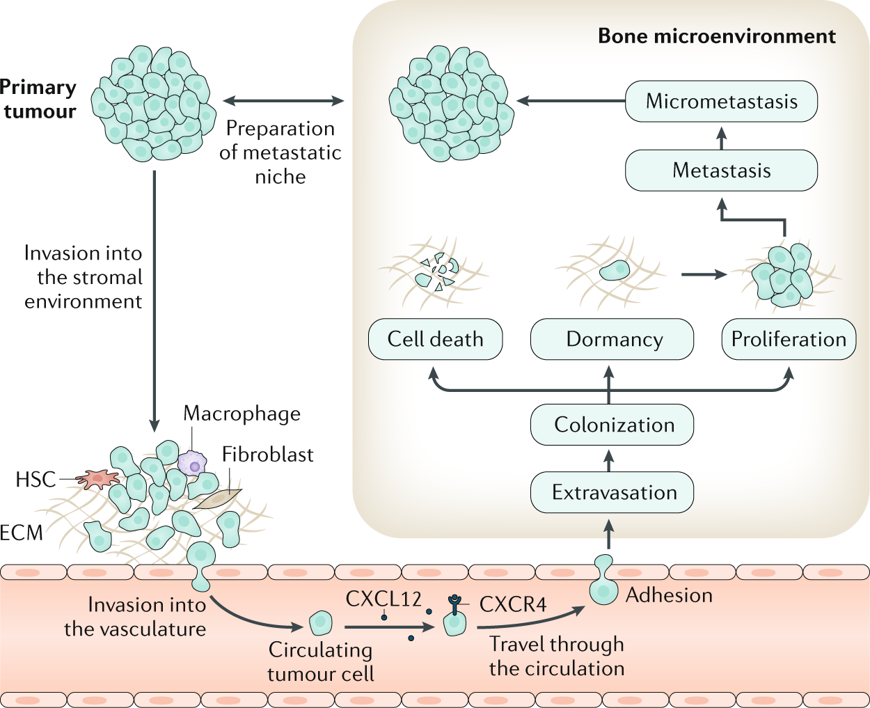
Bone is the most frequent site for metastasis for many cancers, notably for tumours originating in the breast and the prostate. Tumour cells can escape from the primary tumour site and colonize the bone microenvironment. Within the bone, these disseminated tumour cells, as well as those arising in the context of multiple myeloma, may assume a state of dormancy, remaining quiescent for years before resuming proliferation and causing overt metastasis, which causes bone destruction via activation of osteoclast-mediated osteolysis. This structural damage can lead to considerable morbidity, including pain, fractures and impaired quality of life. Although treatment of bone metastases and myeloma bone disease is rarely curative, disease control is often possible for many years through the use of systemic anticancer treatments on a background of multidisciplinary supportive care. This care should include bone-targeted agents to inhibit tumour-associated osteolysis and prevent skeletal morbidity as well as use of appropriate local treatments such as radiation therapy, orthopaedic surgery and specialist palliative care to minimize the impact of metastatic bone disease on physical functioning. In this Primer, we provide an overview of the clinical features, the pathophysiology and the specific treatment approaches to prevent and treat bone metastases from solid tumours as well as myeloma bone disease.
Ten‐Year Simulation of the Effects of Denosumab on Bone Remodeling in Human Biopsies
ABSTRACT Postmenopausal osteoporosis is a disease manifesting in degradation of bone mass and microarchitecture, leading to weakening and increased risk of fracture. Clinical trials are an essential tool for evaluating new treatments and may provide further mechanistic understanding of their effects in vivo. However, the histomorphometry from clinical trials is limited to 2D images and reflects single time points. Biochemical markers of bone turnover give global insight into a drug's action, but not the local dynamics of the bone remodeling process and the cells involved. Additionally, comparative trials necessitate separate treatment groups, meaning only aggregated measures can be compared. In this study, in silico modeling based on histomorphometry and pharmacokinetic data was used to assess the effects of treatment versus control on μCT scans of the same biopsy samples over time, matching the changes in bone volume fraction observed in biopsies from denosumab and placebo groups through year 10 of the FREEDOM Extension trial. In the simulation, treatment decreased osteoclast number, which led to a modest increase in trabecular thickness and osteocyte stress shielding. Long‐term bone turnover suppression led to increased RANKL production, followed by a small increase in osteoclast number at the end of the 6‐month–dosing interval, especially at the end of the Extension study. Lack of treatment led to a significant loss of bone mass and structure. The study's results show how in silico models can generate predictions of denosumab cellular action over a 10‐year period, matching static and dynamic morphometric measures assessed in clinical biopsies. The use of in silico models with clinical trial data can be a method to gain further insight into fundamental bone biology and how treatments can perturb this. With rigorous validation, such models could be used for informing the design of clinical trials, such that the number of participants could be reduced to a minimum to show efficacy. © 2021 The Authors. JBMR Plus published by Wiley Periodicals LLC on behalf of American Society for Bone and Mineral Research.
The Effect of Inflammation on Bone
Bone remodeling is the continual process to renew the adult skeleton through the sequential action of osteoblasts and osteoclasts. Nuclear factor RANK, an osteoclast receptor, and its ligand RANKL, expressed on the surface of osteoblasts, result in coordinated control of bone remodeling. Inflammation, a feature of illness and injury, plays a distinct role in skewing this process toward resorption. It does so via the interaction of inflammatory mediators and their related peptides with osteoblasts and osteoclasts, to alter the expression of RANK and RANKL. Such chemical mediators include TNFa, glucocorticoids, histamine, bradykinin, PGE2, systemic RANKL from immune cells, and interleukins 1 and 6. A thorough understanding of bone response to injury and disease, and ability to detect such biomarkers, as well as imaging to identify early structural and mechanical property changes in bone architecture, is important in improving management and outcomes of bone related pathology. While gut health and vitamin and mineral availability appear vitally important, there are very few intervention strategies currently available to favorably alter bone pathology induced by inflammation. Further research is required in this field to advance early detection and treatments.

Forecasting the critical role of intermittent therapies for the control of bone resorption
Osteoporosis is a chronic metabolic disease characterized by an imbalance of bone resorption and formation, leading to bone fragility and increased susceptibility to fracture. Parathyroid hormone is approved therapy for the treatment of osteoporosis.

The Regulation of Bone Metabolism and Disorders by Wnt Signaling
Wnt, a secreted glycoprotein, has an approximate molecular weight of 40 kDa, and it is a cytokine involved in various biological phenomena including ontogeny, morphogenesis, carcinogenesis, and maintenance of stem cells. The Wnt signaling pathway can be classified into two main pathways: canonical and non-canonical. Of these, the canonical Wnt signaling pathway promotes osteogenesis. Sclerostin produced by osteocytes is an inhibitor of this pathway, thereby inhibiting osteogenesis. Recently, osteoporosis treatment using an anti-sclerostin therapy has been introduced. In this review, the basics of Wnt signaling, its role in bone metabolism and its involvement in skeletal disorders have been covered. Furthermore, the clinical significance and future scopes of Wnt signaling in osteoporosis, osteoarthritis, rheumatoid arthritis and neoplasia are discussed.
Estrogen and the skeleton
Estrogen is the major hormonal regulator of bone metabolism in women and men. Therefore, there is considerable interest in unraveling the pathways by which estrogen exerts its protective effects on bone. Although the major consequence of the loss of estrogen is an increase in bone resorption, estrogen deficiency is associated with a gap between bone resorption and formation, indicating that estrogen is also important for maintaining bone formation at the cellular level. Direct estrogen effects on osteocytes, osteoclasts, and osteoblasts lead to inhibition of bone remodeling, decreased bone resorption, and maintenance of bone formation, respectively.

Critical Role of Estrogens on Bone Homeostasis in Both Male and Female: From Physiology to Medical Implications
Bone is a multi-skilled tissue, protecting major organs, regulating calcium phosphate balance and producing hormones. Its development during childhood determines height and stature as well as resistance against fracture in advanced age. Estrogens are key regulators of bone turnover in both females and males. These hormones play a major role in longitudinal and width growth throughout puberty as well as in the regulation of bone turnover. In women, estrogen deficiency is one of the major causes of postmenopausal osteoporosis. In this review, we will summarize the main clinical and experimental studies reporting the effects of estrogens not only in females but also in males, during different life stages. Effects of estrogens on bone involve either Estrogen Receptor (ER)α or ERβ depending on the type of bone (femur, vertebrae, tibia, mandible), the compartment (trabecular or cortical), cell types involved (osteoclasts, osteoblasts and osteocytes) and sex. Finally, we will discuss new ongoing strategies to increase the benefit/risk ratio of the hormonal treatment of menopause.