Pre-clinical studies have demonstrated that tart cherries, rich in hydroxycinnamic acids and anthocyanins, protect against age-related and inflammation-induced bone loss. This study examined how daily consumption of Montmorency tart cherry juice (TC) alters biomarkers of bone metabolism in older women. Healthy women, aged 65–80 years (n = 27), were randomly assigned to consume ~240 mL (8 fl. oz.) of juice once (TC1X) or twice (TC2X) per day for 90 d. Dual-energy x-ray absorptiometry (DXA) scans were performed to determine bone density at baseline, and pre- and post-treatment serum biomarkers of bone formation and resorption, vitamin D, inflammation, and oxidative stress were assessed. Irrespective of osteoporosis risk, the bone resorption marker, tartrate resistant acid phosphatase type 5b, was significantly reduced with the TC2X dose compared to baseline, but not with the TC1X dose. In terms of indicators of bone formation and turnover, neither serum bone-specific alkaline phosphatase nor osteocalcin were altered. No changes in thiobarbituric acid reactive substances or high sensitivity C-reactive protein were observed in response to either TC1X or TC2X. We conclude that short-term supplementation with the higher dose of tart cherry juice decreased bone resorption from baseline without altering bone formation and turnover biomarkers in this cohort.
Montmorency tart cherry protects against age-related bone loss in female C57BL/6 mice and demonstrates some anabolic effects
Age-related bone loss is a consequence of endocrine and immune changes that disrupt bone remodeling. Functional foods (e.g., tart cherries) with antioxidant, anti-inflammatory and prebiotic activity can potentially counter this age-related phenomenon. The aim of this study was to determine if Montmorency tart cherry protects against early age-related bone loss and the culpable alterations in bone metabolism.

Dried Plum’s Polyphenolic Compounds and Carbohydrates Contribute to Its Osteoprotective Effects and Exhibit Prebiotic Activity in Estrogen Deficient C57BL/6 Mice
Evidence of dried plum’s benefits on bone continues to emerge. This study investigated the contribution of the fruit’s polyphenol (PP) and carbohydrate (CHO) components on a bone model of postmenopausal osteoporosis to explore their prebiotic activity. Osteopenic ovariectomized mice were fed diets supplemented with dried plum, a crude extract of dried plum’s polyphenolic compounds, or the PP or CHO fraction of the crude extract. The effects of treatments on the bone phenotype were assessed at 5 and 10 weeks as well as the prebiotic activity of the different components of dried plum. Both the CHO and PP fractions of the extract contributed to the effects on bone with the CHO suppressing bone formation and resorption, and the PP temporally down-regulating formation. The PP and CHO components also altered the gut microbiota and cecal short chain fatty acids. These findings demonstrate that the CHO as well as the PP components of dried plum have potential prebiotic activity, but they have differential roles in mediating the alterations in bone formation and resorption that protect bone in estrogen deficiency.
Current Status of the Diagnosis and Management of Osteoporosis
Osteoporosis has been defined as the silent disease of the 21st century, becoming a public health risk due to its severity, chronicity and progression and affecting mainly postmenopausal women and older adults. Osteoporosis is characterized by an imbalance between bone resorption and bone production. It is diagnosed through different methods such as bone densitometry and dual X-rays. The treatment of this pathology focuses on different aspects. On the one hand, pharmacological treatments are characterized by the use of anti-resorptive drugs, as well as emerging regenerative medicine treatments such as cell therapies and the use of bioactive hydrogels. On the other hand, non-pharmacological treatments are associated with lifestyle habits that should be incorporated, such as physical activity, diet and the cessation of harmful habits such as a high consumption of alcohol or smoking. This review seeks to provide an overview of the theoretical basis in relation to bone biology, the existing methods for diagnosis and the treatments of osteoporosis, including the development of new strategies.

Postmenopausal osteoporosis
Osteoporosis is a metabolic bone disorder that is characterized by low bone mass and micro-architectural deterioration of bone tissue. Fractures of the proximal femur, the vertebrae and the distal radius are the most frequent osteoporotic fractures, although most fractures in the elderly are probably at least partly related to bone fragility. The incidence of fractures varies greatly by country, but on average up to 50% of women >50 years of age are at risk of fractures. Fractures severely affect the quality of life of an individual and are becoming a major public health problem owing to the ageing population. Postmenopausal osteoporosis, resulting from oestrogen deficiency, is the most common type of osteoporosis. Oestrogen deficiency results in an increase in bone turnover owing to effects on all types of bone cells. The imbalance in bone formation and resorption has effects on trabecular bone (loss of connectivity) and cortical bone (cortical thinning and porosity). Osteoporosis is diagnosed using bone density measurements of the lumbar spine and proximal femur. Preventive strategies to improve bone health include diet, exercise and abstaining from smoking. Fractures may be prevented by reducing falls in high-risk populations. Several drugs are licensed to reduce fracture risk by slowing down bone resorption (such as bisphosphonates and denosumab) or by stimulating bone formation (such as teriparatide). Improved understanding of the cellular basis for osteoporosis has resulted in new drugs targeted to key pathways, which are under development.

Effect of Long-Term Green Tea Polyphenol Supplementation on Bone Architecture, Turnover, and Mechanical Properties in Middle-Aged Ovariectomized Rats
We investigated the effects of 6-month green tea polyphenols (GTP) supplementation on bone architecture, turnover, and mechanical properties in middle-aged ovariectomized (OVX) rats. Female rats were sham-operated (n = 39, 13/group) or OVX (n = 143, 13/group). Sham-control and OVX-control rats (n = 39) receiving no GTP were assigned for sample collection at baseline, 3, or 6 months. The remaining OVX rats (n = 104) were randomized to 0.15%, 0.5%, 1%, and 1.5% (g/dL) GTP for 3 or 6 months. Blood and bone samples were collected. Relative to the OVX-control group, GTP (1% and 1.5%) lowered serum procollagen type 1 N-terminal propeptide at 3 and 6 months, C-terminal telopeptides of type I collagen at 3 months, and insulin-like growth factor-I at 6 months. GTP did not affect bone mineral content and density. At 6 months, no dose of GTP positively affected trabecular bone volume based on microCT, but a higher cortical thickness and improved biomechanical properties of the femur mid-diaphysis was observed in the 1.5% GTP-treated group. At 3 and 6 months, GTP (0.5%, 1%, and 1.5%) had lower rates of trabecular bone formation and resorption than the OVX-control group, but the inhibitory effects of GTP on periosteal and endocortical bone mineralization and formation at the tibial midshaft were only evident at 3 months. GTP at higher doses suppressed bone turnover in the trabecular and cortical bone of OVX rats and resulted in improved cortical bone structural and biomechanical properties, although it was not effective in preventing the ovariectomy-induced dramatic cancellous bone loss.

Disease Systems Analysis of Bone Mineral Density and Bone Turnover Markers in Response to Alendronate, Placebo, and Washout in Postmenopausal Women
A previously established mechanism‐based disease systems model for osteoporosis that is based on a mathematically reduced version of a model describing the interactions between osteoclast (bone removing) and osteoblast (bone forming) cells in bone remodeling has been applied to clinical data from women ( n = 1,379) receiving different doses and treatment regimens of alendronate, placebo, and washout. The changes in the biomarkers, plasma bone‐specific alkaline phosphatase activity (BSAP), urinary N‐telopeptide (NTX), lumbar spine bone mineral density (BMD), and total hip BMD, were linked to the underlying mechanistic core of the model. The final model gave an accurate description of all four biomarkers for the different treatments. Simulations were used to visualize the dynamics of the underlying network and the natural disease progression upon alendronate treatment and discontinuation. These results complement the previous applications of this mechanism‐based disease systems model to data from various treatments for osteoporosis.

Estrogen therapy for osteoporosis in the modern era
Menopause predisposes women to osteoporosis due to declining estrogen levels. This results in a decrease in bone mineral density (BMD) and an increase in fractures. Osteoporotic fractures lead to substantial morbidity and mortality, and are considered one of the largest public health priorities by the World Health Organization (WHO). It is therefore essential for menopausal women to receive appropriate guidance for the prevention and management of osteoporosis. The Women’s Health Initiative (WHI) randomized controlled trial first proved hormonal therapy (HT) reduces the incidence of all osteoporosis-related fractures in postmenopausal women. However, the study concluded that the adverse effects outweighed the potential benefits on bone, leading to a significant decrease in HT use for menopausal symptoms. Additionally, HT was not used as first-line therapy for osteoporosis and fractures. Subsequent studies have challenged these initial conclusions and have shown significant efficacy of HT in various doses, durations, regimens, and routes of administration. These studies support that HT improves BMD and reduces fracture risk in women with and without osteoporosis. Furthermore, the studies suggest that low-dose and transdermal HT are less likely associated with the adverse effects of breast cancer, endometrial hyperplasia, coronary artery disease (CAD), and venous thromboembolism (VTE) previously observed in standard-dose oral HT regimens. Given the need for estrogen in menopausal women and evidence supporting the cost effectiveness, safety, and efficacy of HT, we propose that HT should be considered for the primary prevention and treatment of osteoporosis in appropriate candidates. HT should be individualized and the once “lowest dose for shortest period of time” concept should no longer be used. This review will focus on the prior and current studies for various HT formulations used for the prevention and treatment of osteoporosis, exploring the safety profile of low-dose and transdermal HT that have been shown to be safer than oral standard-dose HT.
Prebiotics Enhance Magnesium Absorption and Inulin‐based Fibers Exert Chronic Effects on Calcium Utilization in a Postmenopausal Rodent Model
Abstract: Age‐related changes in calcium metabolism play a role in the development of osteoporosis. A 4‐wk feeding study was conducted in 5‐mo‐old ovariectomized (OVX) Sprague‐Dawley rats to assess the effect of various dietary fibers on mineral metabolism and bone health parameters. There were 6 treatment groups: sham‐Control, OVX‐Control, OVX rats receiving daily estradiol (E 2 ) injections, and OVX rats receiving an AIN‐93M diet supplement with either an inulin‐based fiber (Synergy1® or Fruitafit HD®) or a novel fiber (polydextrose) at 5% wt. of diet. Calcium and magnesium metabolic balances were performed after early (3 d) and late exposure (4 wk) to dietary treatments. Rats receiving polydextrose had significantly higher net calcium absorption efficiency and retention than all control groups and a trend ( P ≤ 0.10) for higher calcium absorption when compared to inulin‐based fibers after early exposure but the advantage did not persist over long‐term exposure. The inulin‐based fibers had positive chronic effects on calcium metabolism that were related to changes in the gut, that is, production of short chain fatty acids and higher cecal wall weights. All fibers improved magnesium absorption and retention in early and late metabolic balances; effects on magnesium metabolism were more pronounced than for calcium. Practical Application: Steady growth in US middle‐aged and elderly populations has led to higher incidences of several chronic diseases including osteoporosis, a bone disease that primarily affects postmenopausal women. Recent research suggests that certain dietary fibers (prebiotics) enhance mineral absorption and may impart bone health benefits. This work examines the impact of prebiotic supplementation on mineral metabolism and bone health using a postmenopausal rat model. Study findings will aid future investigations in ascertaining the factors related to potential bone health benefits of prebiotic which will aid in developing an effective prebiotics food product/supplement that will address the bone health needs of consumers.

Effect of Bacillus subtilis C-3102 on bone mineral density in healthy postmenopausal Japanese women: a randomized, placebo-controlled, double-blind clinical trial
Gut microbiota influence the host immune system and are associated with various diseases. In recent years, postmenopausal bone loss has been suggested to be related to gut microbiota. In the present study, we investigated the treatment effect of the probiotic Bacillus subtilis C-3102 (C-3102) on bone mineral density (BMD) and its influence on gut microbiota in healthy postmenopausal Japanese women. Seventy-six healthy postmenopausal Japanese women were treated with a placebo or C-3102 spore-containing tablets for 24 weeks. When compared with the placebo, C-3102 significantly increased total hip BMD (placebo = 0.83 ± 0.63%, C-3102 = 2.53 ± 0.52%, p=0.043). There was a significant group-by-time interaction effect for urinary type I collagen cross-linked N-telopeptide (uNTx) (p=0.033), a marker of bone resorption. Specifically, the C-3102 group showed significantly lower uNTx when compared with the placebo group at 12 weeks of treatment (p=0.015). In addition, in the C-3102 group, there was a trend towards a decrease in the bone resorption marker tartrate-resistant acid phosphatase isoform 5b (TRACP-5b) when compared with the placebo group at 12 weeks of treatment (p=0.052). The relative abundance of genus Bifidobacterium significantly increased at 12 weeks of treatment compared with the baseline in the C-3102 group. The relative abundance of genus Fusobacterium was significantly decreased in the C-3102 group at 12 and 24 weeks of treatment compared with the baseline. These data suggested that C-3102 improves BMD by inhibiting bone resorption and modulating gut microbiota in healthy postmenopausal women.

Application of a mechanism-based disease systems model for osteoporosis to clinical data
A recently proposed mechanism-based disease systems model for osteoporosis (Schmidt et al., J Pharmacokinet Pharmacodyn 38:873–900, 2011) was applied to clinical data from post-menopausal women (n = 767) receiving various doses of the selective estrogen receptor modulator tibolone. Plasma bone-specific alkaline phosphatase activity and urinary N-telopeptide were used as biomarkers reflecting the activity of osteoblasts (bone forming cells) and osteoclasts (bone removing cells), respectively. These data were analyzed in conjunction with data on osteocalcin and on bone mineral density (BMD) (both lumbar spine and total hip), which reflect the activity of both cell types. While the dynamics of bone turnover markers changes rapidly, closely following changes in the activity of bone cells, changes in BMD are slower and have their own dynamics. Application of the mechanism-based disease systems model to the clinical data allowed for an adequate description of the data and yielded parameter estimates that are consistent with physiological values reported in the literature (Lemaire et al., J Theor Biol 229:293–309, 2004). The fitted model enabled characterization of (i) the critical time scales involved in disease progression, (ii) the dynamics of the system during onset and offset of the therapeutic intervention, and (iii) the distinction between responders and low-responders to tibolone treatment.

Critical Role of Estrogens on Bone Homeostasis in Both Male and Female: From Physiology to Medical Implications
Bone is a multi-skilled tissue, protecting major organs, regulating calcium phosphate balance and producing hormones. Its development during childhood determines height and stature as well as resistance against fracture in advanced age. Estrogens are key regulators of bone turnover in both females and males. These hormones play a major role in longitudinal and width growth throughout puberty as well as in the regulation of bone turnover. In women, estrogen deficiency is one of the major causes of postmenopausal osteoporosis. In this review, we will summarize the main clinical and experimental studies reporting the effects of estrogens not only in females but also in males, during different life stages. Effects of estrogens on bone involve either Estrogen Receptor (ER)α or ERβ depending on the type of bone (femur, vertebrae, tibia, mandible), the compartment (trabecular or cortical), cell types involved (osteoclasts, osteoblasts and osteocytes) and sex. Finally, we will discuss new ongoing strategies to increase the benefit/risk ratio of the hormonal treatment of menopause.
Nut intake and hyperuricemia risk in young adults
Objective:The relationship between dietary nut intake and hyperuricemia risk remains unclear. The aim of this study was to investigate the relationship between different nut intake and hyperuricemia risk with a cross-sectional study.Design:A semi-quantitative FFQ was adopted to collect dietary information. Biochemical and anthropometric parameters were measured by standard methods. Multivariate-adjusted logistic regression models were implemented to analyse the relationship between individual nut intake and hyperuricemia risk.Setting:Qingdao University in Shandong Province, China.Participants:During 2018–2019, a total of 14 056 undergraduates (6862 males and 7194 females) aged 15–25 years participated in the study.Results:After adjusting for multiple confounding factors, compared with the lowest quartile, the highest quartile intakes of pine nut (95 % CI (0·51, 0·98)) was significantly associated with 29 % reduction in hyperuricemia risk, the highest quartile intake of walnut (OR = 0·78; 95 % CI (0·58, 1·05)) was marginally negatively associated with hyperuricemia risk.Conclusions:The present study showed that the relationships between intakes of different nuts and hyperuricemia risk were different. Increased dietary intakes of walnut and pine nut are negatively associated with the hyperuricemia.

Loss of Bone and Wnt10b Expression in Male Type 1 Diabetic Mice Is Blocked by the Probiotic Lactobacillus reuteri
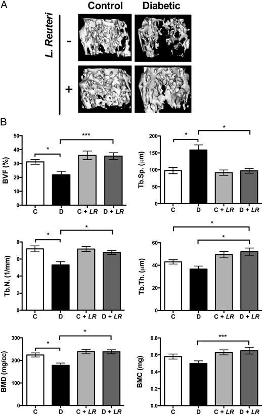
Type 1 diabetes (T1D)–induced osteoporosis is characterized by a predominant suppression of osteoblast number and activity, as well as increased bone marrow adiposity but no change in osteoclast activity. The fundamental mechanisms and alternative anabolic treatments (with few side effects) for T1D bone loss remain undetermined. Recent studies by our laboratory and others indicate that probiotics can benefit bone health. Here, we demonstrate that Lactobacillus reuteri, a probiotic with anti-inflammatory and bone health properties, prevents T1D-induced bone loss and marrow adiposity in mice. We further found that L. reuteri treatment prevented the suppression of Wnt10b in T1D bone. Consistent with a role for attenuated bone Wnt10b expression in T1D osteoporosis, we observed that bone-specific Wnt10b transgenic mice are protected from T1D bone loss. To examine the mechanisms of this protection, we focused on TNF-α, a cytokine up-regulated in T1D that causes suppression of osteoblast Wnt10b expression in vitro. Addition of L. reuteri prevented TNF-α–mediated suppression of Wnt10b and osteoblast maturation markers. Taken together, our findings reveal a mechanism by which T1D causes bone loss and open new avenues for use of probiotics to benefit the bone.
Associations Between Inflammatory Mediators and Bone Outcomes in Postmenopausal Women: A Cross-Sectional Analysis of Baseline Data from the Prune Study
These findings suggest that inflammatory markers may be an important mediator for postmenopausal bone loss.

The Effect of Inflammation on Bone
Bone remodeling is the continual process to renew the adult skeleton through the sequential action of osteoblasts and osteoclasts. Nuclear factor RANK, an osteoclast receptor, and its ligand RANKL, expressed on the surface of osteoblasts, result in coordinated control of bone remodeling. Inflammation, a feature of illness and injury, plays a distinct role in skewing this process toward resorption. It does so via the interaction of inflammatory mediators and their related peptides with osteoblasts and osteoclasts, to alter the expression of RANK and RANKL. Such chemical mediators include TNFa, glucocorticoids, histamine, bradykinin, PGE2, systemic RANKL from immune cells, and interleukins 1 and 6. A thorough understanding of bone response to injury and disease, and ability to detect such biomarkers, as well as imaging to identify early structural and mechanical property changes in bone architecture, is important in improving management and outcomes of bone related pathology. While gut health and vitamin and mineral availability appear vitally important, there are very few intervention strategies currently available to favorably alter bone pathology induced by inflammation. Further research is required in this field to advance early detection and treatments.

The amazing osteocyte
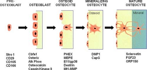
Abstract The last decade has provided a virtual explosion of data on the molecular biology and function of osteocytes. Far from being the “passive placeholder in bone,” this cell has been found to have numerous functions, such as acting as an orchestrator of bone remodeling through regulation of both osteoclast and osteoblast activity and also functioning as an endocrine cell. The osteocyte is a source of soluble factors not only to target cells on the bone surface but also to target distant organs, such as kidney, muscle, and other tissues. This cell plays a role in both phosphate metabolism and calcium availability and can remodel its perilacunar matrix. Osteocytes compose 90% to 95% of all bone cells in adult bone and are the longest lived bone cell, up to decades within their mineralized environment. As we age, these cells die, leaving behind empty lacunae that frequently micropetrose. In aged bone such as osteonecrotic bone, empty lacunae are associated with reduced remodeling. Inflammatory factors such as tumor necrosis factor and glucocorticoids used to treat inflammatory disease induce osteocyte cell death, but by different mechanisms with potentially different outcomes. Therefore, healthy, viable osteocytes are necessary for proper functionality of bone and other organs. © 2011 American Society for Bone and Mineral Research.