

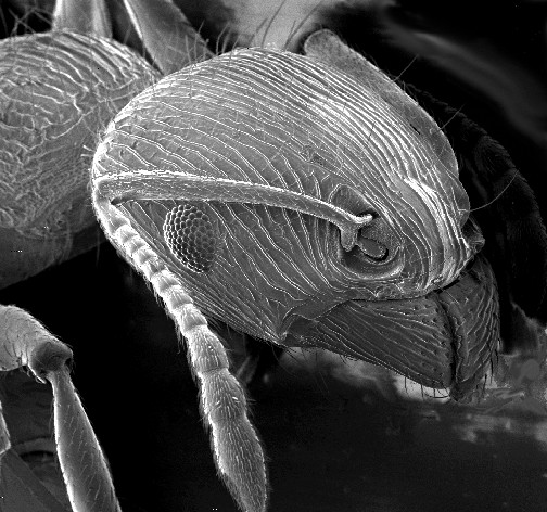
Arthropods are covered with a tough, resilient integument, cuticle or exoskeleton of chitin. Generally the exoskeleton will have thickened areas in which the chitin is reinforced or stiffened by materials such as minerals or hardened proteins. This happens in parts of the body where there is a need for rigidity or elasticity. Typically the mineral crystals, mainly calcium carbonate, are deposited among the chitin and protein molecules in a process called biomineralization. The crystals and fibres interpenetrate and reinforce each other, the minerals supplying the hardness and resistance to compression, while the chitin supplies the tensile strength. Biomineralization occurs mainly in crustaceans. In insects and arachnids, the main reinforcing materials are various proteins hardened by linking the fibres in processes called sclerotisation and the hardened proteins are called sclerotin. The dorsal tergum, ventral sternum, and the lateral pleura form the hardened plates or sclerites of a typical body segment.
Chitin
Chitin (C8H13O5N)n ( KY-tin) is a long-chain polymer of N-acetylglucosamine, an amide derivative of glucose. Chitin is the second most abundant polysaccharide in nature (behind only cellulose); an estimated 1 billion tons of chitin are produced each year in the biosphere. It is a primary component of cell walls in fungi (especially filamentous and mushroom-forming fungi), the exoskeletons of arthropods such as crustaceans and insects, the radulae, cephalopod beaks and gladii of molluscs and in some nematodes and diatoms. It is also synthesised by at least some fish and lissamphibians. Commercially, chitin is extracted from the shells of crabs, shrimps, shellfish and lobsters, which are major by-products of the seafood industry. The structure of chitin is comparable to cellulose, forming crystalline nanofibrils or whiskers. It is functionally comparable to the protein keratin. Chitin has proved useful for several medicinal, industrial and biotechnological purposes.

Exoskeleton
An exoskeleton is a skeleton that is on the exterior of an animal in the form of hardened integument, which both supports the body's shape and protects the internal organs, in contrast to an internal endoskeleton which is enclosed underneath other soft tissues. Some large, hard and non-flexible protective exoskeletons are known as shell or armour.
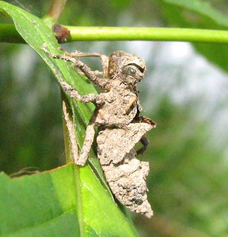
Ecdysis
Ecdysis is the moulting of the cuticle in many invertebrates of the clade Ecdysozoa. Since the cuticle of these animals typically forms a largely inelastic exoskeleton, it is shed during growth and a new, larger covering is formed. The remnants of the old, empty exoskeleton are called exuviae.

A mechano-chemo-biological model for bone remodeling with a new mechano-chemo-transduction approach
Bone remodeling is a fundamental biological process that develops in bone tissue along its whole lifetime. It refers to a continuous bone transformation with new bone formation and old bone resorption that changes the internal microstructure and composition of the tissue. The main objectives of bone remodeling are: repair of the internal microcracks; adaptation of the macroscopic stiffness and strength to the actual changing mechanical demands; and control of the calcium homeostasis. Understanding this process and predicting its evolution is critical to reduce the effects of long-term disuse as happens during periods of reduced mobility. It is also important in the design of bone implants to avoid long-term stress shielding. Many mathematical models have been proposed from the earliest purely phenomenological to the latest that include biological knowledge. However, there still exists a lack of connection between the mechanical driving force and the biochemical and cell processes it triggers. Here, and following previous works that model independently the mechanobiological and biochemical processes in bone remodeling, we present a more complete model, useful for both cortical and trabecular bone, that uses a new mechanotransduction approach based on the effect of strains onto the bonding–unbonding rate of RANK/RANKL/OPG receptor–ligand reactions. We compare the results of this model with previous ones, showing a good agreement in similar conditions. We also apply it to realistic situations such as a femoral bone after implantation of a hip prosthesis, getting similar results to the clinical ones in the final bone density distribution. Finally, we extend this approach to the anisotropic case, getting not only the mean density, but also the directional homogenization of the microstructure. This biochemical approach permits, not only to predict the bone evolution under changes in the mechanical loads, but also, to consider anabolic and catabolic drugs to control bone density, such as those used in osteoporosis.

Symbiotic organs in insects: diversity, functional implications, and terminology
Abstract With over a million described species, insects represent the most successful group of animals on Earth. One of the drivers of insect diversity is their ability to engage in multifold beneficial symbioses with microorganisms, often involving specialized host organs to accommodate intra- or extracellular symbionts. The existence of such organs and their importance for sustaining and transmitting beneficial symbionts has been known for over a century, and specific terms have been established for categorizing organs harbouring intracellular bacteria (bacteriomes) or fungi (mycetomes), or cuticular crypts containing extracellular fungi (mycetangia). For others, however, general terms are lacking, e.g. organs containing extracellular bacteria associated with the cuticle or with the digestive tract. Furthermore, previously established terms have been misused in other contexts. Notably, ‘bacteriome’ has been increasingly employed in the microbiome field to refer to bacterial communities, instead of the term’s original meaning of specialized organs housing intracellular bacterial symbionts. Here, we review and categorize the diversity of symbiotic organs in insects and propose a unified terminology. Our hope is that this common language will facilitate communication and thereby support the field of symbiosis research in unravelling commonalities and differences in the evolution, ecology, development, physiology and molecular basis across symbiotic interactions. This article is part of the theme issue ‘Life in natural microcosms’.

Mandible (insect mouthpart)
Insect mandibles are a pair of appendages near the insect's mouth, and the most anterior of the three pairs of oral appendages. Their function is typically to grasp, crush, or cut the insect's food, or to defend against predators or rivals. Insect mandibles, which appear to be evolutionarily derived from legs, move in the horizontal plane unlike those of vertebrates, which appear to be derived from gill arches and move vertically.

Spiracle (arthropods)
A spiracle or stigma is the opening in the exoskeletons of insects, myriapods, velvet worms and many arachnids that admits air into the respiratory system.

<i>In silico</i> biology of bone modelling and remodelling: adaptation
Modelling and remodelling are the processes by which bone adapts its shape and internal structure to external influences. However, the cellular mechanisms triggering osteoclastic resorption and osteoblastic formation are still unknown. In order to investigate current biological theories, in silico models can be applied. In the past, most of these models were based on the continuum assumption, but some questions related to bone adaptation can be addressed better by models incorporating the trabecular microstructure. In this paper, existing simulation models are reviewed and one of the microstructural models is extended to test the hypothesis that bone adaptation can be simulated without particular knowledge of the local strain distribution in the bone. Validation using an experimental murine loading model showed that this is possible. Furthermore, the experimental model revealed that bone formation cannot be attributed only to an increase in trabecular thickness but also to structural reorganization including the growth of new trabeculae. How these new trabeculae arise is still an unresolved issue and might be better addressed by incorporating other levels of hierarchy, especially the cellular level. The cellular level sheds light on the activity and interplay between the different cell types, leading to the effective change in the whole bone. For this reason, hierarchical multi-scale simulations might help in the future to better understand the biomathematical laws behind bone adaptation.

Instar
An instar is a developmental stage of arthropods, such as insects, which occurs between each moult (ecdysis) until sexual maturity is reached. Arthropods must shed the exoskeleton in order to grow or assume a new form. Differences between instars can often be seen in altered body proportions, colors, patterns, changes in the number of body segments or head width.

Control of Bone Matrix Properties by Osteocytes
Osteocytes make up 90-95% of the cellular content of bone and form a rich dendritic network with a vastly greater surface area than either osteoblasts or osteoclasts. Osteocytes are well positioned to play a role in bone homeostasis by interacting directly with the matrix; however, the ability for these cells to modify bone matrix remains incompletely understood. With techniques for examining the nano- and microstructure of bone matrix components including hydroxyapatite and type I collagen becoming more widespread, there is great potential to uncover novel roles for the osteocyte in maintaining bone quality. In this review, we begin with an overview of osteocyte biology and the lacunar-canalicular system. Next, we describe recent findings from in vitro models of osteocytes, focusing on the transitions in cellular phenotype as they mature. Finally, we describe historical and current research on matrix alteration by osteocytes in vivo, focusing on the exciting potential for osteocytes to directly form, degrade, and modify the mineral and collagen in their surrounding matrix.

A novel mathematical model of bone remodelling cycles for trabecular bone at the cellular level
After an initial phase of growth and development, bone undergoes a continuous cycle of repair, renewal and optimisation by a process called remodelling. This paper describes a novel mathematical model of the trabecular bone remodelling cycle. It is essentially formulated to simulate a remodelling event at a fixed position in the bone, integrating bone removal by osteoclasts and formation by osteoblasts. The model is developed to construct the variation in bone thickness at a particular point during the remodelling event, derived from standard bone histomorphometric analyses. The novelties of the approach are the adoption of a predator–prey model to describe the dynamic interaction between osteoclasts and osteoblasts, using a genetic algorithm–based solution; quantitative reconstruction of the bone remodelling cycle; and the introduction of a feedback mechanism in the bone formation activity to co-regulate bone thickness. The application of the model is first demonstrated by using experimental data recorded for normal (healthy) bone remodelling to predict the temporal variation in the number of osteoblasts and osteoclasts. The simulated histomorphometric data and remodelling cycle characteristics compare well with the specified input data. Sensitivity studies then reveal how variations in the model’s parameters affect its output; it is hoped that these parameters can be linked to specific biochemical factors in the future. Two sample pathological conditions, hypothyroidism and primary hyperparathyroidism, are examined to demonstrate how the model could be applied more broadly, and, for the first time, the osteoblast and osteoclast populations are predicted for these conditions. Further data are required to fully validate the model’s predictive capacity, but this work shows it has potential, especially in the modelling of pathological conditions and the optimisation of the treatment of those conditions.
Integrated Bone Tissue Physiology: Anatomy and Physiology
The Role of Osteocytes in Targeted Bone Remodeling: A Mathematical Model
Until recently many studies of bone remodeling at the cellular level have focused on the behavior of mature osteoblasts and osteoclasts, and their respective precursor cells, with the role of osteocytes and bone lining cells left largely unexplored. This is particularly true with respect to the mathematical modeling of bone remodeling. However, there is increasing evidence that osteocytes play important roles in the cycle of targeted bone remodeling, in serving as a significant source of RANKL to support osteoclastogenesis, and in secreting the bone formation inhibitor sclerostin. Moreover, there is also increasing interest in sclerostin, an osteocyte-secreted bone formation inhibitor, and its role in regulating local response to changes in the bone microenvironment. Here we develop a cell population model of bone remodeling that includes the role of osteocytes, sclerostin, and allows for the possibility of RANKL expression by osteocyte cell populations. We have aimed to give a simple, yet still tractable, model that remains faithful to the underlying system based on the known literature. This model extends and complements many of the existing mathematical models for bone remodeling, but can be used to explore aspects of the process of bone remodeling that were previously beyond the scope of prior modeling work. Through numerical simulations we demonstrate that our model can be used to explore theoretically many of the qualitative features of the role of osteocytes in bone biology as presented in recent literature.
Osteoclasts Provide Coupling Signals to Osteoblast Lineage Cells Through Multiple Mechanisms
Bone remodeling is essential for the repair and replacement of damaged and old bone. The major principle underlying this process is that osteoclast-mediated resorption of a quantum of bone is followed by osteoblast precursor recruitment; these cells differentiate to matrix-producing osteoblasts, which form new bone to replace what was resorbed. Evidence from osteopetrotic syndromes indicate that osteoclasts not only resorb bone, but also provide signals to promote bone formation. Osteoclasts act upon osteoblast lineage cells throughout their differentiation by facilitating growth factor release from resorbed matrix, producing secreted proteins and microvesicles, and expressing membrane-bound factors. These multiple mechanisms mediate the coupling of bone formation to resorption in remodeling. Additional interactions of osteoclasts with osteoblast lineage cells, including interactions with canopy and reversal cells, are required to achieve coordination between bone formation and resorption during bone remodeling.

Isopod Site
Pillbugs, sowbugs, roly-polies and woodlice, collectively referred to as isopods, are land-dwelling crustaceans that feed on decaying organic matter.

Mathematical Model of Bone Remodeling Captures the Antiresorptive and Anabolic Actions of Various Therapies
A better understanding of the molecular pathways regulating the bone remodeling process should help in the development of new antiresorptive regulators and anabolic regulators, that is, regulators of bone resorption and of bone formation. Understanding the mechanisms by which parathyroid hormone (PTH) influences bone formation and how it switches from anabolic to catabolic action is important for treating osteoporosis (Poole and Reeve in Curr Opin Pharmacol 5:612–617, 2005). In this paper we describe a mathematical model of bone remodeling that incorporates, extends, and integrates several models of particular aspects of this biochemical system (Cabal et al. in J Bone Miner Res 28(8):1830–1836, 2013; Lemaire et al. in J Theor Biol 229:293–309, 2004; Peterson and Riggs in Bone 46:49–63, 2010; Raposo et al. in J Clin Endocrinol Metab 87(9):4330–4340, 2002; Ross et al. in J Disc Cont Dyn Sys Series B 17(6):2185–2200, 2012). We plan to use this model as a bone homeostasis platform to develop anabolic and antiresorptive compounds. The model will allow us to test hypotheses about the dynamics of compounds and to test the potential benefits of combination therapies. At the core of the model is the idealized account of osteoclast and osteoblast signaling given by Lemaire et al. (J Theor Biol 229:293–309, 2004). We have relaxed some of their assumptions about the roles of osteoprotegerin, transforming growth factor $$\upbeta $$, and receptor activator of nuclear factor $$\upkappa $$B ligand; we have devised more detailed models of the interactions of these species. We have incorporated a model of the effect of calcium sensing receptor antagonists on remodeling (Cabal et al. in J Bone Miner Res 28(8):1830–1836, 2013). We have also incorporated a basic model of the effects of vitamin D on calcium homeostasis. We have included a simple model of the mechanism proposed by Bellido et al. (2003), Ross et al. (J Disc Cont Dyn Sys Series B 17(6):2185–2200, 2012), of the influence of PTH on osteoblast apoptosis, a mechanism that accounts for the anabolic response to pulsatile PTH administration. Finally, we have devised a simple model of the administration and effects of bisphosphonates. The biomarkers in the model are procollagen type 1 amino-terminal propeptide and C-terminal telopeptide. Bone mineral density is the model’s principal endpoint.
