

MIT neuroscientists have discovered how the brain preferentially forms memories of new locations. A small region of the brainstem, known as the locus coeruleus, releases a flood of dopamine in response to novel sensory stimuli, and this dopamine triggers a certain region of the hippocampus to store a memory of the new location.
How the brain controls our habits: Neuroscientists identify a brain region that can switch between new and old habits
Neuroscientists have found that a small region of the brain's prefrontal cortex, where most thought and planning occurs, is responsible for moment-by-moment control of which habits are switched on at a given time.

The Brain ‘Rotates’ Memories to Save Them From New Sensations | Quanta Magazine
Some populations of neurons simultaneously process sensations and memories. New work shows how the brain rotates those representations to prevent interference.
Neuroscientists show how neurons respond to sequences of familiar objects
The world grows increasingly more chaotic year after year, and our brains are constantly bombarded with images. A new study from Center for the Neural Basis of Cognition (CNBC), a joint project between ...

Thalamus Identified As a Hub for Memory and Brain Plasticity - Neuroscience News
A new study reveals that the thalamus, long overshadowed by the cortex and hippocampus, plays a crucial role in shaping and updating brain connections.
To Remember, the Brain Must Actively Forget | Quanta Magazine
Researchers find evidence that neural systems actively remove memories, suggesting that forgetting may be the default mode of the brain.
How does the brain link events to form a memory? Study reveals unexpected mental processes
A woman walking down the street hears a bang. Several moments later she discovers her boyfriend, who had been walking ahead of her, has been shot. A month later, the woman checks into the emergency room. ...

The Brain Can Recall and Reawaken Past Immune Responses | Quanta Magazine
The brain not only helps to regulate immune responses, but also stores and retrieves “memories” of them.

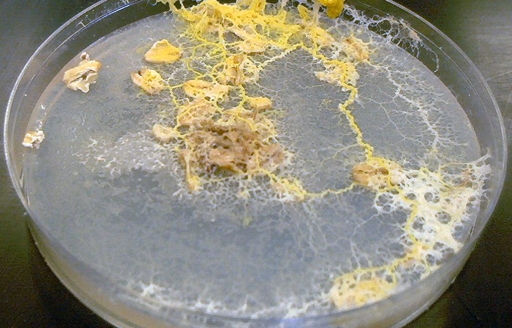
Organism without a brain creates external memories for navigation
The slime mold stores a record of where it's been using, well, slime.
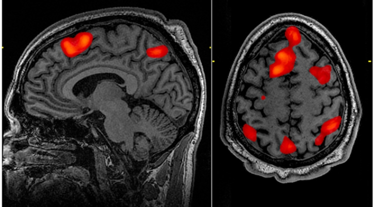
In-brain monitoring shows memory network - Research & Development World
Working with patients with electrodes implanted in their brains, researchers at the University of California, Davis, and The University of Texas Health Science Center at Houston (UTHealth) have shown for the first time that areas of the brain work together at the same time to recall memories. The unique approach promises new insights into how…

Study illuminates the structural features of memory formation at cellular and subcellular levels
NIH-funded study uses cutting-edge imaging techniques to reconstruct features underlying learning and memory in the mouse brain.

On the Neuroscience of Travel Memories
Memory has a habit of expunging linear sequences in favor of moments of strong emotion.

First Brain-Wide Map of Decision-Making Charted In Mice - Slashdot
In a landmark collaboration across 22 labs, neuroscientists have created the first brain-wide map of decision-making in mice, tracking over 620,000 neurons across nearly 280 brain regions. They found that decision-making is distributed much more broadly than previously thought, involving not just "c...
False memories arise because the brain codes similar ideas similarly
Efficiency In neural storage allows false memories to arise.
Memory trace and timing mechanism localized to cerebellar Purkinje cells
The standard view of neural signaling is that a neuron can influence its target cell by exciting or inhibiting it. An important aspect of the standard view is that learning consists of changing the efficacy of synapses, either strengthening (long-term potentiation) or weakening (long-term depression) them. In studying how cerebellar Purkinje cells change their responsiveness to a stimulus during learning of conditioned responses, we have found that these cells can learn the temporal relationship between two paired stimuli. The cells learn to respond at a particular time that reflects the time between the stimuli. This finding radically changes current views of both neural signaling and learning., The standard view of the mechanisms underlying learning is that they involve strengthening or weakening synaptic connections. Learned response timing is thought to combine such plasticity with temporally patterned inputs to the neuron. We show here that a cerebellar Purkinje cell in a ferret can learn to respond to a specific input with a temporal pattern of activity consisting of temporally specific increases and decreases in firing over hundreds of milliseconds without a temporally patterned input. Training Purkinje cells with direct stimulation of immediate afferents, the parallel fibers, and pharmacological blocking of interneurons shows that the timing mechanism is intrinsic to the cell itself. Purkinje cells can learn to respond not only with increased or decreased firing but also with an adaptively timed activity pattern.

In the brain, timing is everything
Study reveals how the brain links memories of events that occur one after the other.

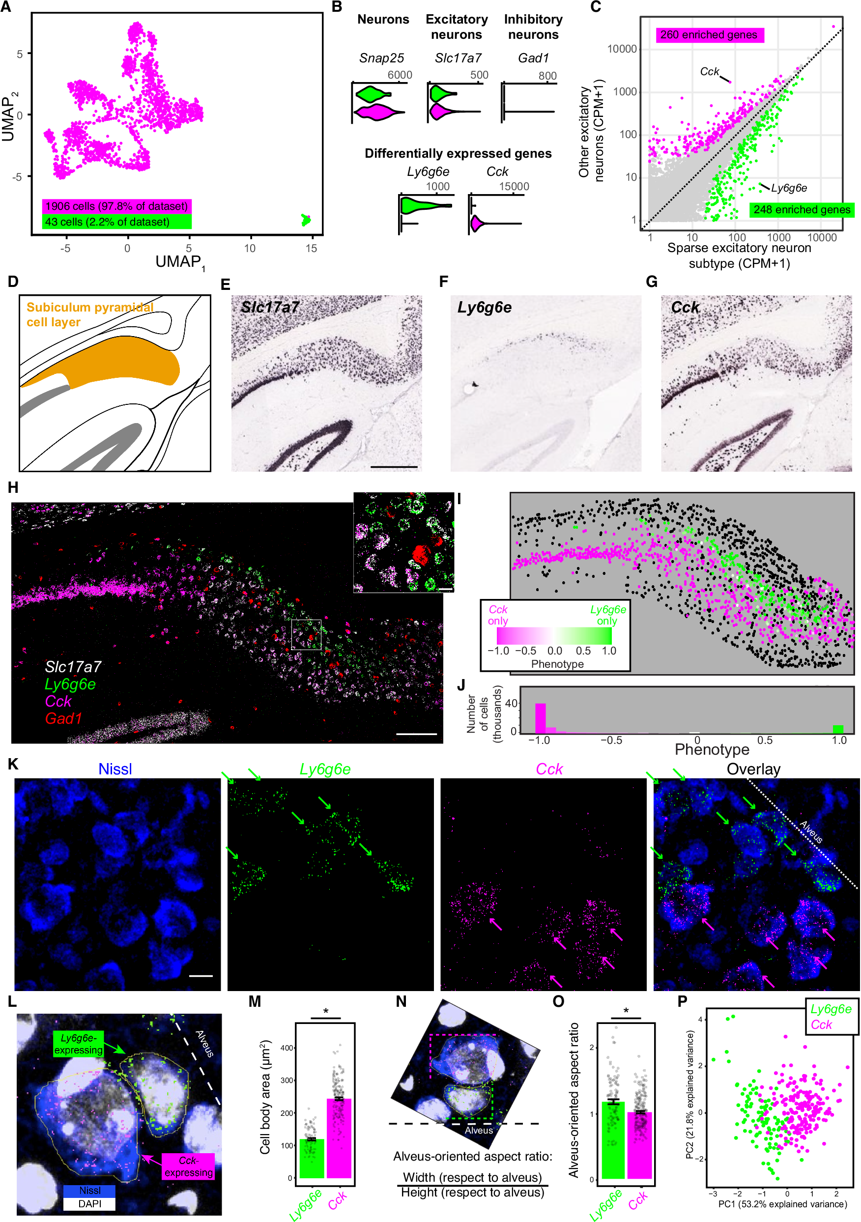
Atypical hippocampal excitatory neurons express and govern object memory
Classically, pyramidal cells of the hippocampus are viewed as flexibly representing spatial and non-spatial information. Recent work has illustrated distinct types of hippocampal excitatory neurons, suggesting that hippocampal representations and functions may be constrained and interpreted by these underlying cell-type identities. In mice, here we reveal a non-pyramidal excitatory neuron type — the “ovoid” neuron — that is spatially adjacent to subiculum pyramidal cells but differs in gene expression, electrophysiology, morphology, and connectivity. Functionally, novel object encounters drive sustained ovoid neuron activity, whereas familiar objects fail to drive activity even months after single-trial learning. Silencing ovoid neurons prevents non-spatial object learning but leaves spatial learning intact, and activating ovoid neurons toggles novel-object seeking to familiar-object seeking. Such function is doubly dissociable from pyramidal neurons, wherein manipulation of pyramidal cells affects spatial assays but not non-spatial learning. Ovoid neurons of the subiculum thus illustrate selective cell-type-specific control of non-spatial memory and behavioral preference.