

The pharyngeal arches are one of the defining features of the vertebrates, with the first arch forming the mandibles of the jaw and the second forming jaw support structures. The cartilaginous elements of each arch are formed from separate migratory neural crest cell streams, which derive from the dorsal aspect of the neural tube. The second and more posterior crest streams are characterized by specific Hox gene expression. The zebrafish has a larger overall number of Hox genes than the tetrapod vertebrates, as the result of a duplication event in its lineage. However, in both zebrafish and mouse, there are just two members of Hox paralogue group 2 (PG2): Hoxa2 and Hoxb2. Here, we show that morpholino-mediated "knock-down" of both zebrafish Hox PG2 genes results in major defects in second pharyngeal arch cartilages, involving replacement of ventral elements with a mirror-image duplication of first arch structures, and accompanying changes to pharyngeal musculature. In the mouse, null mutants of Hoxa2 have revealed that this single Hox gene is required for normal second arch patterning. By contrast, loss-of-function of either zebrafish Hox PG2 gene individually has no phenotypic consequence, showing that these two genes function redundantly to confer proper pattern to the second pharyngeal arch. We have also used hoxb1a mis-expression to induce localized ectopic expression of zebrafish Hox PG2 genes in the first arch; using this strategy, we find that ectopic expression of either Hox PG2 gene can confer second arch identity onto first arch structures, suggesting that the zebrafish Hox PG2 genes act as "selector genes."
Zebrafish Hox Paralogue Group 2 Genes Function Redundantly as Selector Genes to Pattern the Second Pharyngeal Arch
The pharyngeal arches are one of the defining features of the vertebrates, with the first arch forming the mandibles of the jaw and the second forming jaw support structures. The cartilaginous elements of each arch are formed from separate migratory neural crest cell streams, which derive from the dorsal aspect of the neural tube. The second and more posterior crest streams are characterized by specific Hox gene expression. The zebrafish has a larger overall number of Hox genes than the tetrapod vertebrates, as the result of a duplication event in its lineage. However, in both zebrafish and mouse, there are just two members of Hox paralogue group 2 (PG2): Hoxa2 and Hoxb2. Here, we show that morpholino-mediated “knock-down” of both zebrafish Hox PG2 genes results in major defects in second pharyngeal arch cartilages, involving replacement of ventral elements with a mirror-image duplication of first arch structures, and accompanying changes to pharyngeal musculature. In the mouse, null mutants of Hoxa2 have revealed that this single Hox gene is required for normal second arch patterning. By contrast, loss-of-function of either zebrafish Hox PG2 gene individually has no phenotypic consequence, showing that these two genes function redundantly to confer proper pattern to the second pharyngeal arch. We have also used hoxb1a mis-expression to induce localized ectopic expression of zebrafish Hox PG2 genes in the first arch; using this strategy, we find that ectopic expression of either Hox PG2 gene can confer second arch identity onto first arch structures, suggesting that the zebrafish Hox PG2 genes act as “selector genes.”
A Hox regulatory network of hindbrain segmentation is conserved to the base of vertebrates
Nested Hox expression domains are found in jawed vertebrates and in non-vertebrate chordates, but it is unclear whether there is a link between brain regionalization and Hox expression in jawless vertebrates; here, Hox expression is shown to be integrated with hindbrain segmentation in lampreys.

Differences in Krox20-Dependent Regulation of Hoxa2 and Hoxb2 during Hindbrain Development
During hindbrain development, segmental regulation of the paralogous Hoxa2 and Hoxb2 genes in rhombomeres (r) 3 and 5 involves Krox20-dependent enhancers that have been conserved during the duplication of the vertebrate Hox clusters from a common ancestor. Examining these evolutionarily related control regions could provide important insight into the degree to which the basic Krox20-dependent mechanisms, cis-regulatory components, and their organization have been conserved. Toward this goal we have performed a detailed functional analysis of a mouse Hoxa2 enhancer capable of directing reporter expression in r3 and r5. The combined activities of five separate cis-regions, in addition to the conserved Krox20 binding sites, are involved in mediating enhancer function. A CTTT (BoxA) motif adjacent to the Krox20 binding sites is important for r3/r5 activity. The BoxA motif is similar to one (Box1) found in the Hoxb2 enhancer and indicates that the close proximity of these Box motifs to Krox20 sites is a common feature of Krox20 targets in vivo. Two other rhombomeric elements (RE1 and RE3) are essential for r3/r5 activity and share common TCT motifs, indicating that they interact with a similar cofactor(s). TCT motifs are also found in the Hoxb2 enhancer, suggesting that they may be another common feature of Krox20-dependent control regions. The two remaining Hoxa2 cis-elements, RE2 and RE4, are not conserved in the Hoxb2 enhancer and define differences in some of components that can contribute to the Krox20-dependent activities of these enhancers. Furthermore, analysis of regulatory activities of these enhancers in a Krox20 mutant background has uncovered differences in their degree of dependence upon Krox20 for segmental expression. Together, this work has revealed a surprising degree of complexity in the number of cis-elements and regulatory components that contribute to segmental expression mediated by Krox20 and sheds light on the diversity and evolution of Krox20 target sites and Hox regulatory elements in vertebrates.
The zinc finger gene Krox20 regulates HoxB2 (Hox2.8) during hindbrain segmentation
The zinc finger gene Krox20 and many Hox homeobox genes are expressed in segment-restricted domains in the hindbrain. The restricted expression patterns appear before morphological segmentation, suggesting that these transcription factors may play an early role in the establishment and identity of rhombomeric segments. In this paper, we show that the HoxB2 (Hox2.8) gene is normally upregulated in rhombomeres (r) 3, 4, and 5, and we identify an enhancer region upstream of the gene that imposes r3/r5 expression in transgenic mice.

Conserved enhancers control notochord expression of vertebrate Brachyury
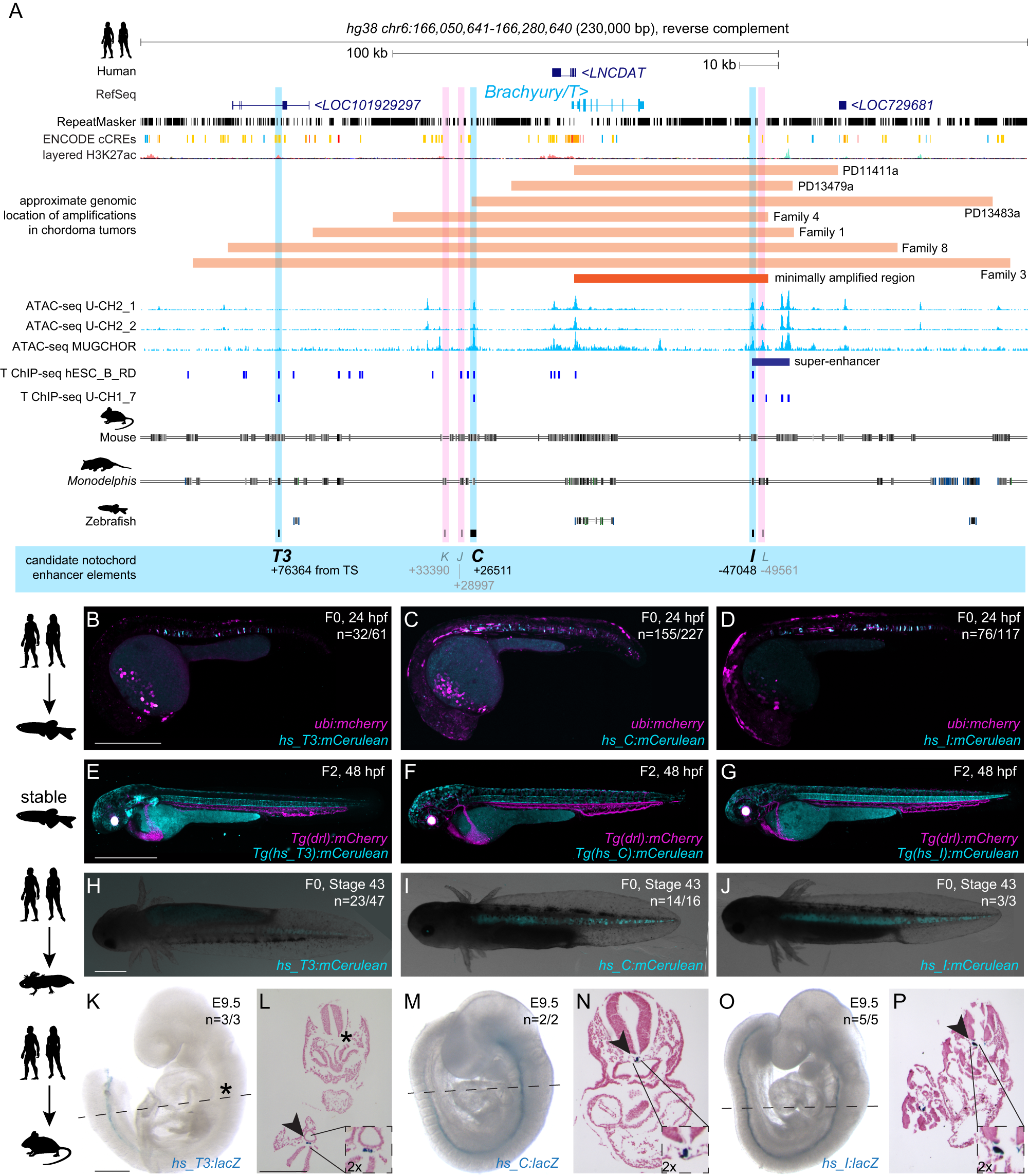
The cell type-specific expression of key transcription factors is central to development and disease. Brachyury/T/TBXT is a major transcription factor for gastrulation, tailbud patterning, and notochord formation; however, how its expression is controlled in the mammalian notochord has remained elusive. Here, we identify the complement of notochord-specific enhancers in the mammalian Brachyury/T/TBXT gene. Using transgenic assays in zebrafish, axolotl, and mouse, we discover three conserved Brachyury-controlling notochord enhancers, T3, C, and I, in human, mouse, and marsupial genomes. Acting as Brachyury-responsive, auto-regulatory shadow enhancers, in cis deletion of all three enhancers in mouse abolishes Brachyury/T/Tbxt expression selectively in the notochord, causing specific trunk and neural tube defects without gastrulation or tailbud defects. The three Brachyury-driving notochord enhancers are conserved beyond mammals in the brachyury/tbxtb loci of fishes, dating their origin to the last common ancestor of jawed vertebrates. Our data define the vertebrate enhancers for Brachyury/T/TBXTB notochord expression through an auto-regulatory mechanism that conveys robustness and adaptability as ancient basis for axis development.
BMP–Smad1/9 signaling plays a critical role in regulating zebrafish PGC proliferation
The germ cell fate in zebrafish is determined by germ plasm, whereas mammalian germ cell fate is induced by bone morphogenetic protein (BMP) signaling. It remains elusive whether BMP signaling is implicated in zebrafish germ cell development. Here, we demonstrate that BMP–Smad1/9 signaling plays a critical role in zebrafish primordial germ cell (PGC) maintenance rather than fate determination. BMP inhibition or smad1/9 knockdown reduces PGC numbers. Furthermore, we generated PGC-specific smad1/9 knockouts using a transgenic approach with PGC-specifically expressed Cas9 and ubiquitously expressed guide RNAs. Smad1/9 deficiency in PGCs leads to impaired PGC proliferation and increased apoptosis, consequently reducing PGC numbers. Transcriptome analysis revealed unchanged PGC-specific gene expression, but a marked upregulation of DNA damage response-related genes, which is validated by ectopic ATR–pChk1 activation in PGCs and PGC restoring by ATR inhibition. Collectively, these findings underscore conserved but functionally distinct roles of BMP signaling in vertebrate PGC development.

Segmental expression of Hoxb-1 is controlled by a highly conserved autoregulatory loop dependent upon exd/pbx
Comparison of Hoxb-1 regulatory regions from different vertebrates identified three related sequence motifs critical for rhombomere 4 (r4) expression in the hindbrain. Functional analysis in transgenic mice and Drosophila embryos demonstrated that the conserved elements are involved in a positive autoregulatory loop dependent on labial (lab) family members. Binding of Hoxb-1 to these elements in vitro requires cofactors, and the motifs closely resemble the consensus binding site for pbxi, a homolog of the Drosophila extradenticle (exd) homeodomain protein.
Osteonal and hemi‐osteonal remodeling: The spatial and temporal framework for signal traffic in adult human bone
Abstract The bone replacement process in the adult skeleton is known as remodeling. When bone is removed by osteoclasts, new bone is laid down by osteoblasts in the same place, because the load bearing requirement is unchanged. Bone is usually replaced because it is too old to carry out its function, which is mainly mechanical in cortical bone and mainly support for homeostasis and hematopoiesis in cancellous bone. Remodeling always begins on a quiescent bone surface, separated from the marrow by flat lining cells that are one of the two modes of terminal differentiation of osteoblasts. Lining cells are gatekeepers, able to be informed of the need for remodeling, and to either execute or mediate all four components of its activation‐selection and preparation of the site, recruitment of mononuclear preosteoclasts, budding of new capillaries, and attraction of preosteoclasts to the chosen site where they fuse into multinucleated osteoclasts. In cortical bone, osteonal remodeling is carried out by a complex and unique structure, the basic multicellular unit (BMU) that comprises a cutting cone of osteoclasts in front, a closing cone lined by osteoblasts following behind, and connective tissue, blood vessels and nerves filling the cavity. The BMU maintains its size, shape and internal organization for many months as it travels through bone in a controlled direction. Individual osteoclast nuclei are short‐lived, turning over about 8% per d, replaced by new preosteoclasts that originated in the bone marrow and travel in the circulation to the site of resorption. Refilling of bone at each successive cross‐sectional location is accomplished by a team of osteoblasts, probably originating from precursors within the local connective tissue, all assembled within a narrow window of time, at the right location, and in the right orientation to the surface. Each osteoblast team forms bone most rapidly at its onset and slows down progressively. Some of the osteoblasts are buried as osteocytes, some die, and the remainder gradually assume the shape of lining cells. Cancellous bone is more accessible to study than cortical bone, but is geometrically complex. Although remodeling conforms to the same sequence of surface activation, resorption and formation, its three‐dimensional organization is difficult to visualize from two‐dimensional histologic sections. But the average sizes of resorption sites, formation sites, and completed structural units increase progressively, as they do in cortical bone, indicating that the cancellous BMU travels across the surface digging a trench rather than a tunnel, but maintaining its size, shape and individual identity by the continuous recruitment of new cells, just as in cortical bone, a process that can be visualized as hemiosteonal remodeling. The conclusion that all remodeling is carried out by individual BMUs has important implications for bone biology, since many questions about how BMUs operate cannot be answered by studying either intact organisms or isolated cell systems. Many different steps in remodeling and many factors that influence each step have been identified, but very little is known about how the process is regulated in vivo to achieve its biologic purposes; most factors studied to date are likely permissive rather than regulatory in nature. Based on the proposed conceptual model of the BMU, much in vitro experimentation is relevant to the growth, modeling and repair of bone, but not to its remodeling in the adult skeleton. Further progress in the understanding of in vivo physiology will require the characterization of gene expression in individual cells to be related to the spatial and temporal organization of the BMU. This is likely to be possible only for osteonal remodeling in cortical bone in which, because of its geometric simplicity, individual BMUs can consistently be observed in two‐dimensional, longitudinal sections. © 1994 Wiley‐Liss, Inc.

Osteoclasts Provide Coupling Signals to Osteoblast Lineage Cells Through Multiple Mechanisms
Bone remodeling is essential for the repair and replacement of damaged and old bone. The major principle underlying this process is that osteoclast-mediated resorption of a quantum of bone is followed by osteoblast precursor recruitment; these cells differentiate to matrix-producing osteoblasts, which form new bone to replace what was resorbed. Evidence from osteopetrotic syndromes indicate that osteoclasts not only resorb bone, but also provide signals to promote bone formation. Osteoclasts act upon osteoblast lineage cells throughout their differentiation by facilitating growth factor release from resorbed matrix, producing secreted proteins and microvesicles, and expressing membrane-bound factors. These multiple mechanisms mediate the coupling of bone formation to resorption in remodeling. Additional interactions of osteoclasts with osteoblast lineage cells, including interactions with canopy and reversal cells, are required to achieve coordination between bone formation and resorption during bone remodeling.

Embryo-scale reverse genetics at single-cell resolution
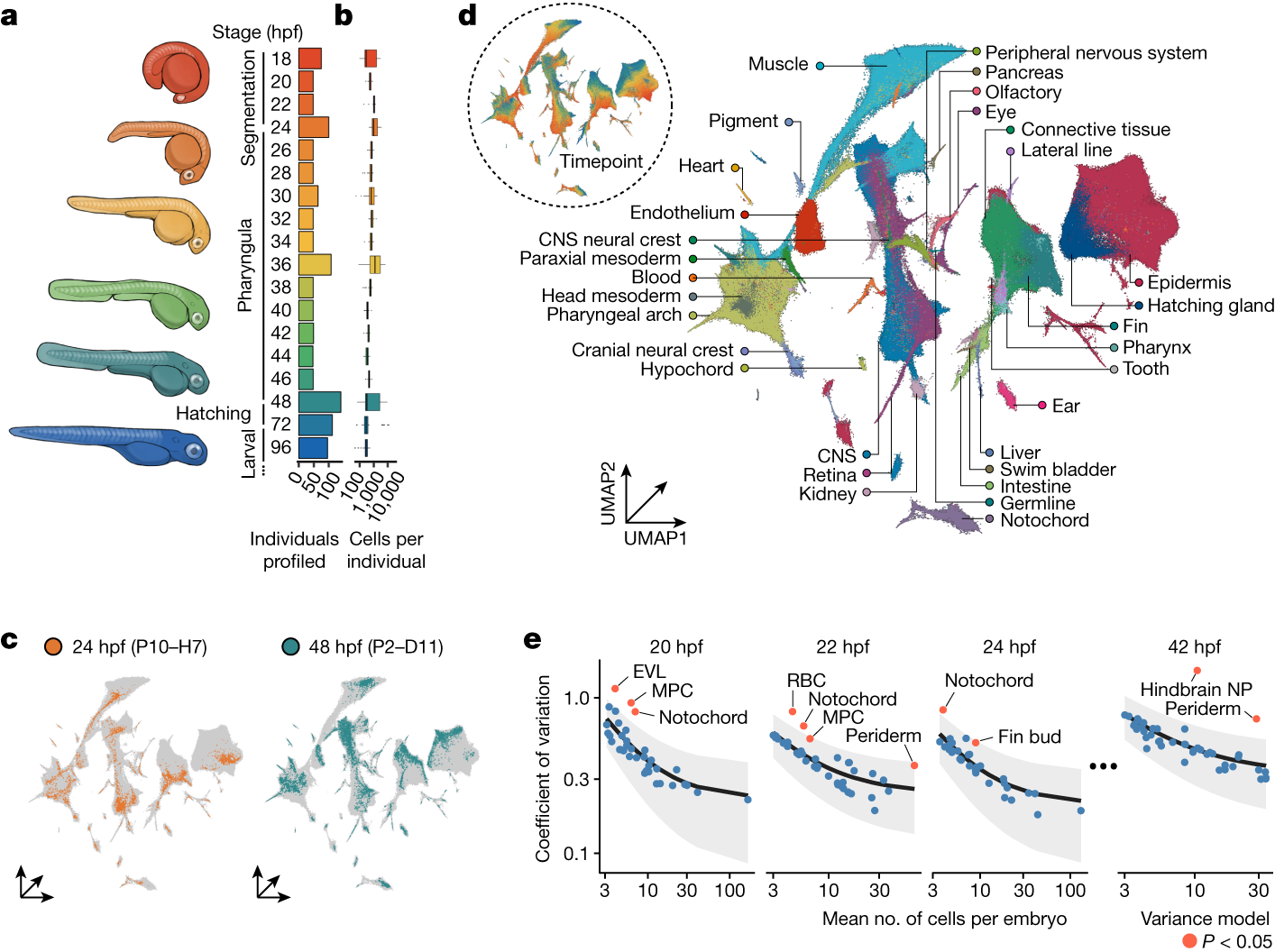
The maturation of single-cell transcriptomic technologies has facilitated the generation of comprehensive cellular atlases from whole embryos1–4. A majority of these data, however, has been collected from wild-type embryos without an appreciation for the latent variation that is present in development. Here we present the ‘zebrafish single-cell atlas of perturbed embryos’: single-cell transcriptomic data from 1,812 individually resolved developing zebrafish embryos, encompassing 19 timepoints, 23 genetic perturbations and a total of 3.2 million cells. The high degree of replication in our study (eight or more embryos per condition) enables us to estimate the variance in cell type abundance organism-wide and to detect perturbation-dependent deviance in cell type composition relative to wild-type embryos. Our approach is sensitive to rare cell types, resolving developmental trajectories and genetic dependencies in the cranial ganglia neurons, a cell population that comprises less than 1% of the embryo. Additionally, time-series profiling of individual mutants identified a group of brachyury-independent cells with strikingly similar transcriptomes to notochord sheath cells, leading to new hypotheses about early origins of the skull. We anticipate that standardized collection of high-resolution, organism-scale single-cell data from large numbers of individual embryos will enable mapping of the genetic dependencies of zebrafish cell types, while also addressing longstanding challenges in developmental genetics, including the cellular and transcriptional plasticity underlying phenotypic diversity across individuals.
Osteoblast-Osteoclast Communication and Bone Homeostasis
Bone remodeling is tightly regulated by a cross-talk between bone-forming osteoblasts and bone-resorbing osteoclasts. Osteoblasts and osteoclasts communicate with each other to regulate cellular behavior, survival and differentiation through direct cell-to-cell contact or through secretory proteins. A direct interaction between osteoblasts and osteoclasts allows bidirectional transduction of activation signals through EFNB2-EPHB4, FASL-FAS or SEMA3A-NRP1, regulating differentiation and survival of osteoblasts or osteoclasts. Alternatively, osteoblasts produce a range of different secretory molecules, including M-CSF, RANKL/OPG, WNT5A, and WNT16, that promote or suppress osteoclast differentiation and development. Osteoclasts also influence osteoblast formation and differentiation through secretion of soluble factors, including S1P, SEMA4D, CTHRC1 and C3. Here we review the current knowledge regarding membrane bound- and soluble factors governing cross-talk between osteoblasts and osteoclasts.

Dynamic Landscape Analysis of cell fate decisions provides predictive models of neural development from single-cell data
Building a mechanistic understanding of cell fate decisions remains a fundamental goal of developmental biology, with implications for stem cell therapies, regenerative medicine and understanding disease mechanisms. Single-cell transcriptomics provides a detailed picture of the cellular states observed during these decisions, but building dynamic and predictive models from these data remains a challenge. Here, we present dynamic landscape analysis (DLA), an integrative framework that applies dynamical systems theory to identify stable cell states, map transition pathways, and generate a predictive cell fate decision landscape from single-cell data. Applying this framework to vertebrate neural tube development revealed that progenitor specification by Sonic Hedgehog (Shh) can be captured in a landscape with an unexpected topology in which initially divergent lineages converge to the same fate through multiple distinct routes. The model accurately predicted cellular responses and cell fate allocation for unseen dynamic signalling regimes. Cross-species validation using human embryonic organoid data demonstrated conservation of this decision-making architecture. By modelling the dynamic responses that drive cell fate decisions, the DLA framework provides a quantitative and generative framework for extracting mechanistic insights from high-dimensional single-cell data.
Sequence Context‐Agnostic TadA‐Derived Cytosine Base Editors for Genome‐Wide Editing in Zebrafish
zTadA-CBEs, developed by introducing key mutations into TadA8e, enable efficient and precise sequence context-agnostic cytosine base editing. Specifically, zTadA-BE4max and zTadA-BEmv provide complem...

Transgene driving GFP expression from the promoter of the zona pellucida gene zpc is expressed in oocytes and provides an early marker for gonad differentiation in zebrafish
Although mechanisms of sex differentiation have been studied intensely in mammals, insects, and worms, little is known about this process in lower vertebrates. To establish a marker for female gonad ...

Rapid generation of maternal mutants via oocyte transgenic expression of CRISPR-Cas9 and sgRNAs in zebrafish
A time-saving and deletion-prone method facilitates functional study of maternal factors in zebrafish.

Osteocyte Wnt/β-Catenin Signaling Is Required for Normal Bone Homeostasis
β-Catenin-dependent canonical Wnt signaling plays an important role in bone metabolism by controlling differentiation of bone-forming osteoblasts and bone-resorbing osteoclasts. To investigate its ...
