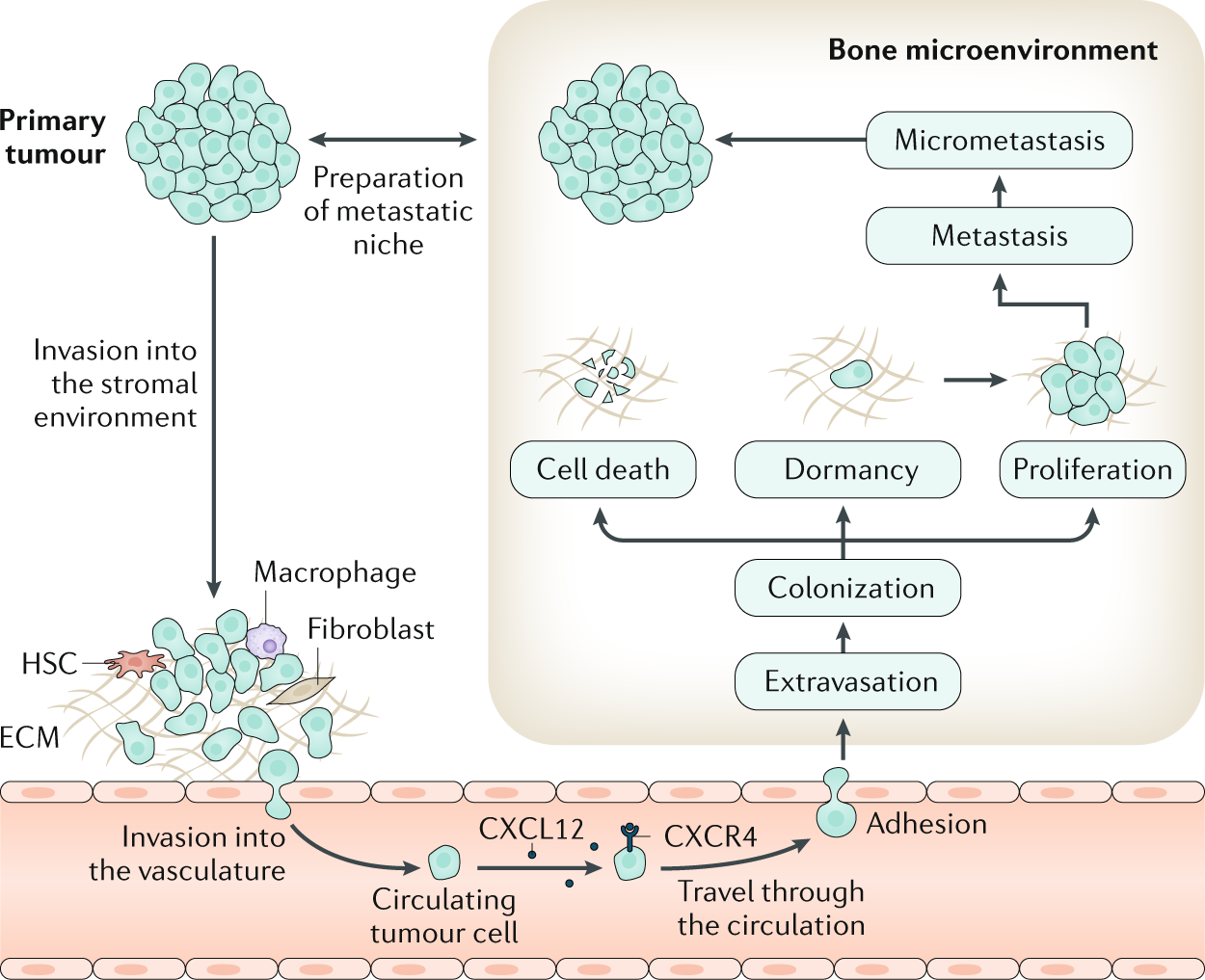
Bone metastases
Bone is the most frequent site for metastasis for many cancers, notably for tumours originating in the breast and the prostate. Tumour cells can escape from the primary tumour site and colonize the bone microenvironment. Within the bone, these disseminated tumour cells, as well as those arising in the context of multiple myeloma, may assume a state of dormancy, remaining quiescent for years before resuming proliferation and causing overt metastasis, which causes bone destruction via activation of osteoclast-mediated osteolysis. This structural damage can lead to considerable morbidity, including pain, fractures and impaired quality of life. Although treatment of bone metastases and myeloma bone disease is rarely curative, disease control is often possible for many years through the use of systemic anticancer treatments on a background of multidisciplinary supportive care. This care should include bone-targeted agents to inhibit tumour-associated osteolysis and prevent skeletal morbidity as well as use of appropriate local treatments such as radiation therapy, orthopaedic surgery and specialist palliative care to minimize the impact of metastatic bone disease on physical functioning. In this Primer, we provide an overview of the clinical features, the pathophysiology and the specific treatment approaches to prevent and treat bone metastases from solid tumours as well as myeloma bone disease.
Long-term use of denosumab and its association with skeletal-related events and osteonecrosis of the jaw
Denosumab, an inhibitor of receptor activator of nuclear factor kappa-B ligand, reduces skeletal-related events (SREs) and is approved for solid tumors with bone metastases. We studied long-term denosumab efficacy and safety because real-world data is scarce. This single-arm, single-center retrospective study included denosumab-treated breast cancer patients with bone metastases. Kaplan–Meier survival curves assessed exposure, SREs, osteonecrosis of the jaw (ONJ), and death. 132 patients were enrolled. The median denosumab exposure was 28.3 months (range 1.0–84.9). In the first year, 11.1% experienced SREs. This increased to 18.6% in the second, 21% in the third, and 35.1% in the fourth year and beyond. The median time to first on-study SRE has not been reached. 10 denosumab users (7.6%) developed ONJ. ONJ incidence was 0.9% in the first year, 6.2% in the second, 13.6% in the third, and 16.2% in subsequent years. The median time to first on-study ONJ has not been reached yet. Seven patients resumed denosumab after careful management of ONJ. Our data suggest that long-term treatment with denosumab may further prevent or postpone SREs at the cost of an increased risk of ONJ. The majority of patients who resumed denosumab did not experience a recurrence of ONJ.

Randomized, Double-Blind Study of Denosumab Versus Zoledronic Acid in the Treatment of Bone Metastases in Patients With Advanced Cancer (Excluding Breast and Prostate Cancer) or Multiple Myeloma
Purpose This study compared denosumab, a fully human monoclonal anti-receptor activator of nuclear factor kappa-B ligand antibody, with zoledronic acid (ZA) for delaying or preventing skeletal-related events (SRE) in patients with advanced cancer and bone metastases (excluding breast and prostate) or myeloma. Patients and Methods Eligible patients were randomly assigned in a double-blind, double-dummy design to receive monthly subcutaneous denosumab 120 mg (n = 886) or intravenous ZA 4 mg (dose adjusted for renal impairment; n = 890). Daily supplemental calcium and vitamin D were strongly recommended. The primary end point was time to first on-study SRE (pathologic fracture, radiation or surgery to bone, or spinal cord compression). Results Denosumab was noninferior to ZA in delaying time to first on-study SRE (hazard ratio, 0.84; 95% CI, 0.71 to 0.98; P = .0007). Although directionally favorable, denosumab was not statistically superior to ZA in delaying time to first on-study SRE (P = .03 unadjusted; P = .06 adjusted for multiplicity) or time to first-and-subsequent (multiple) SRE (rate ratio, 0.90; 95% CI, 0.77 to 1.04; P = .14). Overall survival and disease progression were similar between groups. Hypocalcemia occurred more frequently with denosumab. Osteonecrosis of the jaw occurred at similarly low rates in both groups. Acute-phase reactions after the first dose occurred more frequently with ZA, as did renal adverse events and elevations in serum creatinine based on National Cancer Institute Common Toxicity Criteria for Adverse Events grading. Conclusion Denosumab was noninferior (trending to superiority) to ZA in preventing or delaying first on-study SRE in patients with advanced cancer metastatic to bone or myeloma. Denosumab represents a potential novel treatment option with the convenience of subcutaneous administration and no requirement for renal monitoring or dose adjustment.

Denosumab Compared With Zoledronic Acid for the Treatment of Bone Metastases in Patients With Advanced Breast Cancer: A Randomized, Double-Blind Study
Purpose This randomized study compared denosumab, a fully human monoclonal antibody against receptor activator of nuclear factor κ B (RANK) ligand, with zoledronic acid in delaying or preventing skeletal-related events (SREs) in patients with breast cancer with bone metastases. Patients and Methods Patients were randomly assigned to receive either subcutaneous denosumab 120 mg and intravenous placebo (n = 1,026) or intravenous zoledronic acid 4 mg adjusted for creatinine clearance and subcutaneous placebo (n = 1,020) every 4 weeks. All patients were strongly recommended to take daily calcium and vitamin D supplements. The primary end point was time to first on-study SRE (defined as pathologic fracture, radiation or surgery to bone, or spinal cord compression). Results Denosumab was superior to zoledronic acid in delaying time to first on-study SRE (hazard ratio, 0.82; 95% CI, 0.71 to 0.95; P = .01 superiority) and time to first and subsequent (multiple) on-study SREs (rate ratio, 0.77; 95% CI, 0.66 to 0.89; P = .001). Reduction in bone turnover markers was greater with denosumab. Overall survival, disease progression, and rates of adverse events (AEs) and serious AEs were similar between groups. An excess of renal AEs and acute-phase reactions occurred with zoledronic acid; hypocalcemia occurred more frequently with denosumab. Osteonecrosis of the jaw occurred infrequently (2.0%, denosumab; 1.4%, zoledronic acid; P = .39). Conclusion Denosumab was superior to zoledronic acid in delaying or preventing SREs in patients with breast cancer metastatic to bone and was generally well tolerated. With the convenience of a subcutaneous injection and no requirement for renal monitoring, denosumab represents a potential treatment option for patients with bone metastases.

Systems Modeling of Bortezomib and Dexamethasone Combinatorial Effects on Bone Homeostasis in Multiple Myeloma Patients
Osteolytic bone disease is one of the most debilitating manifestations of multiple myeloma (MM). Bortezomib is a proteasome inhibitor that shows both anticancer and bone anabolic properties and is being evaluated for its positive effects in MM patients with skeletal complications. Dexamethasone is a potent corticosteroid that is often given in combination with bortezomib for its antineoplastic effects; however, bone loss and osteoporosis are major adverse effects of long-term steroid-based therapies.

Denosumab versus zoledronic acid in bone disease treatment of newly diagnosed multiple myeloma: an international, double-blind, double-dummy, randomised, controlled, phase 3 study
In patients with newly diagnosed multiple myeloma, denosumab was non-inferior to zoledronic acid for time to skeletal-related events. The results from this study suggest denosumab could be an additional option for the standard of care for patients with multiple myeloma with bone disease.

Denosumab versus zoledronic acid for treatment of bone metastases in men with castration-resistant prostate cancer: a randomised, double-blind study
Denosumab was better than zoledronic acid for prevention of skeletal-related events, and potentially represents a novel treatment option in men with bone metastases from castration-resistant prostate cancer.

Ten‐Year Simulation of the Effects of Denosumab on Bone Remodeling in Human Biopsies
ABSTRACT Postmenopausal osteoporosis is a disease manifesting in degradation of bone mass and microarchitecture, leading to weakening and increased risk of fracture. Clinical trials are an essential tool for evaluating new treatments and may provide further mechanistic understanding of their effects in vivo. However, the histomorphometry from clinical trials is limited to 2D images and reflects single time points. Biochemical markers of bone turnover give global insight into a drug's action, but not the local dynamics of the bone remodeling process and the cells involved. Additionally, comparative trials necessitate separate treatment groups, meaning only aggregated measures can be compared. In this study, in silico modeling based on histomorphometry and pharmacokinetic data was used to assess the effects of treatment versus control on μCT scans of the same biopsy samples over time, matching the changes in bone volume fraction observed in biopsies from denosumab and placebo groups through year 10 of the FREEDOM Extension trial. In the simulation, treatment decreased osteoclast number, which led to a modest increase in trabecular thickness and osteocyte stress shielding. Long‐term bone turnover suppression led to increased RANKL production, followed by a small increase in osteoclast number at the end of the 6‐month–dosing interval, especially at the end of the Extension study. Lack of treatment led to a significant loss of bone mass and structure. The study's results show how in silico models can generate predictions of denosumab cellular action over a 10‐year period, matching static and dynamic morphometric measures assessed in clinical biopsies. The use of in silico models with clinical trial data can be a method to gain further insight into fundamental bone biology and how treatments can perturb this. With rigorous validation, such models could be used for informing the design of clinical trials, such that the number of participants could be reduced to a minimum to show efficacy. © 2021 The Authors. JBMR Plus published by Wiley Periodicals LLC on behalf of American Society for Bone and Mineral Research.
Bisphosphonates: Mechanism of Action and Role in Clinical Practice
Bisphosphonates are primary agents in the current pharmacological arsenal against osteoclast-mediated bone loss due to osteoporosis, Paget disease of bone, malignancies metastatic to bone, multiple myeloma, and hypercalcemia of malignancy. In addition to currently approved uses, bisphosphonates are commonly prescribed for prevention and treatment of a variety of other skeletal conditions, such as low bone density and osteogenesis imperfecta. However, the recent recognition that bisphosphonate use is associated with pathologic conditions including osteonecrosis of the jaw has sharpened the level of scrutiny of the current widespread use of bisphosphonate therapy.

Physiological and pathophysiological bone turnover — role of the immune system
Receptor activator of NF-κB ligand (RANKL) and osteoprotegerin (OPG) are key downstream effectors of bone resorption; tumour necrosis factor (TNF) might synergize with RANKL to superinduce osteoclastic bone resorptionB cells, regulated by T cells, are a key source of basal OPG whereas activated T cells and B cells are key sources of TNF and RANKL in inflammatory conditionsShort-term antiresorptive therapies might safely prevent bone loss associated with combination antiretroviral therapyAnergic and parathyroid-hormone-treated T cells secrete the protein Wnt-10b, which promotes bone formationNovel therapeutic strategies targeting the immune system might promote bone formation and decrease bone resorption to manage osteoporotic bone loss and prevent fracture
Updates on Osteoimmunology: What's New on the Cross-Talk Between Bone and Immune System
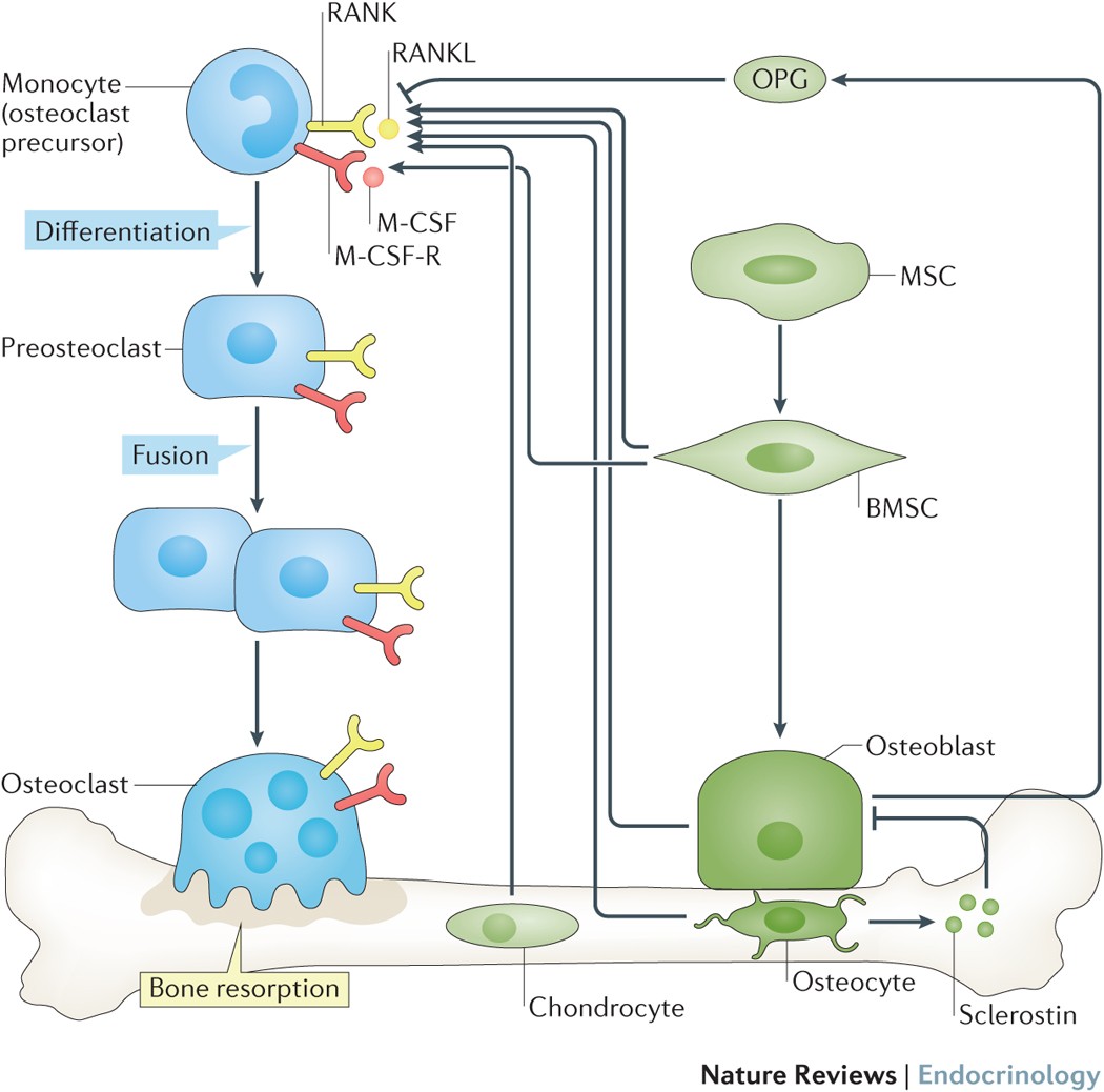
The term osteoimmunology was coined many years ago to describe the research field that deals with the cross-regulation between bone cells and the immune system. As a matter of fact, many factors that are classically considered immune-related, such as interleukins (i.e. IL-6, -11,-17 and -23), tumor necrosis factor (TNF)-α, receptor-activator of nuclear factor kappa B (RANK) and its ligand (RANKL), nuclear factor of activated t-cell, cytoplasmatic-1 (NFATc1) and others have all been found to be crucial in osteoclast and osteoblast biology. Conversely, bone cells, which we used to think would only regulate each other and take care of remodeling bone, actually regulate immune cells, by creating the so-called “endosteal niche”. Both osteoblasts and osteoclasts participate to this niche, either by favoring engraftment, or mobilization of hematopoietic stem cells (HSCs). In this review, we will describe the main milestones at the base of the osteoimmunology and present the key cellular players of the bone-immune system cross-talk, including HSCs, osteoblasts, osteoclasts, bone marrow macrophages, osteomacs, T- and B-lymphocytes, dendritic cells and neutrophils. We will also briefly describe some pathological conditions in which the bone-immune system cross-talk plays a crucial role, with the final aim to portray the state of the art in the mechanisms regulating the bone-immune system interplay, and some of the latest molecular players in the field. This is important to encourage investigation in this field, to identify new targets in the treatment of bone and immune diseases.

Prompting My Way Through Cancer
ChatGPT didn’t replace my care team, but it did make me a better patient.
Forecasting the critical role of intermittent therapies for the control of bone resorption
Osteoporosis is a chronic metabolic disease characterized by an imbalance of bone resorption and formation, leading to bone fragility and increased susceptibility to fracture. Parathyroid hormone is approved therapy for the treatment of osteoporosis.

Integrated model for denosumab and ibandronate pharmacodynamics in postmenopausal women
ABSTRACT This study aims to characterize the pharmacodynamic properties of denosumab, a RANK ligand inhibitor, and ibandronate, a bisphosphonate, using an integrated bone homeostasis model in postmenopausal women. Mean temporal profiles of denosumab, serum and urine N‐telopeptide (sNTX, uNTX), lumbar spine bone mineral density (BMD) following denosumab administration, and urine C‐telopeptide (uCTX) and lumbar spine BMD upon ibandronate administration were extracted from the literature. A mechanistic model was developed that integrates denosumab pharmacokinetics with binding to RANK ligand and ibandronate inhibition of osteoclast precursor differentiation to active osteoclasts (AOC). Biomarker concentrations were linked to the AOC pool. The BMD was characterized by a turnover model with stimulation of bone formation and degradation by AOB (active osteoblasts) and AOC pools. The estimated basal sNTX, uNTX and uCTX concentrations were 7.24 n m , 14.4 nmol/mmolCr and 31 µg/mmolCr. The BMD degradation rate was 0.00161 day ‐1 with stimulation constants associated with AOB and AOC of 1214 and 790 p m ‐1 . The plasma ibandronate concentration producing 50% of maximum inhibition of osteoclast differentiation was 522 ng/l. The integrated model, which incorporates multiple pathways of therapeutic intervention, quantitatively describes changes in clinical biomarkers of bone turnover and BMD after denosumab and ibandronate exposures in postmenopausal women. Copyright © 2011 John Wiley & Sons, Ltd.

Estrogen therapy for osteoporosis in the modern era
Menopause predisposes women to osteoporosis due to declining estrogen levels. This results in a decrease in bone mineral density (BMD) and an increase in fractures. Osteoporotic fractures lead to substantial morbidity and mortality, and are considered one of the largest public health priorities by the World Health Organization (WHO). It is therefore essential for menopausal women to receive appropriate guidance for the prevention and management of osteoporosis. The Women’s Health Initiative (WHI) randomized controlled trial first proved hormonal therapy (HT) reduces the incidence of all osteoporosis-related fractures in postmenopausal women. However, the study concluded that the adverse effects outweighed the potential benefits on bone, leading to a significant decrease in HT use for menopausal symptoms. Additionally, HT was not used as first-line therapy for osteoporosis and fractures. Subsequent studies have challenged these initial conclusions and have shown significant efficacy of HT in various doses, durations, regimens, and routes of administration. These studies support that HT improves BMD and reduces fracture risk in women with and without osteoporosis. Furthermore, the studies suggest that low-dose and transdermal HT are less likely associated with the adverse effects of breast cancer, endometrial hyperplasia, coronary artery disease (CAD), and venous thromboembolism (VTE) previously observed in standard-dose oral HT regimens. Given the need for estrogen in menopausal women and evidence supporting the cost effectiveness, safety, and efficacy of HT, we propose that HT should be considered for the primary prevention and treatment of osteoporosis in appropriate candidates. HT should be individualized and the once “lowest dose for shortest period of time” concept should no longer be used. This review will focus on the prior and current studies for various HT formulations used for the prevention and treatment of osteoporosis, exploring the safety profile of low-dose and transdermal HT that have been shown to be safer than oral standard-dose HT.
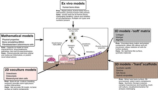
Modeling the Human Bone–Tumor Niche: Reducing and Replacing the Need for Animal Data
ABSTRACT Bone is the most common site for cancer metastasis. Understanding the interactions within the complex, heterogeneous bone–tumor microenvironment is essential for the development of new therapeutics. Various animal models of tumor‐induced bone disease are routinely used to provide valuable information on the relationship between cancer cells and the skeleton. However, new model systems exist that offer an alternative approach to the use of animals and might more accurately reveal the cellular interactions occurring within the human bone–tumor niche. This review highlights replacement models that mimic the bone microenvironment and where cancer metastases and tumor growth might be assessed alongside bone turnover. Such culture models include the use of calcified regions of animal tissue and scaffolds made from bone mineral hydroxyapatite, synthetic polymers that can be manipulated during manufacture to create structures resembling trabecular bone surfaces, gel composites that can be modified for stiffness and porosity to resemble conditions in the tumor–bone microenvironment. Possibly the most accurate model system involves the use of fresh human bone samples, which can be cultured ex vivo in the presence of human tumor cells and demonstrate similar cancer cell–bone cell interactions as described in vivo. In addition, the use of mathematical modeling and computational biology approaches provide an alternative to preliminary animal testing. The use of such models offers the capacity to mimic significant elements of the human bone–tumor environment, and complement, refine, or replace the use of preclinical models. © 2020 The Authors. JBMR Plus published by Wiley Periodicals, Inc. on behalf of American Society for Bone and Mineral Research.