


An anonymous reader writes "For the first time, there is no need to chemically fix, stain or cut cells in order to study them. Instead, whole living cells are fast-frozen and studied in their natural environment. The new method delivers an immediate 3-D image, thereby closing a gap between conventio...
Your Textbooks Are Wrong, This Is What Cells Actually Look Like
Allen Integrated Cell is a powerful tool for visualizing biology in 3D | TechCrunch
What does a cell look like? If you had to draw one, you'd probably do the usual thing: a sort of fried egg with a nucleus yolk and a couple ribosomes peppered around, maybe a rough endoplasmic reticulum if you're fancy. But cells are vastly more complicated than that, not to mention 3-dimensional. Allen Integrated Cell is a new tool that lets anyone visualize cells the way they actually exist in the body.

For the First Time, a Cell Built From Scratch Grows and Divides | Quanta Magazine
Scientists built a synthetic cell that combines more lifelike properties than ever before — proof of concept that it’s possible to bring nonliving materials to life, or something close to it, in the lab.
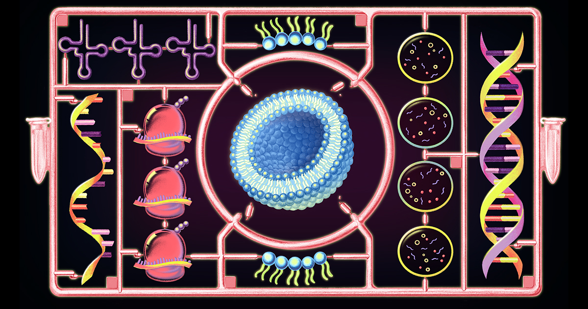
For the First Time, a Cell Built From Scratch Grows and Divides | Quanta Magazine
Scientists built a synthetic cell that combines more lifelike properties than ever before — proof of concept that it’s possible to bring nonliving materials to life, or something close to it, in the lab.
Researchers 3D-Print Heart From Human Patient's Cells - Slashdot
Researchers have 3D-printed a heart using a patient's cells, providing hope that the technique could be used to heal hearts or engineer new ones for transplants. "This is the first time anyone anywhere has successfully engineered and printed an entire heart replete with cells, blood vessels, ventric...

ConvNeXt Microscope — Medium · 7.06M — Neural Networks Are Already Clean
The evolving concept of cell identity in the single cell era
Summary: This Spotlight explores emerging technologies that are enabling the systematic and unbiased quantification of cell identity, and how these efforts will enable the construction of high-resolution, dynamic cell atlases.

Cease-fire at the leading edge: new perspectives on actin filament branching, debranching and cross-linking
Membrane protrusion at the leading edge of migrating cells is driven by the polymerization of actin. Recent studies using advanced imaging techniques raised a lively controversy about the morphology of these filaments; however, common ground between ...

The Open Microscopy Environment
A consortium of universities, research labs, industry and developers producing open-source software and format standards for microscopy data.

Researchers Have Successfully Grown Electrodes In Living Tissue - Slashdot
Researchers at Linkoping, Lund and Gothenburg universities in Sweden have successfully grown electrodes in living tissue using the body's molecules as triggers. The result, published in the journal Science, paves the way for the formation of fully integrated electronic circuits in living organisms. ...

A multimodal perturbation atlas defines the phenotypic resolution of cellular morphology
Because cells are complex dynamical systems, modeling cellular behaviors requires methods that capture how cells evolve across time, environments, and interventions. Microscopy is uniquely suited to this goal in that it can be applied to living cells in their native context. However, the phenotypic resolving power of live-cell microscopy remains incompletely characterized, particularly relative to molecular assays. Here, we present a multimodal perturbation atlas of 1,000 pooled CRISPR knockouts in A549 cells, profiled by fluorescence microscopy (39 live, 13 fixed markers), label-free phase imaging of the same live cells, and single-cell RNA sequencing (scRNA-seq). Totaling ∼57 million single-cell profiles, our data yield rich cell-biological signatures that map individual gene function. We find that phase imaging matches — and, with sufficient cell coverage, exceeds — the phenotypic resolution of fluorescence imaging and scRNA-seq, while capturing higher-order pathway organization that scRNA-seq does not resolve. These results establish intrinsic morphology as a high-precision readout of cellular state, and lay a foundation for live-cell profiling of phenotypic trajectories. ### Competing Interest Statement The authors have declared no competing interest. Biohub, Redwoood City, CA, USA

A periodic table of cell types
Summary: This Hypothesis proposes a periodic table that aligns cell types according to their developmental stages, connecting them to one another according to the universal axis from stem cells to differentiated cells.

A Cell So Minimal That It Challenges Definitions of Life | Quanta Magazine
The newly described microbe represents a world of parasitic, intercellular biodiversity only beginning to be revealed by genome sequencing.
Open-source software reveals complete 3D architecture of brain cells
The neurons in our brain that underlie thought connect to each other using tiny branch-like structures on their surfaces known as dendritic spines. Now scientists at Columbia's Zuckerman Institute and ...

These Science Photos Are So Beautiful They're Basically Art
The winners of the 2015 FASEB BioArt Image and Video Competition have been announced, and they’re amazing. Chosen from a diverse cross-section of biology,
