


Background Pseudo-bulk RNA-seq, generated by aggregating single-cell profiles, is widely used for benchmarking deconvolution methods because it globally approximates bulk transcriptomes and provides known cell-type proportions as ground truth. However, pseudo-bulk inherits single-cell specific measurement properties, and the extent to which these differ from real bulk RNA-seq remains difficult to quantify in the absence of paired data. In practice, deconvolution studies also commonly rely on large external single-cell references, yet the value of small, protocol-matched in-study references has not been systematically evaluated under real bulk conditions. These gaps motivate a paired benchmark that jointly examines pseudo-bulk fidelity, reference design, and their consequences for deconvolution accuracy. Results We establish a fully paired benchmarking framework using split-sample, donor-matched bulk and single-cell RNA-seq (scRNA-seq) from human bronchoalveolar lavage (BAL). Embedded within a reverse five-fold cross-validation design and evaluated on real bulk RNA-seq data, this framework benchmarks 15 deconvolution algorithms published in 2013-2025 across 3 cell-type resolutions and 5 single-cell references, including three published BAL datasets, a harmonized lung BAL atlas, and an in-study reference derived from paired aliquots. We show that real bulk and matched pseudo-bulk profiles exhibit systematic gene-level differences, identifying 557 reproducibly discordant genes (|log2 FC| > 1, FDR< 0.05 by LIMMA), including cell-type informative features. These discrepancies reflect technology-specific effects and violate the linear mixing assumption underlying most deconvolution methods. We demonstrate that a protocol-matched in-study reference constructed from only six donors consistently outperforms substantially larger external references, with the advantage that increases at finer cell-type resolution. Moreover, paired samples enable the identification and selective removal of discordant genes, which further improves deconvolution accuracy for many algorithms, particularly in high-resolution settings. These findings are robust across methods, references, and evaluation criteria and extend beyond compositional accuracy to improving recovery of disease-associated cell-type differences in clinical applications. Conclusions Our study provides the first fully paired benchmark of transcriptomic deconvolution on real bulk RNA-seq data of human BAL samples and demonstrates that reference design and data compatibility are as influential as algorithm choice. Beyond benchmarking, we introduce a practical and cost-effective protocol for deconvolution studies: generate single-cell data for a minimal subset of bulk samples (pilot pairing), use these data to construct an in-study reference and identify discordant genes, and apply the resulting insights to the full cohort. This strategy requires limited additional experimental effort yet yields substantial gains in accuracy and stability, offering actionable guidance for future bulk RNA-seq deconvolution studies across tissues and platforms. One-sentence summary Systematic benchmarking reveals pseudo-bulk biases and provides practical fixes for accurate RNA deconvolution.
Rectangle: robust and scalable multiscale deconvolution informed by single-cell RNA sequencing data
Bulk RNA-seq enables effective profiling of large cohorts and complex experimental designs, but current single-cell-informed deconvolution methods incompletely resolve closely related cell phenotypes, do not scale efficiently to large single-cell datasets, or fail to account for cellular content not represented in the reference. Here, we present Rectangle, an scverse Python framework for single-cell-informed deconvolution of bulk RNA-seq data.
An atlas-scale generative model for unified representation learning of bulk RNA-seq data
Public bulk RNA-seq repositories contain hundreds of thousands of samples, creating opportunities for large-scale representation learning, but integration across studies remains challenging because of heterogeneous annotations, experimental protocols, and technical variation. While pre-trained foundation models are now widely available for single-cell RNA-seq, comparable resources for bulk RNA-seq remain scarce, motivating a model that learns a unified, tissue-aware representation directly from bulk data. We trained a supervised variational autoencoder (VAE) on a compendium of 118,263 bulk RNA-seq samples that we assembled from TCGA, GTEx, and ARCHS4 and mapped to 42 tissue categories. The model classifies tissue of origin at 94.9% balanced accuracy (weighted F1 96.2%) and compresses 16,115 genes into a 121-dimensional latent space. Tissue identity is the primary organizing axis of the latent space, while source effects remain secondary. To assess the impact of data volume, we constructed training sets at three different scales (38K, 75K, and 118K samples). Our results demonstrated that reconstruction fidelity improved incrementally with each expansion of the dataset, but with diminishing returns. We validated the model on an independent cohort of 734 paediatric tumour samples from TARGET, achieving 84.6% agreement with the expected tissue of origin. The trained model and code are available at GitHub ([https://github.com/BIMSBbioinfo/flexynesis\_tissue\_vae_manuscript][1]) with an interactive web application. ### Competing Interest Statement The authors have declared no competing interest. [1]: https://github.com/BIMSBbioinfo/flexynesis_tissue_vae_manuscript
Realistic in silico generation and augmentation of single cell RNA-seq data using Generative Adversarial Neural Networks
A fundamental problem in biomedical research is the low number of observations available, mostly due to a lack of available biosamples, prohibitive costs, or ethical reasons. Augmenting few real observations with generated in silico samples could lead to more robust analysis results and a higher reproducibility rate. Here we propose the use of conditional single cell Generative Adversarial Neural Networks (cscGANs) for the realistic generation of single cell RNA-seq data. cscGANs learn non-linear gene-gene dependencies from complex, multi cell type samples and use this information to generate realistic cells of defined types. Augmenting sparse cell populations with cscGAN generated cells improves downstream analyses such as the detection of marker genes, the robustness and reliability of classifiers, the assessment of novel analysis algorithms, and might reduce the number of animal experiments and costs in consequence. cscGANs outperform existing methods for single cell RNA-seq data generation in quality and hold great promise for the realistic generation and augmentation of other biomedical data types. *
Hierarchical classification of immune cell transcriptomes at population-scale
Accurate immune cell classification is essential for interpreting single-cell RNA sequencing (scRNA-seq) data. However, progress is constrained by the lack of independent, high-resolution benchmarks, as the routine integration of datasets introduces statistical dependencies that artificially inflate model generalizability. Here, we present the single-cell universal classification omnibus (Suco), a resource of independent, uniform expert annotations, and Compocyte, a modular hierarchical classifier. Together, they establish a framework designed for the scale of human population immunology. This approach substantially outperforms existing classifiers while facilitating expert review of ambiguous annotations. Applying Compocyte across 50 studies, including three newly generated datasets, we classified 15.6 million leukocytes from 3,965 patients. Within this expansive cohort, we identified a new tumor-associated resorptive macrophage phenotype, a non-canonical monocyte subtype in subclinical cytokine release syndrome, and the programmatic erosion of T cell memory stemness across metastatic sites. Suco and Compocyte thus provide a generalizable architecture and benchmark capable of sustaining high-resolution annotation across massive clinical cohorts. ### Competing Interest Statement CMR has consulted regarding oncology drug development with Amgen, AstraZeneca, Daiichi Sankyo, Genentech, Merck, and Novartis, and has received licensing and royalty payments for DLL3-directed therapeutics. T.W. reports stock ownership for Roche, Astra Zeneca, Bayer, Innate Pharma, Kyntra, Illumina, 10x Genomics, and Merck KGaA as well as research funding from Atrandi Biosciences, Vilnius, Lithuania; CanVirex AG, Basel Switzerland; and Institut fuer Klinische Krebsforschung GmbH, Frankfurt, Germany, and travel funding from Roche, Basel, Switzerland. S.Z. reports advisory board membership and honoraria from Amgen, Astellas, AstraZeneca, Bayer, Bristol-Myers Squibb, Daiichi Sankyo, Eisai, EUSA, Gilead, Ipsen, Johnson&Johnson, Lilly, MedSir, Medtoday, Merck, MSD, Novartis, Pfizer, Roche, Sanofi Aventis, StreamedUp, Urotrials, Urotube, Zentiva and resarch funding from Eisai. S.Z. reports clinical trial support from Amgen, AstraZeneca, AVEO, Bayer, Biontech, Bristol-Myers Squibb, Calithera, Exelixis, Gilead, Lilly, MSD, Novartis, Pfizer, Roche, Seagen/Astellas, Urotrials and travels & conference support from Amgen, Astellas, AstraZeneca, Bayer, EISAI, Ipsen, Johnson&Johnson, Merck, MSD, Pfizer. All remaining authors declare no relevant competing interests. Spanish Association Against Cancer, PI049999 Federal Ministry of Research, Technology and Space, 001001KT2322 National Cancer Institute, R35 CA263816 National Cancer Institute, U24 CA213274 National Cancer Institute, P30 CA008748 Research Council of Lithuania, P-MIP-24-93
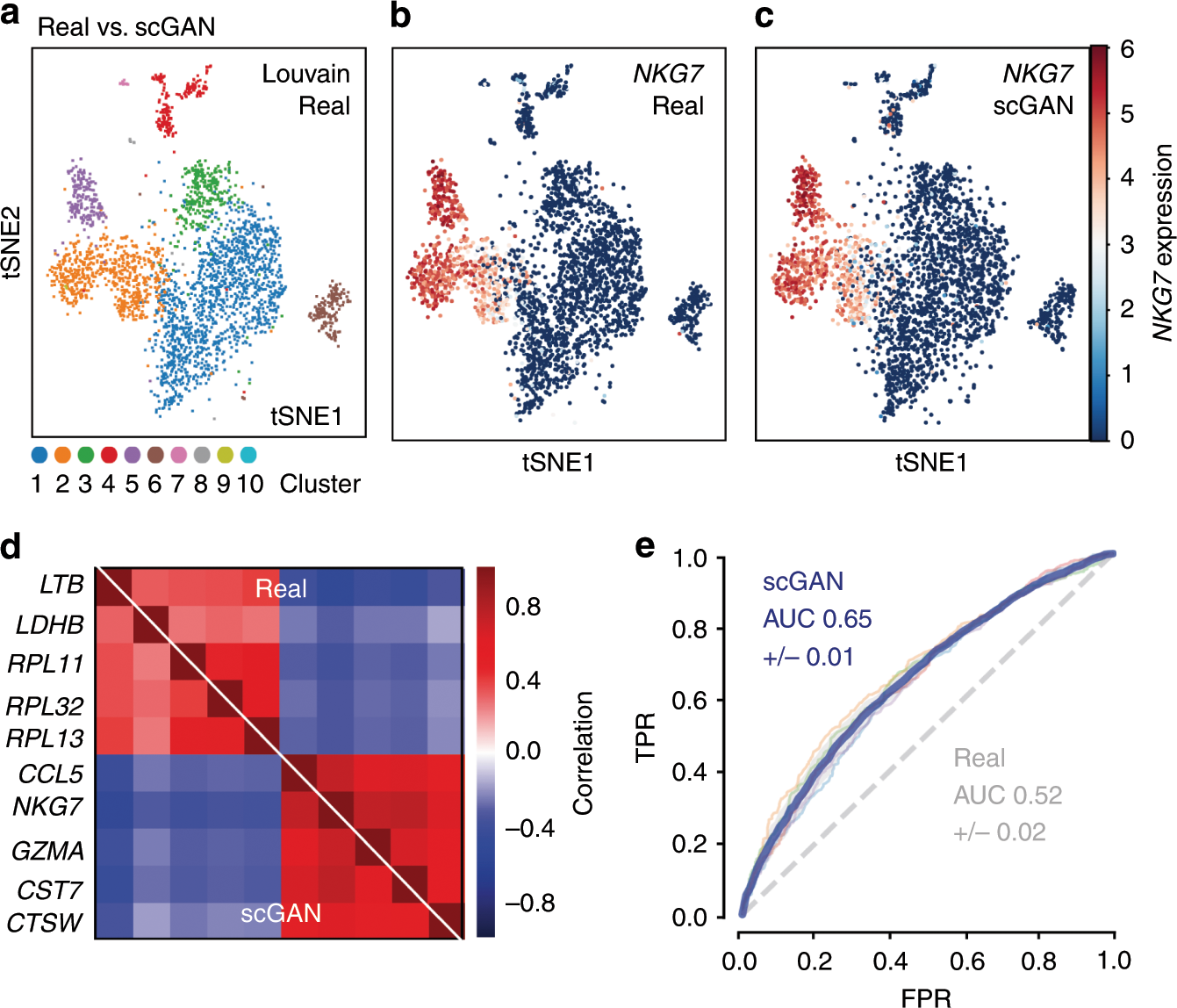
Realistic in silico generation and augmentation of single-cell RNA-seq data using generative adversarial networks
A fundamental problem in biomedical research is the low number of observations available, mostly due to a lack of available biosamples, prohibitive costs, or ethical reasons. Augmenting few real observations with generated in silico samples could lead to more robust analysis results and a higher reproducibility rate. Here, we propose the use of conditional single-cell generative adversarial neural networks (cscGAN) for the realistic generation of single-cell RNA-seq data. cscGAN learns non-linear gene–gene dependencies from complex, multiple cell type samples and uses this information to generate realistic cells of defined types. Augmenting sparse cell populations with cscGAN generated cells improves downstream analyses such as the detection of marker genes, the robustness and reliability of classifiers, the assessment of novel analysis algorithms, and might reduce the number of animal experiments and costs in consequence. cscGAN outperforms existing methods for single-cell RNA-seq data generation in quality and hold great promise for the realistic generation and augmentation of other biomedical data types.
Benchmarking gene expression reconstruction from single-cell latent representations
Single-cell transcriptomics is typically modeled in low-dimensional latent representations that improve the signal-to-noise ratio of the data. Such representations underpin data integration, cell state discovery, and perturbation prediction, with applications ranging from large-scale organ atlases to latent trajectory modeling. Recent virtual cell approaches further leverage these representations to predict cellular responses as distributional shifts in latent space. Each of these applications ultimately requires faithful gene expression reconstruction from latent spaces for biological interpretation, enabling gene-level analysis of predicted perturbed or batch-corrected cells. Yet representation choice is typically treated as an implementation detail rather than a primary modeling decision, with no systematic evaluation of how well latent representations support gene expression reconstruction. Here, we introduce ReconEval, a benchmark for evaluating gene expression reconstruction from single-cell latent spaces. We benchmark two classes of latent representations: end-to-end trained models such as PCA, autoencoders, and variational autoencoders, and pretrained single-cell foundation model embeddings coupled to newly trained decoders. Reconstruction is evaluated both directly and after latent-space perturbation prediction. Across perturbational and observational datasets totaling over 100 million cells, our metric suite quantifies statistical fidelity; biological signal preservation, including differential expression, coexpression, cell-cycle structure, cytokine response and pathway activity; and perturbation-specific effects. We find that autoencoders achieve the strongest stand-alone reconstruction at low dimensionality, while variational regularization does not improve generalization in reconstruction. Frozen foundation model embeddings retain recoverable gene-level information, with reconstruction quality depending strongly on decoder architecture and pretraining objective. In latent perturbation modeling, high-dimensional PCA matches foundation model embeddings, while low-dimensional AE embeddings are optimal for flow-based generative models. Overall, reconstruction depends critically on the interplay between representation and downstream model, and simpler representations can outperform complex alternatives given appropriate capacity. Our benchmark establishes reconstruction as a critical evaluation axis for single-cell foundation models. We envision it improving the biological interpretability of latent-space modeling, a prerequisite for future virtual cell models to be validated by domain experts and grounded in biology. ### Competing Interest Statement F.J.T. consults for Immunai, CytoReason, Valinor Industries, Bioturing and Phylo Inc., and has ownership interest in RN.AI Therapeutics, Dermagnostix, and Cellarity. The remaining authors declare no competing interests.
Comparing phenotypic manifolds with Kompot: Detecting differential abundance and gene expression at single-cell resolution
Single-cell studies are frequently designed to compare across conditions such as health and disease. However, existing computational approaches typically rely on grouping cells into discrete populations before making comparisons, which can limit resolution for detecting state-dependent changes. Here, we introduce Kompot, a statistical framework for comparative analysis of multi-condition single-cell data. Kompot quantifies both differential abundance, capturing how cells redistribute across the phenotypic space, and differential expression, identifying condition-specific transcriptional changes that may be localized, heterogeneous, or oppositely regulated across states. By modeling cell density and gene expression as continuous functions over a shared cell-state representation, Kompot enables single-cell–resolution inference with principled uncertainty estimates, without requiring predefined clusters or cell types. Applying Kompot to aging murine bone marrow, we identified a continuum of shifts in hematopoietic stem cell and mature cell states, transcriptional remodeling of monocytes independent of compositional changes, and divergent regulation of oxidative stress response genes across cell types. We demonstrate the utility of Kompot in disease settings by identifying cell-state and gene expression changes associated with improved efficacy of combinatorial immunotherapy in melanoma. Additionally, Kompot enables multi-sample comparative analysis by accounting for sample-to-sample heterogeneity. By capturing both global and cell-state–specific effects of perturbation, the Kompot framework is broadly applicable to dissecting condition-specific effects in complex single-cell landscapes. ### Competing Interest Statement The authors have declared no competing interest. National Institutes of Health, R35GM147125, R01CA292932, T32GM136534, S10OD028685 The Mark Foundation for Cancer Research, https://ror.org/00v7th354, Endeavor Award Edward P. Evans Foundation, https://ror.org/03h22gm35, Discovery Research Grant Brotman Baty Institute, https://ror.org/03jxvbk42, Pilot Award
Designing RNA sequencing experiments: A practical guide to reproducible gene expression analysis
RNA sequencing (RNA-seq) has become a cornerstone of modern biotechnology, offering a comprehensive and high-resolution view of gene expression that enables the discovery of novel transcripts across diverse biological systems. Its applications extend beyond basic transcriptomics, providing powerful tools for uncovering molecular mechanisms underlying disease, environmental responses, and chemical toxicity. In biotechnology and biomedical research, RNA-seq facilitates the identification of regulatory networks and biomarkers that inform therapeutic development, risk assessment, and precision medicine.

A multimodal perturbation atlas defines the phenotypic resolution of cellular morphology
Because cells are complex dynamical systems, modeling cellular behaviors requires methods that capture how cells evolve across time, environments, and interventions. Microscopy is uniquely suited to this goal in that it can be applied to living cells in their native context. However, the phenotypic resolving power of live-cell microscopy remains incompletely characterized, particularly relative to molecular assays. Here, we present a multimodal perturbation atlas of 1,000 pooled CRISPR knockouts in A549 cells, profiled by fluorescence microscopy (39 live, 13 fixed markers), label-free phase imaging of the same live cells, and single-cell RNA sequencing (scRNA-seq). Totaling ∼57 million single-cell profiles, our data yield rich cell-biological signatures that map individual gene function. We find that phase imaging matches — and, with sufficient cell coverage, exceeds — the phenotypic resolution of fluorescence imaging and scRNA-seq, while capturing higher-order pathway organization that scRNA-seq does not resolve. These results establish intrinsic morphology as a high-precision readout of cellular state, and lay a foundation for live-cell profiling of phenotypic trajectories. ### Competing Interest Statement The authors have declared no competing interest. Biohub, Redwoood City, CA, USA
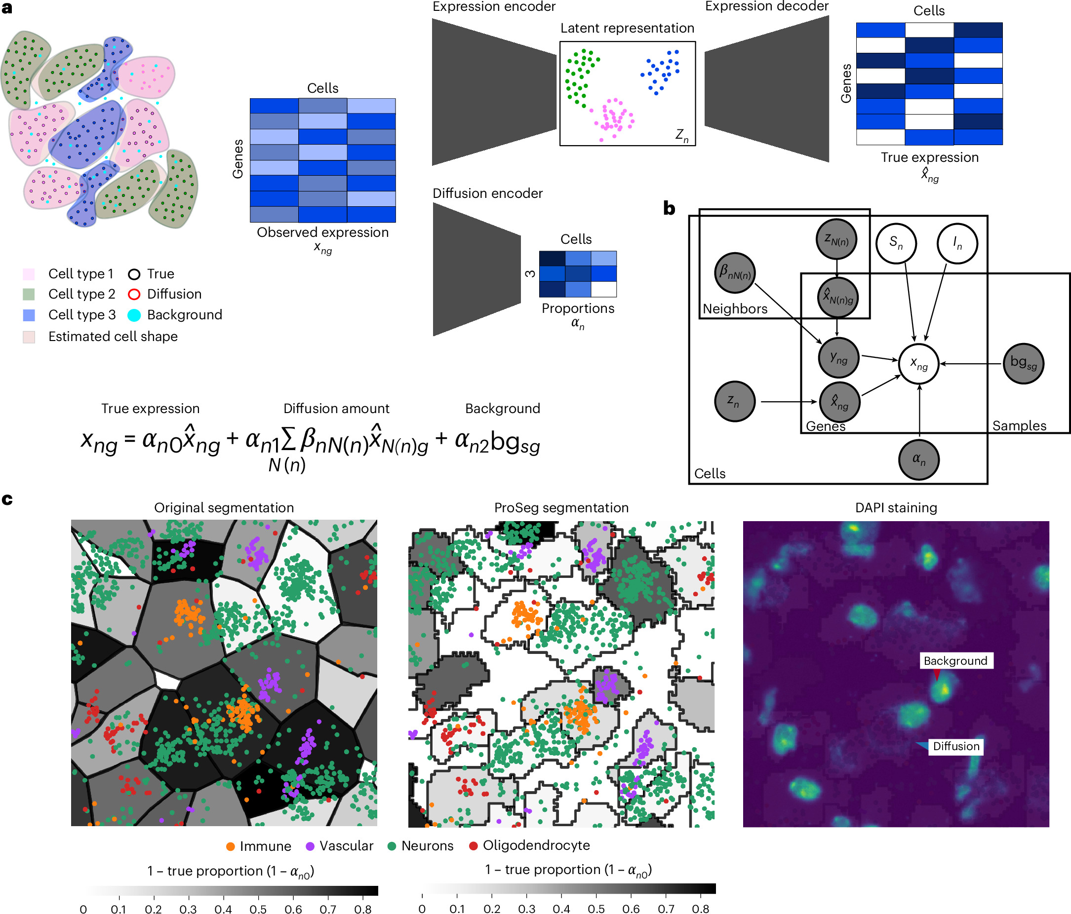
ResolVI: addressing noise and bias in spatial transcriptomics
Technologies for estimating RNA expression at high throughput, in intact tissue slices and with high spatial resolution (spatial transcriptomics) shed new light on how cells communicate and tissues function. A fundamental step in analyzing data generated by subcellular resolution spatial transcriptomics technologies is quantification, namely, segmenting the plane into regions, each approximating a cell, and then collating the molecules inside each region to estimate the cellular expression profile. Despite many advances in this area, a persistent problem is that of the incorrect assignment of molecules to cells, which limits many current applications to the level of a priori-defined cell subsets and complicates the discovery of novel cell states. Here we develop resolVI, a model that operates downstream of any segmentation algorithm to generate a probabilistic representation, correcting for the misassignment of molecules, as well as for batch effects and other nuisance factors. We demonstrate that resolVI improves our ability to distinguish between cell states, identify subtle expression changes in space and perform integrated analysis across datasets. ResolVI is available as open source software within scvi-tools.
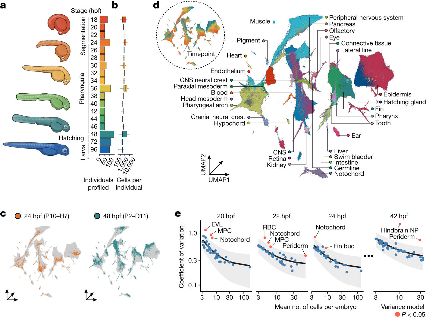
Embryo-scale reverse genetics at single-cell resolution
The maturation of single-cell transcriptomic technologies has facilitated the generation of comprehensive cellular atlases from whole embryos1–4. A majority of these data, however, has been collected from wild-type embryos without an appreciation for the latent variation that is present in development. Here we present the ‘zebrafish single-cell atlas of perturbed embryos’: single-cell transcriptomic data from 1,812 individually resolved developing zebrafish embryos, encompassing 19 timepoints, 23 genetic perturbations and a total of 3.2 million cells. The high degree of replication in our study (eight or more embryos per condition) enables us to estimate the variance in cell type abundance organism-wide and to detect perturbation-dependent deviance in cell type composition relative to wild-type embryos. Our approach is sensitive to rare cell types, resolving developmental trajectories and genetic dependencies in the cranial ganglia neurons, a cell population that comprises less than 1% of the embryo. Additionally, time-series profiling of individual mutants identified a group of brachyury-independent cells with strikingly similar transcriptomes to notochord sheath cells, leading to new hypotheses about early origins of the skull. We anticipate that standardized collection of high-resolution, organism-scale single-cell data from large numbers of individual embryos will enable mapping of the genetic dependencies of zebrafish cell types, while also addressing longstanding challenges in developmental genetics, including the cellular and transcriptional plasticity underlying phenotypic diversity across individuals.
Spatial Transcriptomics As Rasterized Image Tensors (STARIT) characterizes cell states with subcellular molecular heterogeneity
Imaging-based spatially resolved transcriptomics (imSRT) technologies provide high-throughput molecular-resolution spatial characterization of genes within cells. Conventional analysis methods to identify cell-types and states in imSRT data rely on gene count matrices derived from tallying the number of mRNA molecules detected for each gene per segmented cell, thereby overlooking subcellular heterogeneity that can be useful in defining cell states. To take advantage of the molecular-resolution information in imSRT data and potentially identify cell-states based on subcellular heterogeneity, we developed STARIT (Spatial Transcriptomics As Rasterized Image Tensors). STARIT converts transcripts within segmented cells in imSRT data into an image-based tensor representation that can be combined with deep learning computer vision models for downstream analysis. Using simulated and real imSRT data, we demonstrate that STARIT distinguishes transcriptionally distinct cell-types and further separates cell states based on subcellular transcript localization, which conventional gene count analysis fails to capture. By providing a standardized framework to encode subcellular molecular information in imSRT data, STARIT will enable deeper insights into subcellular heterogeneity and enhance the identification and characterization of cell-types and states that are overlooked by gene count representations.
The evolving concept of cell identity in the single cell era
Summary: This Spotlight explores emerging technologies that are enabling the systematic and unbiased quantification of cell identity, and how these efforts will enable the construction of high-resolution, dynamic cell atlases.
Dynamic Landscape Analysis of cell fate decisions provides predictive models of neural development from single-cell data
Building a mechanistic understanding of cell fate decisions remains a fundamental goal of developmental biology, with implications for stem cell therapies, regenerative medicine and understanding disease mechanisms. Single-cell transcriptomics provides a detailed picture of the cellular states observed during these decisions, but building dynamic and predictive models from these data remains a challenge. Here, we present dynamic landscape analysis (DLA), an integrative framework that applies dynamical systems theory to identify stable cell states, map transition pathways, and generate a predictive cell fate decision landscape from single-cell data. Applying this framework to vertebrate neural tube development revealed that progenitor specification by Sonic Hedgehog (Shh) can be captured in a landscape with an unexpected topology in which initially divergent lineages converge to the same fate through multiple distinct routes. The model accurately predicted cellular responses and cell fate allocation for unseen dynamic signalling regimes. Cross-species validation using human embryonic organoid data demonstrated conservation of this decision-making architecture. By modelling the dynamic responses that drive cell fate decisions, the DLA framework provides a quantitative and generative framework for extracting mechanistic insights from high-dimensional single-cell data.
Relationships between type 2 diabetes, cell dysfunction, and redox signaling: A meta‐analysis of single‐cell gene expression of human pancreatic α‐ and β‐cells
Abstract Background Type 2 diabetes mellitus (T2DM) is a chronic disease characterized by insulin resistance and failure of β‐cells to meet the metabolic demand for insulin. Recent advances in single‐cell RNA sequencing (sc‐RNA‐Seq) have allowed for in‐depth studies to further understand the underlying cellular mechanisms of T2DM. In β‐cells, redox signaling is critical for insulin production. A meta‐analysis of human pancreas islet sc‐RNA‐Seq data was conducted to evaluate how T2DM may modify the transcriptomes of α‐ and β‐cells. Methods Annotated sc‐RNA‐Seq data from six studies of human pancreatic islets from metabolically healthy and donors with T2DM were collected. α‐ and β‐cells, subpopulations of proliferating α‐cells, immature, and senescent β‐cells were identified based on expression levels of key marker genes. Each dataset was analyzed individually before combining, using weighted comparisons. Pathways of significant genes and individual redox‐related gene expression were then evaluated to further understand the role that redox signaling may play in T2DM‐induced β‐cell dysfunction. Results α‐ and β‐cells from T2DM donors modified genes involved in energy metabolism, immune response, autophagy, and cellular stress. α‐ and β‐cells also had an increased nuclear factor erythroid 2‐related factor 2 (NFE2L2)‐mediated antioxidant response in T2DM donors. The proportion of immature and senescent β‐cells increased in T2DM donors, and in immature and senescent β‐cells, genes regulated by NFE2L2 were further upregulated. Conclusions These findings suggest that NFE2L2 plays a role in β‐cell maturation and dysfunction. Redox singling maybe a key pathway for β‐cell restoration and T2DM therapeutics. , 摘要 背景 2型糖尿病(T2D)是一种以胰岛素抵抗和β细胞无法满足胰岛素代谢需求为特征的慢性疾病。近年来在单细胞RNA测序(sc‐RNA‐Seq)方面的进展为进一步深入研究T2D的细胞机制奠定了基础。在β细胞中, 氧化还原信号对胰岛素的产生至关重要。本研究对人胰岛sc‐RNA‐Seq数据进行 meta 分析, 以评估T2D如何调控α‐和β‐细胞的转录组。 方法 收集6项研究的sc‐RNA‐Seq数据, 这些数据来自代谢正常的人胰岛和T2D供者。根据关键标记基因的表达水平鉴定α‐细胞和β‐细胞、增殖中的α‐细胞亚群、未成熟和衰老的β‐细胞。在使用加权比较进行组合之前, 每个数据集都被单独分析。然后评估重要基因的通路和单个氧化还原相关基因的表达, 以进一步了解氧化还原信号在T2D诱导的β‐细胞功能障碍中可能扮演的角色 结果 来自T2D供者的α‐和β‐细胞修饰了与能量代谢、免疫反应、自噬和细胞应激相关的基因, 在T2D供者中α‐和β‐细胞NFE2L2介导的抗氧化反应增强。在T2D供者中, 未成熟和衰老的β‐细胞比例增加, 并且在未成熟和衰老的β‐细胞中, 受NFE2L2调控的基因进一步上调。 结论 这些发现提示NFE2L2在β‐细胞成熟和功能障碍中起作用。氧化还原信号通路可能是β‐细胞修复和T2D治疗的关键途径。

Accelerating scientific discovery with Co-Scientist
Scientific discovery is driven by scientists generating novel hypotheses for complex problems that undergo rigorous experimental validation. To augment this process, we introduce Co-Scientist, a multi-agent AI system built on Gemini for structured scientific thinking and hypothesis generation. Co-Scientist aims to help scientists discover new original knowledge. Conditioned on their research objectives and prior scientific evidence, it formulates demonstrably novel research hypotheses for experimental verification. The system's design involves agents continuously generating, critiquing and refining hypotheses accelerated by scaling test-time compute. Key contributions include: (1) a multi-agent architecture with an asynchronous task execution framework for flexible compute scaling; (2) a tournament evolution process for self-improving hypotheses generation. Automated evaluations show continued benefits of test-time compute scaling, improving hypothesis quality over time. While general purpose, we focus the validation in three biomedical applications: drug repurposing, novel target discovery, and explaining mechanisms of anti-microbial resistance. Specifically, Co-Scientist helped identify new drug repurposing candidates and synergistic combination therapies for acute myeloid leukemia, which were validated through in vitro experiments. These real-world validations demonstrate the potential of Co-Scientist to accelerate scientific discovery and usher in an era of AI empowered scientists.
