


Accurate immune cell classification is essential for interpreting single-cell RNA sequencing (scRNA-seq) data. However, progress is constrained by the lack of independent, high-resolution benchmarks, as the routine integration of datasets introduces statistical dependencies that artificially inflate model generalizability. Here, we present the single-cell universal classification omnibus (Suco), a resource of independent, uniform expert annotations, and Compocyte, a modular hierarchical classifier. Together, they establish a framework designed for the scale of human population immunology. This approach substantially outperforms existing classifiers while facilitating expert review of ambiguous annotations. Applying Compocyte across 50 studies, including three newly generated datasets, we classified 15.6 million leukocytes from 3,965 patients. Within this expansive cohort, we identified a new tumor-associated resorptive macrophage phenotype, a non-canonical monocyte subtype in subclinical cytokine release syndrome, and the programmatic erosion of T cell memory stemness across metastatic sites. Suco and Compocyte thus provide a generalizable architecture and benchmark capable of sustaining high-resolution annotation across massive clinical cohorts.
Comparing phenotypic manifolds with Kompot: Detecting differential abundance and gene expression at single-cell resolution
Single-cell studies are frequently designed to compare across conditions such as health and disease. However, existing computational approaches typically rely on grouping cells into discrete populations before making comparisons, which can limit resolution for detecting state-dependent changes. Here, we introduce Kompot, a statistical framework for comparative analysis of multi-condition single-cell data. Kompot quantifies both differential abundance, capturing how cells redistribute across the phenotypic space, and differential expression, identifying condition-specific transcriptional changes that may be localized, heterogeneous, or oppositely regulated across states. By modeling cell density and gene expression as continuous functions over a shared cell-state representation, Kompot enables single-cell–resolution inference with principled uncertainty estimates, without requiring predefined clusters or cell types. Applying Kompot to aging murine bone marrow, we identified a continuum of shifts in hematopoietic stem cell and mature cell states, transcriptional remodeling of monocytes independent of compositional changes, and divergent regulation of oxidative stress response genes across cell types. We demonstrate the utility of Kompot in disease settings by identifying cell-state and gene expression changes associated with improved efficacy of combinatorial immunotherapy in melanoma. Additionally, Kompot enables multi-sample comparative analysis by accounting for sample-to-sample heterogeneity. By capturing both global and cell-state–specific effects of perturbation, the Kompot framework is broadly applicable to dissecting condition-specific effects in complex single-cell landscapes. ### Competing Interest Statement The authors have declared no competing interest. National Institutes of Health, R35GM147125, R01CA292932, T32GM136534, S10OD028685 The Mark Foundation for Cancer Research, https://ror.org/00v7th354, Endeavor Award Edward P. Evans Foundation, https://ror.org/03h22gm35, Discovery Research Grant Brotman Baty Institute, https://ror.org/03jxvbk42, Pilot Award
Accurate and scalable multi-disease classification from adaptive immune repertoires
Summary BackgroundMachine learning models trained on paratope-similarity networks have shown superior accuracy compared with clonotype-based models in binary disease classification. However, the computational demands of paratope networks hinder their use on large datasets and multi-disease classification.MethodsWe reanalyzed publicly available T cell receptor (TCR) repertoire data from 1,421 donors across 15 disease groups and a large control group, encompassing approximately 81 million TCR sequences.
Rectangle: robust and scalable multiscale deconvolution informed by single-cell RNA sequencing data
Bulk RNA-seq enables effective profiling of large cohorts and complex experimental designs, but current single-cell-informed deconvolution methods incompletely resolve closely related cell phenotypes, do not scale efficiently to large single-cell datasets, or fail to account for cellular content not represented in the reference. Here, we present Rectangle, an scverse Python framework for single-cell-informed deconvolution of bulk RNA-seq data.
An atlas-scale generative model for unified representation learning of bulk RNA-seq data
Public bulk RNA-seq repositories contain hundreds of thousands of samples, creating opportunities for large-scale representation learning, but integration across studies remains challenging because of heterogeneous annotations, experimental protocols, and technical variation. While pre-trained foundation models are now widely available for single-cell RNA-seq, comparable resources for bulk RNA-seq remain scarce, motivating a model that learns a unified, tissue-aware representation directly from bulk data. We trained a supervised variational autoencoder (VAE) on a compendium of 118,263 bulk RNA-seq samples that we assembled from TCGA, GTEx, and ARCHS4 and mapped to 42 tissue categories. The model classifies tissue of origin at 94.9% balanced accuracy (weighted F1 96.2%) and compresses 16,115 genes into a 121-dimensional latent space. Tissue identity is the primary organizing axis of the latent space, while source effects remain secondary. To assess the impact of data volume, we constructed training sets at three different scales (38K, 75K, and 118K samples). Our results demonstrated that reconstruction fidelity improved incrementally with each expansion of the dataset, but with diminishing returns. We validated the model on an independent cohort of 734 paediatric tumour samples from TARGET, achieving 84.6% agreement with the expected tissue of origin. The trained model and code are available at GitHub ([https://github.com/BIMSBbioinfo/flexynesis\_tissue\_vae_manuscript][1]) with an interactive web application. ### Competing Interest Statement The authors have declared no competing interest. [1]: https://github.com/BIMSBbioinfo/flexynesis_tissue_vae_manuscript
Realistic in silico generation and augmentation of single cell RNA-seq data using Generative Adversarial Neural Networks
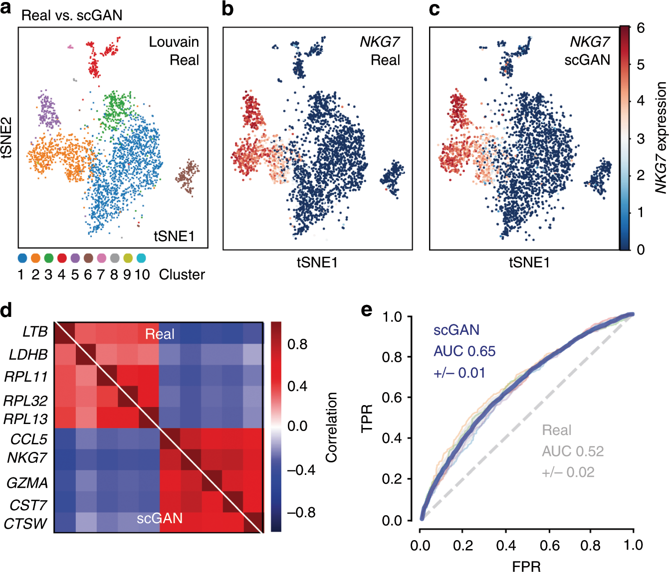
A fundamental problem in biomedical research is the low number of observations available, mostly due to a lack of available biosamples, prohibitive costs, or ethical reasons. Augmenting few real observations with generated in silico samples could lead to more robust analysis results and a higher reproducibility rate. Here we propose the use of conditional single cell Generative Adversarial Neural Networks (cscGANs) for the realistic generation of single cell RNA-seq data. cscGANs learn non-linear gene-gene dependencies from complex, multi cell type samples and use this information to generate realistic cells of defined types. Augmenting sparse cell populations with cscGAN generated cells improves downstream analyses such as the detection of marker genes, the robustness and reliability of classifiers, the assessment of novel analysis algorithms, and might reduce the number of animal experiments and costs in consequence. cscGANs outperform existing methods for single cell RNA-seq data generation in quality and hold great promise for the realistic generation and augmentation of other biomedical data types. *
Realistic in silico generation and augmentation of single-cell RNA-seq data using generative adversarial networks
A fundamental problem in biomedical research is the low number of observations available, mostly due to a lack of available biosamples, prohibitive costs, or ethical reasons. Augmenting few real observations with generated in silico samples could lead to more robust analysis results and a higher reproducibility rate. Here, we propose the use of conditional single-cell generative adversarial neural networks (cscGAN) for the realistic generation of single-cell RNA-seq data. cscGAN learns non-linear gene–gene dependencies from complex, multiple cell type samples and uses this information to generate realistic cells of defined types. Augmenting sparse cell populations with cscGAN generated cells improves downstream analyses such as the detection of marker genes, the robustness and reliability of classifiers, the assessment of novel analysis algorithms, and might reduce the number of animal experiments and costs in consequence. cscGAN outperforms existing methods for single-cell RNA-seq data generation in quality and hold great promise for the realistic generation and augmentation of other biomedical data types.
Dynamic Landscape Analysis of cell fate decisions provides predictive models of neural development from single-cell data
Building a mechanistic understanding of cell fate decisions remains a fundamental goal of developmental biology, with implications for stem cell therapies, regenerative medicine and understanding disease mechanisms. Single-cell transcriptomics provides a detailed picture of the cellular states observed during these decisions, but building dynamic and predictive models from these data remains a challenge. Here, we present dynamic landscape analysis (DLA), an integrative framework that applies dynamical systems theory to identify stable cell states, map transition pathways, and generate a predictive cell fate decision landscape from single-cell data. Applying this framework to vertebrate neural tube development revealed that progenitor specification by Sonic Hedgehog (Shh) can be captured in a landscape with an unexpected topology in which initially divergent lineages converge to the same fate through multiple distinct routes. The model accurately predicted cellular responses and cell fate allocation for unseen dynamic signalling regimes. Cross-species validation using human embryonic organoid data demonstrated conservation of this decision-making architecture. By modelling the dynamic responses that drive cell fate decisions, the DLA framework provides a quantitative and generative framework for extracting mechanistic insights from high-dimensional single-cell data.
A unified network systems approach uncovers a core program underlying T follicular helper cell differentiation
Characterizing multi-scale processes underlying immune-state progression is critical for defining their function. This becomes pertinent for functionally diverse and plastic immune cells, such as T follicular helper (Tfh) cells. Here, we adopt a multi-scale network-systems approach that incorporates both regulatory and protein-protein interactions. This approach integrates diverse data types, captures regulation across levels of immune system organization, and recapitulates known Tfh differentiation drivers. Further, we present CoreNet, a core Tfh gene set that is conserved between humans and mice, across tissue types and disease contexts, and is consistent across data modalities. Using CoreNet, we implicate NR3C1 and interleukin (IL)-12 in the regulation of Tfh differentiation. Notably, IL-12 is permissive for differentiation of Tfh precursors but blocks differentiation into germinal center Tfh cells. Overall, this work elucidates networks with unexplored roles governing Tfh differentiation across species and tissues, while providing a generalizable framework. CoreNet is accessible through an interactive web server: https://pitt-csi.shinyapps.io/tfhcorenet/.
ResolVI: addressing noise and bias in spatial transcriptomics
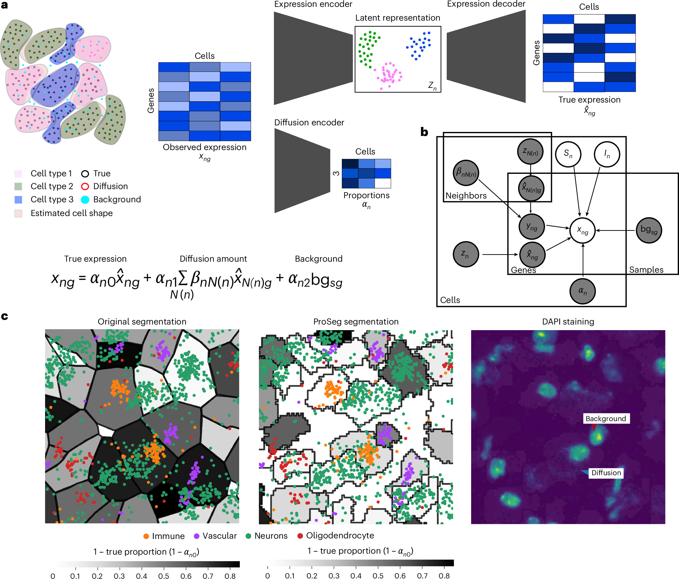
Technologies for estimating RNA expression at high throughput, in intact tissue slices and with high spatial resolution (spatial transcriptomics) shed new light on how cells communicate and tissues function. A fundamental step in analyzing data generated by subcellular resolution spatial transcriptomics technologies is quantification, namely, segmenting the plane into regions, each approximating a cell, and then collating the molecules inside each region to estimate the cellular expression profile. Despite many advances in this area, a persistent problem is that of the incorrect assignment of molecules to cells, which limits many current applications to the level of a priori-defined cell subsets and complicates the discovery of novel cell states. Here we develop resolVI, a model that operates downstream of any segmentation algorithm to generate a probabilistic representation, correcting for the misassignment of molecules, as well as for batch effects and other nuisance factors. We demonstrate that resolVI improves our ability to distinguish between cell states, identify subtle expression changes in space and perform integrated analysis across datasets. ResolVI is available as open source software within scvi-tools.
Temporal tissue dynamics from a spatial snapshot
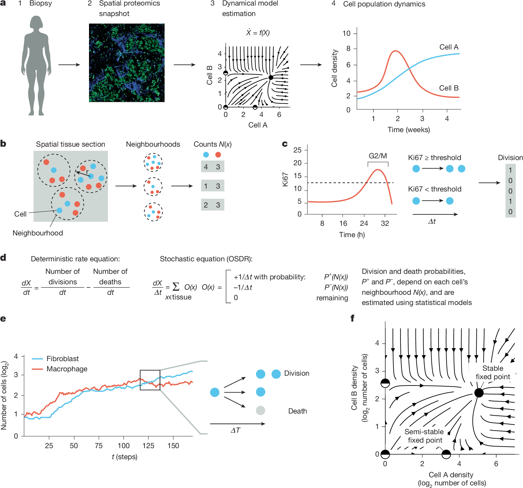
Physiological and pathological processes such as inflammation and cancer emerge from interactions between cells over time1. However, methods to follow cell populations over time within the native context of a human tissue are lacking because a biopsy offers only a single snapshot. Here we present one-shot tissue dynamics reconstruction (OSDR), an approach to estimate a dynamical model of cell populations based on a single tissue sample. OSDR uses spatial proteomics to learn how the composition of cellular neighbourhoods influences division rate, providing a dynamical model of cell population change over time. We apply OSDR to human breast cancer data2–4, and reconstruct two fixed points of fibroblasts and macrophage interactions5,6. These fixed points correspond to hot and cold fibrosis7, in agreement with co-culture experiments that measured these dynamics directly8. We then use OSDR to discover a pulse-generating excitable circuit of T and B cells in the tumour microenvironment, suggesting temporal flares of anticancer immune responses. Finally, we study longitudinal biopsies from a triple-negative breast cancer clinical trial3, in which OSDR predicts the collapse of the tumour cell population in responders but not in non-responders, based on early-treatment biopsies. OSDR can be applied to a wide range of spatial proteomics assays to enable analysis of tissue dynamics based on patient biopsies.
Mathematical Discovery of Potential Therapeutic Targets: Application to Rare Melanomas
Patients with rare types of melanoma such as acral, mucosal, or uveal melanoma, have lower survival rates than patients with cutaneous melanoma; these lower survival rates reflect the lower objective response rates to immunotherapy compared to cutaneous melanoma. Understanding tumor-immune dynamics in rare melanomas is critical for the development of new therapies and for improving response rates to current cancer therapies. Progress has been hindered by the lack of clinical data and the need for better preclinical models of rare melanomas. Canine melanoma provides a valuable comparative oncology model for rare types of human melanomas. We analyzed RNA sequencing data from canine melanoma patients and combined this with literature information to create a novel mechanistic mathematical model of melanoma-immune dynamics. Sensitivity analysis of the mathematical model indicated influential pathways in the dynamics, providing support for potential new therapeutic targets and future combinations of therapies. We share our learnings from this work, to help enable the application of this proof-of-concept workflow to other rare disease settings with sparse available data.

Benchmarking gene expression reconstruction from single-cell latent representations
Single-cell transcriptomics is typically modeled in low-dimensional latent representations that improve the signal-to-noise ratio of the data. Such representations underpin data integration, cell state discovery, and perturbation prediction, with applications ranging from large-scale organ atlases to latent trajectory modeling. Recent virtual cell approaches further leverage these representations to predict cellular responses as distributional shifts in latent space. Each of these applications ultimately requires faithful gene expression reconstruction from latent spaces for biological interpretation, enabling gene-level analysis of predicted perturbed or batch-corrected cells. Yet representation choice is typically treated as an implementation detail rather than a primary modeling decision, with no systematic evaluation of how well latent representations support gene expression reconstruction. Here, we introduce ReconEval, a benchmark for evaluating gene expression reconstruction from single-cell latent spaces. We benchmark two classes of latent representations: end-to-end trained models such as PCA, autoencoders, and variational autoencoders, and pretrained single-cell foundation model embeddings coupled to newly trained decoders. Reconstruction is evaluated both directly and after latent-space perturbation prediction. Across perturbational and observational datasets totaling over 100 million cells, our metric suite quantifies statistical fidelity; biological signal preservation, including differential expression, coexpression, cell-cycle structure, cytokine response and pathway activity; and perturbation-specific effects. We find that autoencoders achieve the strongest stand-alone reconstruction at low dimensionality, while variational regularization does not improve generalization in reconstruction. Frozen foundation model embeddings retain recoverable gene-level information, with reconstruction quality depending strongly on decoder architecture and pretraining objective. In latent perturbation modeling, high-dimensional PCA matches foundation model embeddings, while low-dimensional AE embeddings are optimal for flow-based generative models. Overall, reconstruction depends critically on the interplay between representation and downstream model, and simpler representations can outperform complex alternatives given appropriate capacity. Our benchmark establishes reconstruction as a critical evaluation axis for single-cell foundation models. We envision it improving the biological interpretability of latent-space modeling, a prerequisite for future virtual cell models to be validated by domain experts and grounded in biology. ### Competing Interest Statement F.J.T. consults for Immunai, CytoReason, Valinor Industries, Bioturing and Phylo Inc., and has ownership interest in RN.AI Therapeutics, Dermagnostix, and Cellarity. The remaining authors declare no competing interests.
Quantifying cell-state densities in single-cell phenotypic landscapes using Mellon
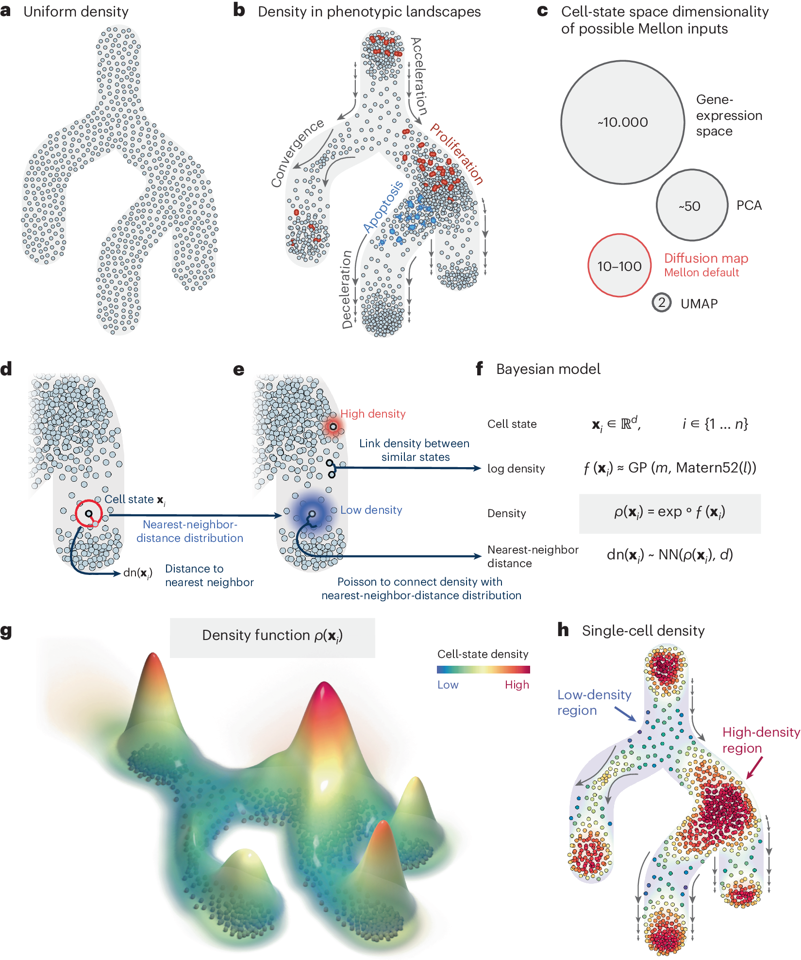
Cell-state density characterizes the distribution of cells along phenotypic landscapes and is crucial for unraveling the mechanisms that drive diverse biological processes. Here, we present Mellon, an algorithm for estimation of cell-state densities from high-dimensional representations of single-cell data. We demonstrate Mellon’s efficacy by dissecting the density landscape of differentiating systems, revealing a consistent pattern of high-density regions corresponding to major cell types intertwined with low-density, rare transitory states. We present evidence implicating enhancer priming and the activation of master regulators in emergence of these transitory states. Mellon offers the flexibility to perform temporal interpolation of time-series data, providing a detailed view of cell-state dynamics during developmental processes. Mellon facilitates density estimation across various single-cell data modalities, scaling linearly with the number of cells. Our work underscores the importance of cell-state density in understanding the differentiation processes, and the potential of Mellon to provide insights into mechanisms guiding biological trajectories.
Single-cell multiregion dissection of Alzheimer’s disease
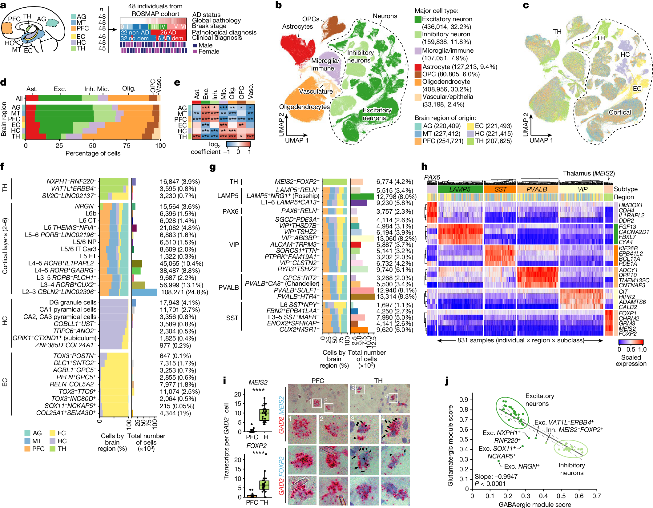
Alzheimer’s disease is the leading cause of dementia worldwide, but the cellular pathways that underlie its pathological progression across brain regions remain poorly understood1–3. Here we report a single-cell transcriptomic atlas of six different brain regions in the aged human brain, covering 1.3 million cells from 283 post-mortem human brain samples across 48 individuals with and without Alzheimer’s disease. We identify 76 cell types, including region-specific subtypes of astrocytes and excitatory neurons and an inhibitory interneuron population unique to the thalamus and distinct from canonical inhibitory subclasses. We identify vulnerable populations of excitatory and inhibitory neurons that are depleted in specific brain regions in Alzheimer’s disease, and provide evidence that the Reelin signalling pathway is involved in modulating the vulnerability of these neurons. We develop a scalable method for discovering gene modules, which we use to identify cell-type-specific and region-specific modules that are altered in Alzheimer’s disease and to annotate transcriptomic differences associated with diverse pathological variables. We identify an astrocyte program that is associated with cognitive resilience to Alzheimer’s disease pathology, tying choline metabolism and polyamine biosynthesis in astrocytes to preserved cognitive function late in life. Together, our study develops a regional atlas of the ageing human brain and provides insights into cellular vulnerability, response and resilience to Alzheimer’s disease pathology.
Patient-derived model capturing hypoxia and extracellular matrix remodelling of immunologically cold high-grade serous tumours
High-grade serous carcinoma tumours present poor survival rates, often associated with immunologically excluded environments driven by hypoxia and extensive extracellular matrix remodelling that disrupt tumour-stromal-immune interactions. Current experimental models fail to fully capture these microenvironmental features, limiting understanding of tumour-immune dynamics and drug development. Here, we present bioengineered patient-derived tumour-immune models to mimic physiologically relevant oxygen levels and extracellular matrix remodelling. Cancer cells are co-cultured with cancer-associated fibroblasts within human plasma-3D matrices or grown on decellularized human ovaries. Immune cells are either included within the 3D constructs to study multi-cellular interactions or challenged to infiltrate the matrices. We demonstrate that intratumoural hypoxia acts as a friend and a foe enhancing the activation and cytotoxicity of CD8 + T cells while inducing stromal/matrix dysregulation associated with impaired immune infiltration. Targeting TGF-β signalling attenuates the hypoxia-driven stromal-mediated immune exclusion. These relevant models may aid the development of targeted therapies to transform immunologically cold tumours into immunogenic to benefit female patients.
Self-Driving Datasets: From 20 Million Papers to Nuanced Biomedical Knowledge at Scale
Manually curated biomedical repositories -- spanning bioactivity, genomics, and chemistry -- are expensive to maintain, lag behind primary literature, and discard experimental context, obscuring nuances needed to assess data correctness and coverage. We show that PubMed itself can be autonomously and cost-effectively turned into structured datasets that are larger, more nuanced, and more accurate than the curated databases they replace. We present three coupled contributions: (1) an LLM-based entity-tagging pipeline, grounded in nine biomedical ontologies, that tags 4.5B entities across 19 categories in a 22.5M-paper, 2.5T-token PubMed corpus; (2) hybrid sparse-dense retrieval supporting entity-filtered semantic queries over the tagged corpus; and (3) Starling, a multi-agent deep research system that, given only a natural-language task description, designs precision- and recall-targeted retrieval filters, induces an extraction schema, and emits structured records with nuance-rich fields and supporting passages. Across six tasks -- blood-brain barrier permeability, oral bioavailability, acute toxicity (LD50), gene-disease associations, protein subcellular localization, and chemical reactions -- Starling produces ~6.3M records (91K-3M per task); several are, to our knowledge, the largest public datasets for their property. Frontier-model rejection of our extractions is 0.6-7.7% across tasks, far below error rates we measure on widely used curated counterparts (e.g., 16.5% on BBB_Martins, 7.3% on Bioavailability_Ma). Beyond scale and accuracy, the supporting passages carry nuance tabular databases discard -- e.g., oral bioavailability may depend on fed vs. fasted state. Together, the corpus, retrieval, and agent establish a foundation for AI-driven therapeutic design. Code and datasets: https://github.com/starling-labs/starling.