A family of rationally designed time-resolved fluorescent proteins with controllable lifetimes across the visible spectrum enables simultaneous multiplexed live imaging, super-resolution microscopy, and protein stoichiometry quantification, offering a transformative toolset for advancing biological research with enhanced complexity and quantitative precision.
A multimodal perturbation atlas defines the phenotypic resolution of cellular morphology
Because cells are complex dynamical systems, modeling cellular behaviors requires methods that capture how cells evolve across time, environments, and interventions. Microscopy is uniquely suited to this goal in that it can be applied to living cells in their native context. However, the phenotypic resolving power of live-cell microscopy remains incompletely characterized, particularly relative to molecular assays. Here, we present a multimodal perturbation atlas of 1,000 pooled CRISPR knockouts in A549 cells, profiled by fluorescence microscopy (39 live, 13 fixed markers), label-free phase imaging of the same live cells, and single-cell RNA sequencing (scRNA-seq). Totaling ∼57 million single-cell profiles, our data yield rich cell-biological signatures that map individual gene function. We find that phase imaging matches — and, with sufficient cell coverage, exceeds — the phenotypic resolution of fluorescence imaging and scRNA-seq, while capturing higher-order pathway organization that scRNA-seq does not resolve. These results establish intrinsic morphology as a high-precision readout of cellular state, and lay a foundation for live-cell profiling of phenotypic trajectories. ### Competing Interest Statement The authors have declared no competing interest. Biohub, Redwoood City, CA, USA

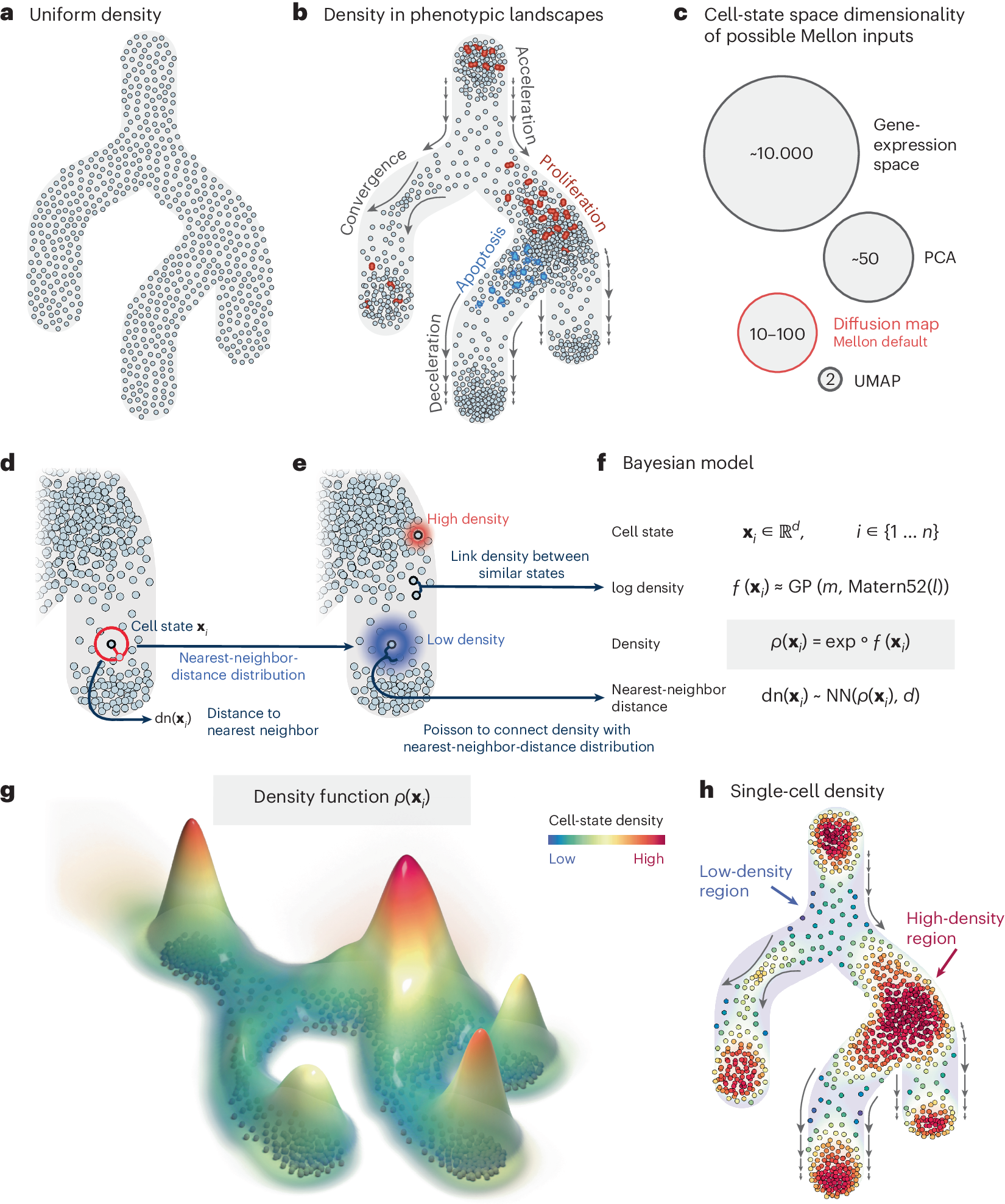
Quantifying cell-state densities in single-cell phenotypic landscapes using Mellon
Cell-state density characterizes the distribution of cells along phenotypic landscapes and is crucial for unraveling the mechanisms that drive diverse biological processes. Here, we present Mellon, an algorithm for estimation of cell-state densities from high-dimensional representations of single-cell data. We demonstrate Mellon’s efficacy by dissecting the density landscape of differentiating systems, revealing a consistent pattern of high-density regions corresponding to major cell types intertwined with low-density, rare transitory states. We present evidence implicating enhancer priming and the activation of master regulators in emergence of these transitory states. Mellon offers the flexibility to perform temporal interpolation of time-series data, providing a detailed view of cell-state dynamics during developmental processes. Mellon facilitates density estimation across various single-cell data modalities, scaling linearly with the number of cells. Our work underscores the importance of cell-state density in understanding the differentiation processes, and the potential of Mellon to provide insights into mechanisms guiding biological trajectories.
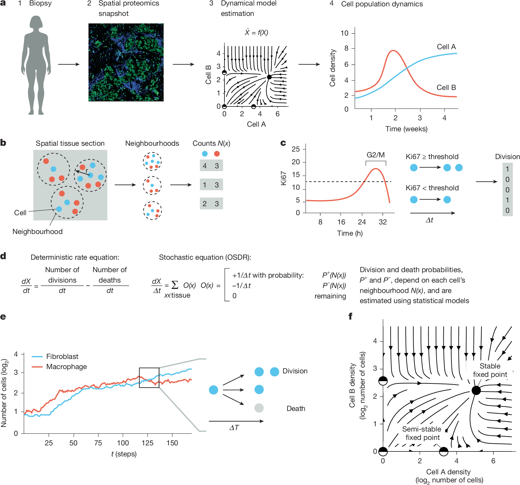
Temporal tissue dynamics from a spatial snapshot
Physiological and pathological processes such as inflammation and cancer emerge from interactions between cells over time1. However, methods to follow cell populations over time within the native context of a human tissue are lacking because a biopsy offers only a single snapshot. Here we present one-shot tissue dynamics reconstruction (OSDR), an approach to estimate a dynamical model of cell populations based on a single tissue sample. OSDR uses spatial proteomics to learn how the composition of cellular neighbourhoods influences division rate, providing a dynamical model of cell population change over time. We apply OSDR to human breast cancer data2–4, and reconstruct two fixed points of fibroblasts and macrophage interactions5,6. These fixed points correspond to hot and cold fibrosis7, in agreement with co-culture experiments that measured these dynamics directly8. We then use OSDR to discover a pulse-generating excitable circuit of T and B cells in the tumour microenvironment, suggesting temporal flares of anticancer immune responses. Finally, we study longitudinal biopsies from a triple-negative breast cancer clinical trial3, in which OSDR predicts the collapse of the tumour cell population in responders but not in non-responders, based on early-treatment biopsies. OSDR can be applied to a wide range of spatial proteomics assays to enable analysis of tissue dynamics based on patient biopsies.
Claude accelerates protein design and analytical chemistry
Two results show how Claude helps life sciences researchers speed up their work: designing protein binders from scratch and analyzing NMR and LC-MS data to speed chemical analysis.

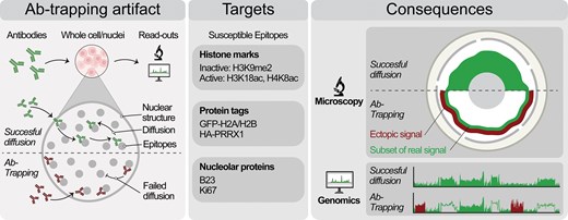
Antibody-trapping presents a widespread pitfall for microscopy and genomics in the nucleus
Abstract. Chromatin has a complex 3D structure and diverse binding proteins that coordinate the genome’s most essential functions. Many microscopy and geno
The evolving concept of cell identity in the single cell era
Summary: This Spotlight explores emerging technologies that are enabling the systematic and unbiased quantification of cell identity, and how these efforts will enable the construction of high-resolution, dynamic cell atlases.

Are biological systems poised at criticality?
Many of life's most fascinating phenomena emerge from interactions among many elements--many amino acids determine the structure of a single protein, many genes determine the fate of a cell, many neurons are involved in shaping our thoughts and memories. Physicists have long hoped that these collective behaviors could be described using the ideas and methods of statistical mechanics. In the past few years, new, larger scale experiments have made it possible to construct statistical mechanics models of biological systems directly from real data. We review the surprising successes of this "inverse" approach, using examples form families of proteins, networks of neurons, and flocks of birds. Remarkably, in all these cases the models that emerge from the data are poised at a very special point in their parameter space--a critical point. This suggests there may be some deeper theoretical principle behind the behavior of these diverse systems.

AlphaFold2 @ CASP14: “It feels like one’s child has left home.”
The past week was a momentous occasion for protein structure prediction, structural biology at large, and in due time, may prove to be so for the whole of life sciences. CASP14, the conference for …

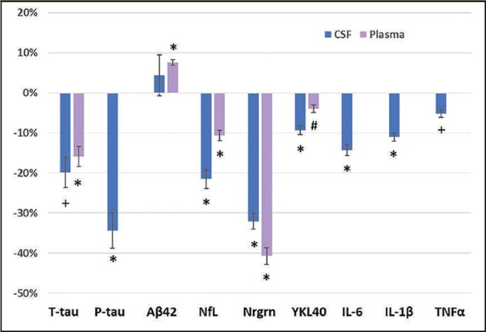
PTI-125 Reduces Biomarkers of Alzheimer’s Disease in Patients
The most common dementia worldwide, Alzheimer’s disease is often diagnosed via biomarkers in cerebrospinal fluid, including reduced levels of Aβ1-42, and increases in total tau and phosphorylated tau-181. Here we describe results of a Phase 2a study of a promising new drug candidate that significantly reversed all measured biomarkers of Alzheimer’s disease, neurodegeneration and neuroinflammation. PTI-125 is an oral small molecule drug candidate that binds and reverses an altered conformation of the scaffolding protein filamin A found in Alzheimer’s disease brain. Altered filamin A links to the α7-nicotinic acetylcholine receptor to allow Aβ42’s toxic signaling through this receptor to hyperphosphorylate tau. Altered filamin A also links to toll-like receptor 4 to enable Aβ-induced persistent activation of this receptor and inflammatory cytokine release. Restoring the native shape of filamin A prevents or reverses filamin A’s linkages to the α7-nicotinic acetylcholine receptor and tolllike receptor 4, thereby blocking Aβ42’s activation of these receptors. The result is reduced tau hyperphosphorylation and neuroinflammation, with multiple functional improvements demonstrated in transgenic mice and postmortem Alzheimer’s disease brain.
Precision Medicine in Neuroscience: Tools, Translation, and Implementation: A Workshop
Precision medicine approaches are rapidly transforming neuroscience, driven by advances in genetics, neuroimaging, biomarkers, and data science. These tools enable more refined disease classification, improved diagnosis, and treatments tailored to individual patients across neurological and psychiatric disorders. However, challenges remain in translating these advances into routine research and clinical practice. On March 4–5, the National Academies’ Forum on Neuroscience and Nervous System Disorders, in collaboration with the Forum on Drug Discovery, Development, and Translation and the Roundtable on Genomics and Precision Health, will host a workshop exploring opportunities, challenges, and strategies for integrating precision medicine into neuroscience research and care.

Integrated computational and in vivo models reveal Key Insights into macrophage behavior during bone healing
Myeloid-derived monocyte and macrophages are key cells in the bone that contribute to remodeling and injury repair. However, their temporal polarization status and control of bone-resorbing osteoclasts and bone-forming osteoblasts responses is largely unknown. In this study, we focused on two aspects of monocyte/macrophage dynamics and polarization states over time: 1) the injury-triggered pro- and anti-inflammatory monocytes/macrophages temporal profiles, 2) the contributions of pro- versus anti-inflammatory monocytes/macrophages in coordinating healing response. Bone healing is a complex multicellular dynamic process. While traditional in vitro and in vivo experimentation may capture the behavior of select populations with high resolution, they cannot simultaneously track the behavior of multiple populations. To address this, we have used an integrated coupled ordinary differential equations (ODEs)-based framework describing multiple cellular species to in vivo bone injury data in order to identify and test various hypotheses regarding bone cell populations dynamics. Our approach allowed us to infer several biological insights including, but not limited to,: 1) anti-inflammatory macrophages are key for early osteoclast inhibition and pro-inflammatory macrophage suppression, 2) pro-inflammatory macrophages are involved in osteoclast bone resorptive activity, whereas osteoblasts promote osteoclast differentiation, 3) Pro-inflammatory monocytes/macrophages rise during two expansion waves, which can be explained by the anti-inflammatory macrophages-mediated inhibition phase between the two waves. In addition, we further tested the robustness of the mathematical model by comparing simulation results to an independent experimental dataset. Taken together, this novel comprehensive mathematical framework allowed us to identify biological mechanisms that best recapitulate bone injury data and that explain the coupled cellular population dynamics involved in the process. Furthermore, our hypothesis testing methodology could be used in other contexts to decipher mechanisms in complex multicellular processes.
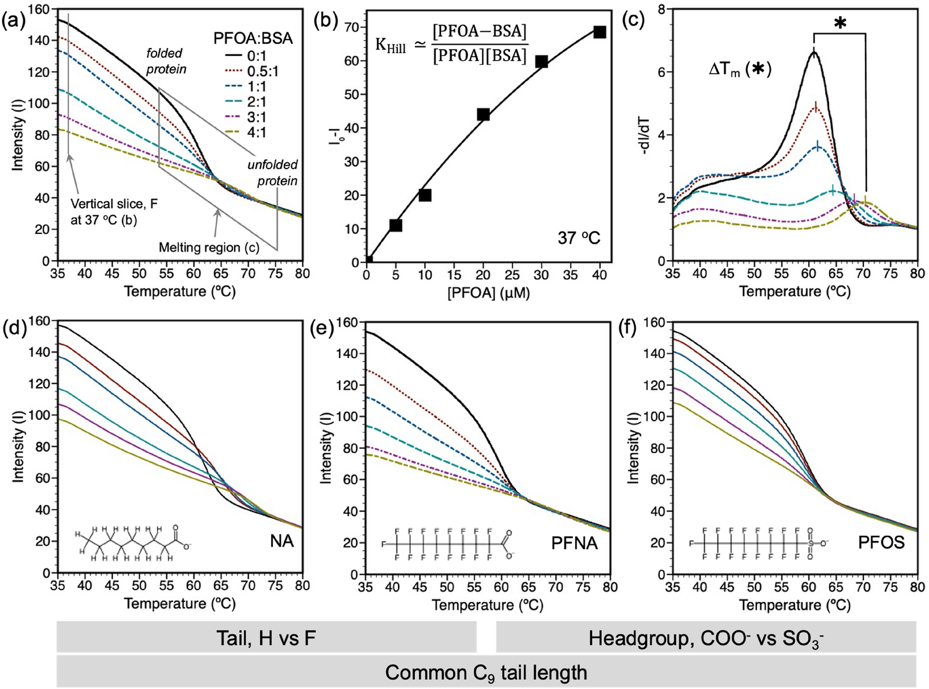
Differential scanning fluorimetry to assess PFAS binding to bovine serum albumin protein
The rapid screening of protein binding affinity for poly- and perfluoroalkyl substances (PFAS) benefits risk assessment and fate and transport modelling. PFAS are known to bioaccumulate in livestock through contaminated food and water. One excretion pathway is through milk, which may be facilitated by binding to milk proteins such as bovine serum albumin (BSA). We report a label-free differential scanning fluorimetry approach to determine PFAS–BSA binding over a broad temperature range. This method utilizes the tryptophan residue within the protein binding pocket as an intrinsic fluorophore, eliminating the need for fluorophore labels that may influence binding. BSA association constants were determined by (a) an equilibrium-based model at the melting temperature of BSA and (b) the Hill adsorption model to account for temperature dependent binding and binding cooperativity. Differences in binding between PFAS and fatty acid analogs revealed that a combination of size and hydrophobicity drives PFAS binding.
Comparing phenotypic manifolds with Kompot: Detecting differential abundance and gene expression at single-cell resolution
Single-cell studies are frequently designed to compare across conditions such as health and disease. However, existing computational approaches typically rely on grouping cells into discrete populations before making comparisons, which can limit resolution for detecting state-dependent changes. Here, we introduce Kompot, a statistical framework for comparative analysis of multi-condition single-cell data. Kompot quantifies both differential abundance, capturing how cells redistribute across the phenotypic space, and differential expression, identifying condition-specific transcriptional changes that may be localized, heterogeneous, or oppositely regulated across states. By modeling cell density and gene expression as continuous functions over a shared cell-state representation, Kompot enables single-cell–resolution inference with principled uncertainty estimates, without requiring predefined clusters or cell types. Applying Kompot to aging murine bone marrow, we identified a continuum of shifts in hematopoietic stem cell and mature cell states, transcriptional remodeling of monocytes independent of compositional changes, and divergent regulation of oxidative stress response genes across cell types. We demonstrate the utility of Kompot in disease settings by identifying cell-state and gene expression changes associated with improved efficacy of combinatorial immunotherapy in melanoma. Additionally, Kompot enables multi-sample comparative analysis by accounting for sample-to-sample heterogeneity. By capturing both global and cell-state–specific effects of perturbation, the Kompot framework is broadly applicable to dissecting condition-specific effects in complex single-cell landscapes. ### Competing Interest Statement The authors have declared no competing interest. National Institutes of Health, R35GM147125, R01CA292932, T32GM136534, S10OD028685 The Mark Foundation for Cancer Research, https://ror.org/00v7th354, Endeavor Award Edward P. Evans Foundation, https://ror.org/03h22gm35, Discovery Research Grant Brotman Baty Institute, https://ror.org/03jxvbk42, Pilot Award
Even a Single Bacterial Cell Can Sense the Seasons Changing | Quanta Magazine
Though they live only a few hours before dividing, bacteria can anticipate the approach of cold weather and prepare for it. The discovery suggests that seasonal tracking is fundamental to life.

Even a Single Bacterial Cell Can Sense the Seasons Changing | Quanta Magazine
Though they live only a few hours before dividing, bacteria can anticipate the approach of cold weather and prepare for it. The discovery suggests that seasonal tracking is fundamental to life.
Precision Medicine in Neuroscience: Tools, Translation, and Implementation: A Workshop
Join the web’s most supportive community of creators and get high-quality tools for hosting, sharing, and streaming videos in gorgeous HD with no ads.