Hypermobility involves excessive flexibility and systemic manifestations of connective tissue fragility. We propose a folate-dependent hypermobility syndrome model based on clinical observations, and through a review of existing literature, we raise the possibility that hypermobility presentation may be dependent on folate status. In our model, decreased methylenetetrahydrofolate reductase (MTHFR) activity disrupts the regulation of the ECM-specific proteinase matrix metalloproteinase 2 (MMP-2), leading to high levels of MMP-2 and elevated MMP-2-mediated cleavage of the proteoglycan decorin.
Red Blood Cell Folate Modifies the Association between Serum Per- and Polyfluoroalkyl Substances and Antibody Concentrations in U.S. Adolescents
Abstract. Per- and polyfluoroalkyl substances (PFAS) exposure has been associated with reduced antibody levels. Higher red blood cell (RBC) folate was prev

Navigating the Odyssey of Refractory Hypoglycemia: A Diagnostic and Therapeutic Puzzle in Ehlers-Danlos Syndrome Managed With Octreotide
Author Block: Mehwish Zeb, Michigan State University, Yarub Al-Alousi, St. Joseph University Medical Center, Syed Farasat Ali Shah, Institute of Diabetes and Endocrinology
Molecular Signaling Pathways and Essential Metabolic Elements in Bone Remodeling: An Implication of Therapeutic Targets for Bone Diseases
Bone is one of the dynamic tissues in the human body that undergoes continuous remodelling through subsequent actions of bone cells, osteoclasts, and osteoblasts. Several signal transduction pathways are involved in the transition of mesenchymal stem cells into osteoblasts. These primarily include Runx2, ATF4, Wnt signaling and sympathetic signalling. The differentiation of osteoclasts is controlled by M-CSF, RANKL, and costimulatory signalling. It is well known that bone remodelling is regulated through receptor activator of nuclear factor-kappa B ligand followed by the binding to RANK, which eventually induces the differentiation of osteoclasts. The resorbing osteoclasts secrete TRAP, cathepsin K, MMP-9 and gelatinase to digest the proteinaceous matrix of type I collagen and form a saucer-shaped lacuna along with resorption tunnels in the trabecular bone. Osteoblasts secrete a soluble decoy receptor, osteoprotegerin that prevents the binding of RANK/RANKL and thus moderating osteoclastogenesis. Moreover, bone homeostasis is also regulated by several growth factors, cytokines, calciotropic hormones, parathyroid hormone and sex steroids. The current review presents a correlation of the probable molecular targets underlying the regulation of bone mass and the role of essential metabolic elements in bone remodelling. Targeting these signaling pathways may help design newer therapies for treating bone diseases.

AI model detects very early normally ‘invisible’ tissue changes of pancreatic cancer
An AI model (REDMOD) can pick up the very early subtle tissue changes of pancreatic ductal adenocarcinoma, the most common form of pancreatic cancer, which conventional imaging and the human eye find difficult to detect, finds research published online in the journal Gut. As such, it offers the potential to shift an all too common late stage, terminal disease diagnosis to one that is at an early stage (stage 0) and treatable, say the researchers. While REDMOD was more accurate than experienced radiologists, it requires testing in high risk patients, defined as those with unexpected weight loss and newly diagnosed diabetes, before it can be widely used in clinical practice, they add.
The bone remodelling cycle
The bone remodelling cycle replaces old and damaged bone and is a highly regulated, lifelong process essential for preserving bone integrity and maintaining mineral homeostasis. During the bone remodelling cycle, osteoclastic resorption is tightly coupled to osteoblastic bone formation. The remodelling cycle occurs within the basic multicellular unit and comprises five co-ordinated steps; activation, resorption, reversal, formation and termination. These steps occur simultaneously but asynchronously at multiple different locations within the skeleton. Study of rare human bone disease and animal models have helped to elucidate the cellular and molecular mechanisms that regulate the bone remodelling cycle. The key signalling pathways controlling osteoclastic bone resorption and osteoblastic bone formation are receptor activator of nuclear factor-κB (RANK)/RANK ligand/osteoprotegerin and canonical Wnt signalling. Cytokines, growth factors and prostaglandins act as paracrine regulators of the cycle, whereas endocrine regulators include parathyroid hormone, vitamin D, calcitonin, growth hormone, glucocorticoids, sex hormones, and thyroid hormone. Disruption of the bone remodelling cycle and any resulting imbalance between bone resorption and formation leads to metabolic bone disease, most commonly osteoporosis. The advances in understanding the cellular and molecular mechanisms underlying bone remodelling have also provided targets for pharmacological interventions which include antiresorptive and anabolic therapies. This review will describe the remodelling process and its regulation, discuss osteoporosis and summarize the commonest pharmacological interventions used in its management.

TRANCE Is a Novel Ligand of the Tumor Necrosis Factor Receptor Family That Activates c-Jun N-terminal Kinase in T Cells *
A novel member of the tumor necrosis factor (TNF) cytokine family, designated TRANCE, was cloned during a search for apoptosis-regulatory genes using a somatic cell genetic approach in T cell hybridomas. The TRANCEgene encodes a type II membrane protein of 316 amino acids with a predicted molecular mass of 35 kDa. Its extracellular domain is most closely related to TRAIL, FasL, and TNF. TRANCE is an immediate early gene up-regulated by TCR stimulation and is controlled by calcineurin-regulated transcription factors.

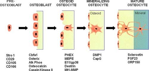
The amazing osteocyte
Abstract The last decade has provided a virtual explosion of data on the molecular biology and function of osteocytes. Far from being the “passive placeholder in bone,” this cell has been found to have numerous functions, such as acting as an orchestrator of bone remodeling through regulation of both osteoclast and osteoblast activity and also functioning as an endocrine cell. The osteocyte is a source of soluble factors not only to target cells on the bone surface but also to target distant organs, such as kidney, muscle, and other tissues. This cell plays a role in both phosphate metabolism and calcium availability and can remodel its perilacunar matrix. Osteocytes compose 90% to 95% of all bone cells in adult bone and are the longest lived bone cell, up to decades within their mineralized environment. As we age, these cells die, leaving behind empty lacunae that frequently micropetrose. In aged bone such as osteonecrotic bone, empty lacunae are associated with reduced remodeling. Inflammatory factors such as tumor necrosis factor and glucocorticoids used to treat inflammatory disease induce osteocyte cell death, but by different mechanisms with potentially different outcomes. Therefore, healthy, viable osteocytes are necessary for proper functionality of bone and other organs. © 2011 American Society for Bone and Mineral Research.
Disease Systems Analysis of Bone Mineral Density and Bone Turnover Markers in Response to Alendronate, Placebo, and Washout in Postmenopausal Women
A previously established mechanism‐based disease systems model for osteoporosis that is based on a mathematically reduced version of a model describing the interactions between osteoclast (bone removing) and osteoblast (bone forming) cells in bone remodeling has been applied to clinical data from women ( n = 1,379) receiving different doses and treatment regimens of alendronate, placebo, and washout. The changes in the biomarkers, plasma bone‐specific alkaline phosphatase activity (BSAP), urinary N‐telopeptide (NTX), lumbar spine bone mineral density (BMD), and total hip BMD, were linked to the underlying mechanistic core of the model. The final model gave an accurate description of all four biomarkers for the different treatments. Simulations were used to visualize the dynamics of the underlying network and the natural disease progression upon alendronate treatment and discontinuation. These results complement the previous applications of this mechanism‐based disease systems model to data from various treatments for osteoporosis.

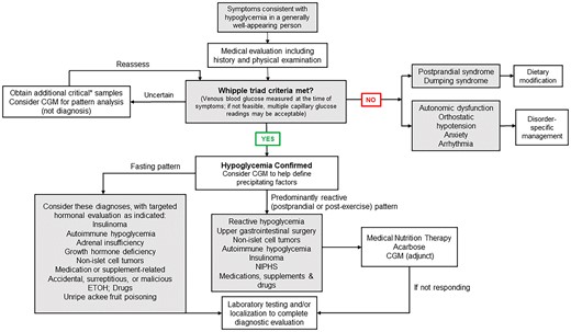
Hypoglycemia Associated With Hypermobile Ehlers-Danlos Syndrome
Abstract. Hypoglycemia in the absence of diabetes is often multifactorial and challenging to diagnose definitively. We present a case report and an expande
RETRACTED ARTICLE: Efficacy of oral folinicacid supplementation in children with autism spectrum disorder: a randomizeddouble-blind, placebo-controlled trial
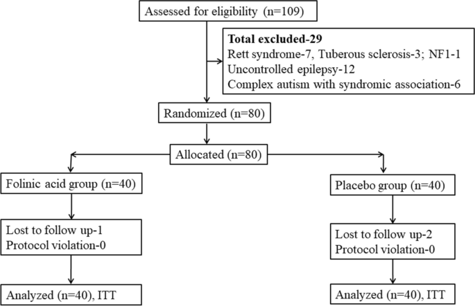
Oral folinic acid has shown potential to improve symptoms in childrenwith autism spectrum disorder (ASD). However, randomized controlled trials (RCTs)are limited. This double-blind, placebo-controlled RCT aimed to compare changes inChildhood Autism Rating Scale (CARS) scores in children with ASD aged 2–10 years,among folinic acid (2 mg/kg/day, maximum of 50 mg/day) and placebo groups at24 weeks, in comparison with baseline. Both the groups received standard care (ABAand sensory integration therapy). Secondary objectives included changes inbehavioral problems measured by the Child Behavior Checklist (CBCL) and serum levelsof anti-folate receptor autoantibodies and folic acid, correlated with changes inautism symptom severity. Out of the 40 participants recruited in each group, 39 and38 participants completed the 24-week follow-up in the folinic acid and placebogroups, respectively. The change in CARS score was higher in the folinic acid group(3.6 ± 0.8) compared to the placebo group (2.4 ± 0.7, p < 0.001). Changes in CBCL total score and CBCL internalizingscore were also better in the folinic acid group (19.7 ± 9.5 vs. 12.6 ± 8.4 and15.4 ± 7.8 vs. 8.5 ± 5.7, p < 0.001 for both).High-titer anti-folate receptor autoantibodies were positive in 32/40 and 33/40cases in the folinic acid and placebo groups, respectively (p = 0.78). In the placebo group, improvement in CARS score wascomparable regardless of autoantibody status (p = 0.11), but in the folinic acid group, improvement was morepronounced in the high-titer autoantibody group (p = 0.03). No adverse reactions were reported in either group.
Patient-derived model capturing hypoxia and extracellular matrix remodelling of immunologically cold high-grade serous tumours
High-grade serous carcinoma tumours present poor survival rates, often associated with immunologically excluded environments driven by hypoxia and extensive extracellular matrix remodelling that disrupt tumour-stromal-immune interactions. Current experimental models fail to fully capture these microenvironmental features, limiting understanding of tumour-immune dynamics and drug development. Here, we present bioengineered patient-derived tumour-immune models to mimic physiologically relevant oxygen levels and extracellular matrix remodelling. Cancer cells are co-cultured with cancer-associated fibroblasts within human plasma-3D matrices or grown on decellularized human ovaries. Immune cells are either included within the 3D constructs to study multi-cellular interactions or challenged to infiltrate the matrices. We demonstrate that intratumoural hypoxia acts as a friend and a foe enhancing the activation and cytotoxicity of CD8 + T cells while inducing stromal/matrix dysregulation associated with impaired immune infiltration. Targeting TGF-β signalling attenuates the hypoxia-driven stromal-mediated immune exclusion. These relevant models may aid the development of targeted therapies to transform immunologically cold tumours into immunogenic to benefit female patients.
(PDF) Neuroendocrine, Autonomic and Metabolic Challenges in Hypermobile Ehlers-Danlos Syndrome- A Case Study on Hypoglycemia in a patient with Craniocervical Instability.. Medical Research Archives. [Online] 12-5
PDF | This report presents a compelling case of hypoglycemia in a 20-year-old female with craniocervical instability receiving chronic total parenteral... | Find, read and cite all the research you need on ResearchGate

The role of WNT10B in physiology and disease: A 10-year update
WNT10B, a member of the WNT family of secreted glycoproteins, activates the WNT/β-CATENIN signaling cascade to control proliferation, stemness, pluripotency, and cell fate decisions. WNT10B has roles in many tissues, including bone, adipocytes, skin, hair, muscle, placenta, and the immune system. Aberrant WNT10B signaling leads to diseases such as osteoporosis, obesity, split-hand/foot malformation, fibrosis, dental anomalies, and cancer. We previously reviewed WNT10B ten years ago, and here we provide a comprehensive update to the field. Novel research on WNT10B has expanded to many more tissues and diseases. WNT10B polymorphisms and mutations correlate with many phenotypes, including bone mineral density, obesity, pig litter size, dog elbow dysplasia, and cow body size. In addition, the field has focused on the regulation of WNT10B through upstream mediators such as microRNAs (miRNAs) and long non-coding RNAs (lncRNAs). We also discuss the therapeutic implications of WNT10B regulation. In summary, research from 2012-2022 has revealed many new, diverse functions in the role of WNT10B in physiology and disease.

In Utero Metabolic Disruption—A Preventable Antecedent of Adult Disease?
The early manifestation of metabolic syndrome (MetS) in children, characterized by the premature onset of visceral obesity, elevated blood pressure, and the progression to dyslipidemia and type 2 diabetes, is associated with adult cardiovascular disease. Tackling the rising epidemic of obesity and...

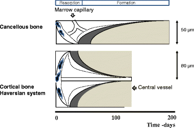
Cellular mechanisms of bone remodeling
Bone remodeling is a tightly regulated process securing repair of microdamage (targeted remodeling) and replacement of old bone with new bone through sequential osteoclastic resorption and osteoblastic bone formation. The rate of remodeling is regulated by a wide variety of calcitropic hormones (PTH, thyroid hormone, sex steroids etc.). In recent years we have come to appreciate that bone remodeling proceeds in a specialized vascular structure,—the Bone Remodeling Compartment (BRC). The outer lining of this compartment is made up of flattened cells, displaying all the characteristics of lining cells in bone including expression of OPG and RANKL. Reduced bone turnover leads to a decrease in the number of BRCs, while increased turnover causes an increase in the number of BRCs. The secretion of regulatory factors inside a confined space separated from the bone marrow would facilitate local regulation of the remodeling process without interference from growth factors secreted by blood cells in the marrow space. The BRC also creates an environment where cells inside the structure are exposed to denuded bone, which may enable direct cellular interactions with integrins and other matrix factors known to regulate osteoclast/osteoblast activity. However, the denuded bone surface inside the BRC also constitutes an ideal environment for the seeding of bone metastases, known to have high affinity for bone matrix. Circulating osteoclast- and osteoblast precursor cells have been demonstrated in peripheral blood. The dominant pathway regulating osteoclast recruitment is the RANKL/OPG system, while many different factors (RUNX, Osterix) are involved in osteoblast differentiation. Both pathways are modulated by calcitropic hormones.
Binding of Per- and Polyfluoroalkyl Substances (PFAS) to Serum Proteins: Implications for Toxicokinetics in Humans
Abstract. Per- and polyfluoroalkyl substances (PFAS) are a diverse class of highly persistent anthropogenic chemicals that are detectable in the serum of m