

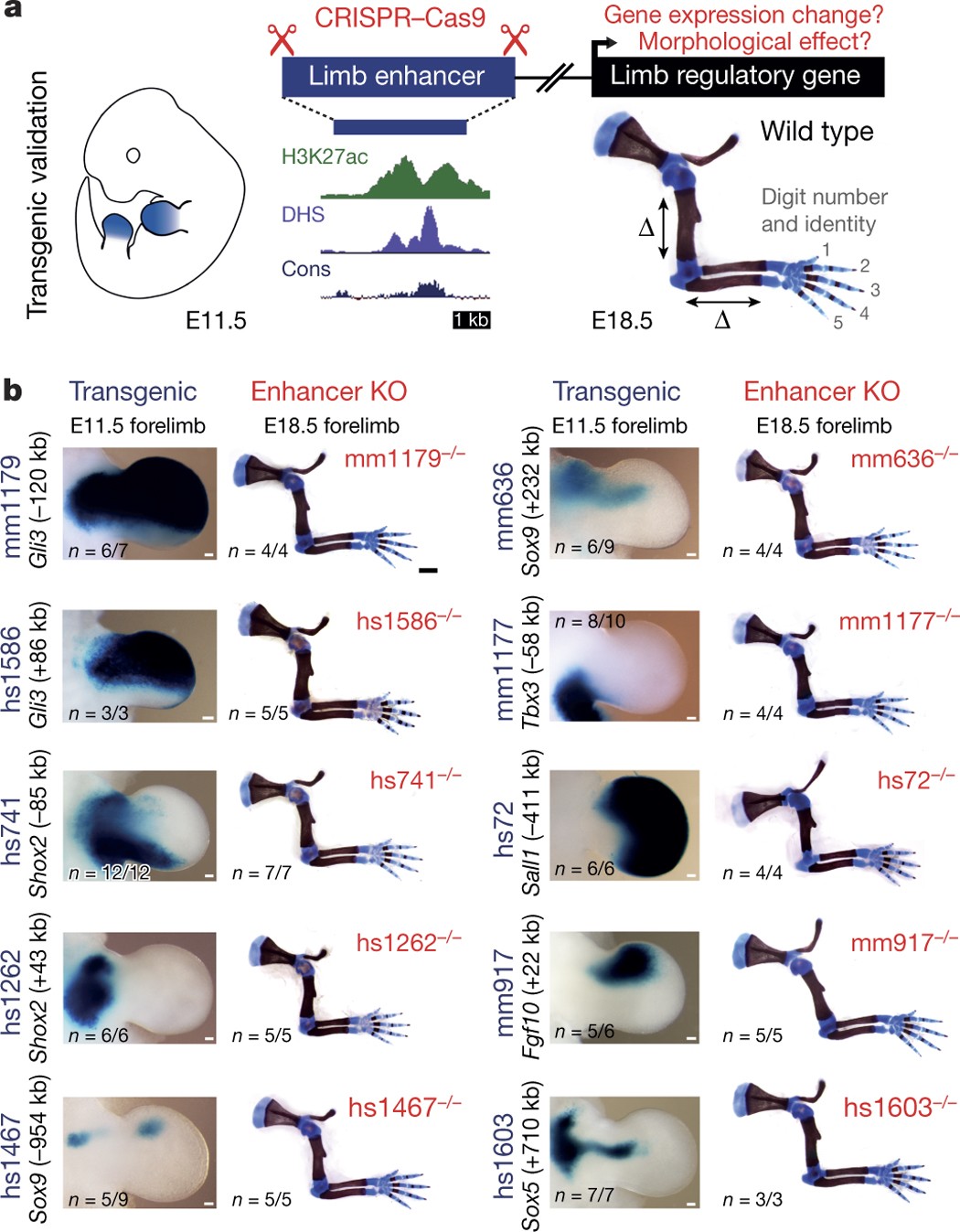
Gene enhancer knockout phenotypes and analysis of enhancer activity patterns show that developmental genes are regulated by multiple redundant enhancers in mouse embryos.
Shadow Enhancers Are Pervasive Features of Developmental Regulatory Networks
Cannavò et al. examine redundant (shadow) enhancers genome wide, finding that the majority of loci have more than two elements with similar activity. Evolutionary analyses show evidence of pervasive stabilizing selection and an ability to buffer mutations, suggesting that shadow enhancers have complex and fundamental roles in developmental networks

Differences in Krox20-Dependent Regulation of Hoxa2 and Hoxb2 during Hindbrain Development
During hindbrain development, segmental regulation of the paralogous Hoxa2 and Hoxb2 genes in rhombomeres (r) 3 and 5 involves Krox20-dependent enhancers that have been conserved during the duplication of the vertebrate Hox clusters from a common ancestor. Examining these evolutionarily related control regions could provide important insight into the degree to which the basic Krox20-dependent mechanisms, cis-regulatory components, and their organization have been conserved. Toward this goal we have performed a detailed functional analysis of a mouse Hoxa2 enhancer capable of directing reporter expression in r3 and r5. The combined activities of five separate cis-regions, in addition to the conserved Krox20 binding sites, are involved in mediating enhancer function. A CTTT (BoxA) motif adjacent to the Krox20 binding sites is important for r3/r5 activity. The BoxA motif is similar to one (Box1) found in the Hoxb2 enhancer and indicates that the close proximity of these Box motifs to Krox20 sites is a common feature of Krox20 targets in vivo. Two other rhombomeric elements (RE1 and RE3) are essential for r3/r5 activity and share common TCT motifs, indicating that they interact with a similar cofactor(s). TCT motifs are also found in the Hoxb2 enhancer, suggesting that they may be another common feature of Krox20-dependent control regions. The two remaining Hoxa2 cis-elements, RE2 and RE4, are not conserved in the Hoxb2 enhancer and define differences in some of components that can contribute to the Krox20-dependent activities of these enhancers. Furthermore, analysis of regulatory activities of these enhancers in a Krox20 mutant background has uncovered differences in their degree of dependence upon Krox20 for segmental expression. Together, this work has revealed a surprising degree of complexity in the number of cis-elements and regulatory components that contribute to segmental expression mediated by Krox20 and sheds light on the diversity and evolution of Krox20 target sites and Hox regulatory elements in vertebrates.
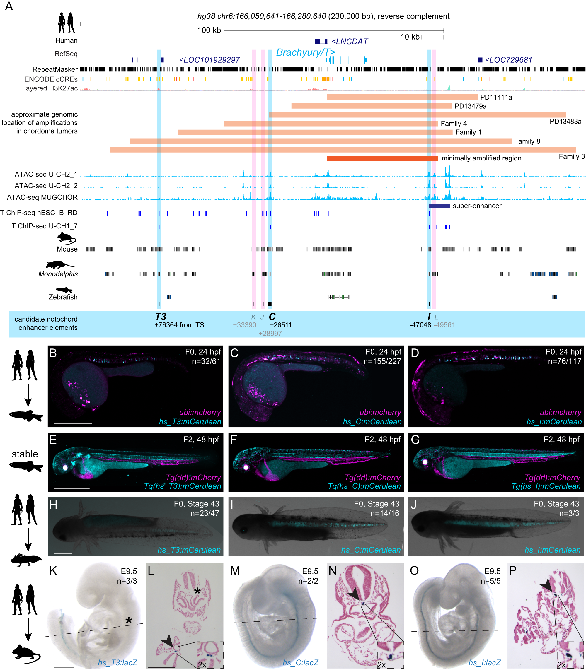
Conserved enhancers control notochord expression of vertebrate Brachyury
The cell type-specific expression of key transcription factors is central to development and disease. Brachyury/T/TBXT is a major transcription factor for gastrulation, tailbud patterning, and notochord formation; however, how its expression is controlled in the mammalian notochord has remained elusive. Here, we identify the complement of notochord-specific enhancers in the mammalian Brachyury/T/TBXT gene. Using transgenic assays in zebrafish, axolotl, and mouse, we discover three conserved Brachyury-controlling notochord enhancers, T3, C, and I, in human, mouse, and marsupial genomes. Acting as Brachyury-responsive, auto-regulatory shadow enhancers, in cis deletion of all three enhancers in mouse abolishes Brachyury/T/Tbxt expression selectively in the notochord, causing specific trunk and neural tube defects without gastrulation or tailbud defects. The three Brachyury-driving notochord enhancers are conserved beyond mammals in the brachyury/tbxtb loci of fishes, dating their origin to the last common ancestor of jawed vertebrates. Our data define the vertebrate enhancers for Brachyury/T/TBXTB notochord expression through an auto-regulatory mechanism that conveys robustness and adaptability as ancient basis for axis development.
The zinc finger gene Krox20 regulates HoxB2 (Hox2.8) during hindbrain segmentation
The zinc finger gene Krox20 and many Hox homeobox genes are expressed in segment-restricted domains in the hindbrain. The restricted expression patterns appear before morphological segmentation, suggesting that these transcription factors may play an early role in the establishment and identity of rhombomeric segments. In this paper, we show that the HoxB2 (Hox2.8) gene is normally upregulated in rhombomeres (r) 3, 4, and 5, and we identify an enhancer region upstream of the gene that imposes r3/r5 expression in transgenic mice.

Highly Efficient CRISPR-Cas9-Based Methods for Generating Deletion Mutations and F0 Embryos that Lack Gene Function in Zebrafish
Cas9 RNP complexes consisting of synthetic crRNA:tracrRNA duplex guide RNAs consistently induce mutations in virtually all copies of a targeted gene in zebrafish embryos. Hoshijima et al. show these tools allow effective screening of individual or combinations of gene function in F0 embryos and the facile induction of deletion mutations.

Targeted mutagenesis of specific genomic DNA sequences in animals for the in vivo generation of variant libraries
Understanding how the number, placement and affinity of transcription factor binding sites dictates gene regulatory programs remains a major unsolved challenge in biology, particularly in the context of multicellular organisms. To uncover these rules, it is first necessary to find the binding sites within a regulatory region with high precision, and then to systematically modulate this binding site arrangement while simultaneously measuring the effect of this modulation on output gene expression. Massively parallel reporter assays (MPRAs), where the gene expression stemming from 10,000s of in vitro-generated regulatory sequences is measured, have made this feat possible in high-throughput in single cells in culture. However, because of lack of technologies to incorporate DNA libraries, MPRAs are limited in whole organisms. To enable MPRAs in multicellular organisms, we generated tools to create a high degree of mutagenesis in specific genomic loci in vivo using base editing. Targeting GFP integrated in the genome of Drosophila cell culture and whole animals as a case study, we show that the base editor AIDevoCDA1 stemming from sea lamprey fused to nCas9 is highly mutagenic. Surprisingly, longer gRNAs increase mutation efficiency and expand the mutating window, which can allow the introduction of mutations in previously untargetable sequences. Finally, we demonstrate arrays of >20 gRNAs that can efficiently introduce mutations along a 200bp sequence, making it a promising tool to test enhancer function in vivo in a high throughput manner.

Embryo-scale reverse genetics at single-cell resolution
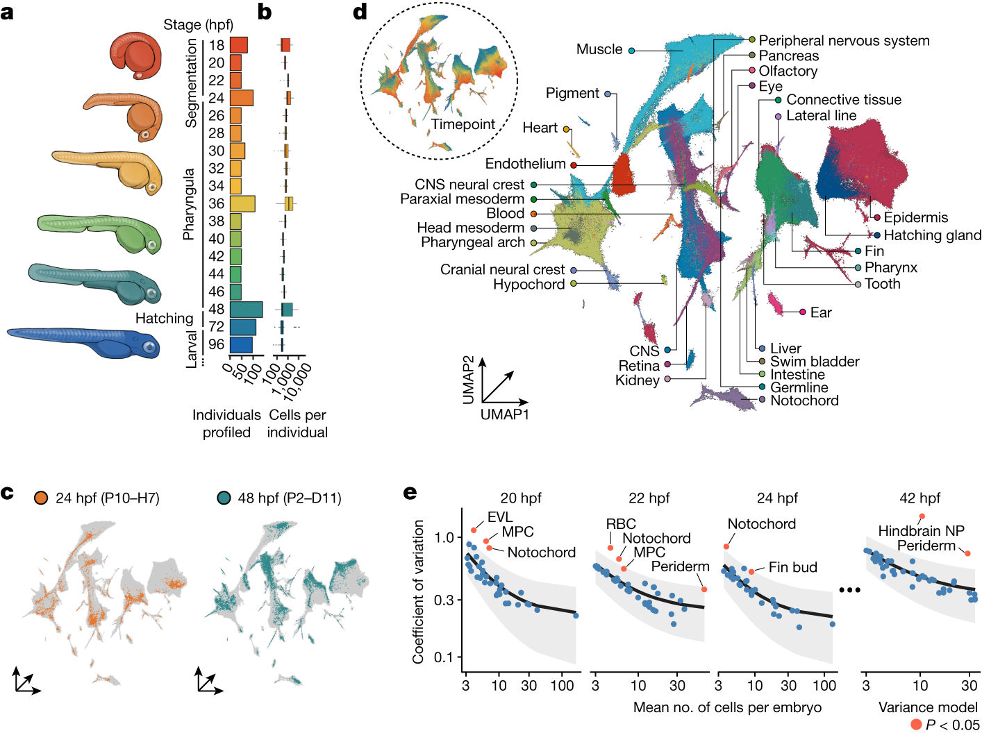
The maturation of single-cell transcriptomic technologies has facilitated the generation of comprehensive cellular atlases from whole embryos1–4. A majority of these data, however, has been collected from wild-type embryos without an appreciation for the latent variation that is present in development. Here we present the ‘zebrafish single-cell atlas of perturbed embryos’: single-cell transcriptomic data from 1,812 individually resolved developing zebrafish embryos, encompassing 19 timepoints, 23 genetic perturbations and a total of 3.2 million cells. The high degree of replication in our study (eight or more embryos per condition) enables us to estimate the variance in cell type abundance organism-wide and to detect perturbation-dependent deviance in cell type composition relative to wild-type embryos. Our approach is sensitive to rare cell types, resolving developmental trajectories and genetic dependencies in the cranial ganglia neurons, a cell population that comprises less than 1% of the embryo. Additionally, time-series profiling of individual mutants identified a group of brachyury-independent cells with strikingly similar transcriptomes to notochord sheath cells, leading to new hypotheses about early origins of the skull. We anticipate that standardized collection of high-resolution, organism-scale single-cell data from large numbers of individual embryos will enable mapping of the genetic dependencies of zebrafish cell types, while also addressing longstanding challenges in developmental genetics, including the cellular and transcriptional plasticity underlying phenotypic diversity across individuals.
Zebrafish hox paralogue group 2 genes function redundantly as selector genes to pattern the second pharyngeal arch
The pharyngeal arches are one of the defining features of the vertebrates, with the first arch forming the mandibles of the jaw and the second forming jaw support structures. The cartilaginous elements of each arch are formed from separate migratory neural crest cell streams, which derive from the dorsal aspect of the neural tube. The second and more posterior crest streams are characterized by specific Hox gene expression. The zebrafish has a larger overall number of Hox genes than the tetrapod vertebrates, as the result of a duplication event in its lineage. However, in both zebrafish and mouse, there are just two members of Hox paralogue group 2 (PG2): Hoxa2 and Hoxb2. Here, we show that morpholino-mediated "knock-down" of both zebrafish Hox PG2 genes results in major defects in second pharyngeal arch cartilages, involving replacement of ventral elements with a mirror-image duplication of first arch structures, and accompanying changes to pharyngeal musculature. In the mouse, null mutants of Hoxa2 have revealed that this single Hox gene is required for normal second arch patterning. By contrast, loss-of-function of either zebrafish Hox PG2 gene individually has no phenotypic consequence, showing that these two genes function redundantly to confer proper pattern to the second pharyngeal arch. We have also used hoxb1a mis-expression to induce localized ectopic expression of zebrafish Hox PG2 genes in the first arch; using this strategy, we find that ectopic expression of either Hox PG2 gene can confer second arch identity onto first arch structures, suggesting that the zebrafish Hox PG2 genes act as "selector genes."
Rapid generation of maternal mutants via oocyte transgenic expression of CRISPR-Cas9 and sgRNAs in zebrafish
A time-saving and deletion-prone method facilitates functional study of maternal factors in zebrafish.

Zebrafish Hox Paralogue Group 2 Genes Function Redundantly as Selector Genes to Pattern the Second Pharyngeal Arch
The pharyngeal arches are one of the defining features of the vertebrates, with the first arch forming the mandibles of the jaw and the second forming jaw support structures. The cartilaginous elements of each arch are formed from separate migratory neural crest cell streams, which derive from the dorsal aspect of the neural tube. The second and more posterior crest streams are characterized by specific Hox gene expression. The zebrafish has a larger overall number of Hox genes than the tetrapod vertebrates, as the result of a duplication event in its lineage. However, in both zebrafish and mouse, there are just two members of Hox paralogue group 2 (PG2): Hoxa2 and Hoxb2. Here, we show that morpholino-mediated “knock-down” of both zebrafish Hox PG2 genes results in major defects in second pharyngeal arch cartilages, involving replacement of ventral elements with a mirror-image duplication of first arch structures, and accompanying changes to pharyngeal musculature. In the mouse, null mutants of Hoxa2 have revealed that this single Hox gene is required for normal second arch patterning. By contrast, loss-of-function of either zebrafish Hox PG2 gene individually has no phenotypic consequence, showing that these two genes function redundantly to confer proper pattern to the second pharyngeal arch. We have also used hoxb1a mis-expression to induce localized ectopic expression of zebrafish Hox PG2 genes in the first arch; using this strategy, we find that ectopic expression of either Hox PG2 gene can confer second arch identity onto first arch structures, suggesting that the zebrafish Hox PG2 genes act as “selector genes.”
Single-Nucleus Multi-Omic Atlas Maps Regulatory Architecture and Non-Coding Variant Effects across Adult Human Tissues
Diverse human cell types establish specialized functions through lineage- and context-specific regulatory programs. Interpreting non-coding genetic risk requires integrated multi-omic reference maps that directly connect regulatory DNA to cellular expression across human tissues.
Dynamic Landscape Analysis of cell fate decisions provides predictive models of neural development from single-cell data
Building a mechanistic understanding of cell fate decisions remains a fundamental goal of developmental biology, with implications for stem cell therapies, regenerative medicine and understanding disease mechanisms. Single-cell transcriptomics provides a detailed picture of the cellular states observed during these decisions, but building dynamic and predictive models from these data remains a challenge. Here, we present dynamic landscape analysis (DLA), an integrative framework that applies dynamical systems theory to identify stable cell states, map transition pathways, and generate a predictive cell fate decision landscape from single-cell data. Applying this framework to vertebrate neural tube development revealed that progenitor specification by Sonic Hedgehog (Shh) can be captured in a landscape with an unexpected topology in which initially divergent lineages converge to the same fate through multiple distinct routes. The model accurately predicted cellular responses and cell fate allocation for unseen dynamic signalling regimes. Cross-species validation using human embryonic organoid data demonstrated conservation of this decision-making architecture. By modelling the dynamic responses that drive cell fate decisions, the DLA framework provides a quantitative and generative framework for extracting mechanistic insights from high-dimensional single-cell data.
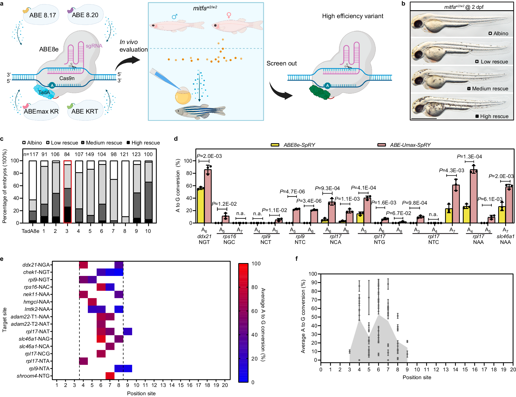
ABE-ultramax for high-efficiency biallelic adenine base editing in zebrafish
Advancements in CRISPR technology, particularly the development of base editors, revolutionize genetic variant research. When combined with model organisms like zebrafish, base editors significantly accelerate and refine in vivo analysis of genetic variations. However, base editors are restricted by protospacer adjacent motif (PAM) sequences and specific editing windows, hindering their applicability to a broad spectrum of genetic variants. Additionally, base editors can introduce unintended mutations and often exhibit reduced efficiency in living organisms compared to cultured cell lines. Here, we engineer a suite of adenine base editors (ABEs) called ABE-Ultramax (Umax), demonstrating high editing efficiency and low rates of insertions and deletions (indels) in zebrafish. The ABE-Umax suite of editors includes ABEs with shifted, narrowed, or broadened editing windows, reduced bystander mutation frequency, and highly flexible PAM sequence requirements. These advancements have the potential to address previous challenges in disease modeling and advance gene therapy applications.
BE4max and AncBE4max Are Efficient in Germline Conversion of C:G to T:A Base Pairs in Zebrafish
The ease of use and robustness of genome editing by CRISPR/Cas9 has led to successful use of gene knockout zebrafish for disease modeling. However, it still remains a challenge to precisely edit the zebrafish genome to create single-nucleotide substitutions, which account for ~60% of human disease-causing mutations. Recently developed base editing nucleases provide an excellent alternate to CRISPR/Cas9-mediated homology dependent repair for generation of zebrafish with point mutations. A new set of cytosine base editors, termed BE4max and AncBE4max, demonstrated improved base editing efficiency in mammalian cells but have not been evaluated in zebrafish. Therefore, we undertook this study to evaluate their efficiency in converting C:G to T:A base pairs in zebrafish by somatic and germline analysis using highly active sgRNAs to twist and ntl genes. Our data demonstrated that these improved BE4max set of plasmids provide desired base substitutions at similar efficiency and without any indels compared to the previously reported BE3 and Target-AID plasmids in zebrafish. Our data also showed that AncBE4max produces fewer incorrect and bystander edits, suggesting that it can be further improved by codon optimization of its components for use in zebrafish.

A multimodal perturbation atlas defines the phenotypic resolution of cellular morphology
Because cells are complex dynamical systems, modeling cellular behaviors requires methods that capture how cells evolve across time, environments, and interventions. Microscopy is uniquely suited to this goal in that it can be applied to living cells in their native context. However, the phenotypic resolving power of live-cell microscopy remains incompletely characterized, particularly relative to molecular assays. Here, we present a multimodal perturbation atlas of 1,000 pooled CRISPR knockouts in A549 cells, profiled by fluorescence microscopy (39 live, 13 fixed markers), label-free phase imaging of the same live cells, and single-cell RNA sequencing (scRNA-seq). Totaling ∼57 million single-cell profiles, our data yield rich cell-biological signatures that map individual gene function. We find that phase imaging matches — and, with sufficient cell coverage, exceeds — the phenotypic resolution of fluorescence imaging and scRNA-seq, while capturing higher-order pathway organization that scRNA-seq does not resolve. These results establish intrinsic morphology as a high-precision readout of cellular state, and lay a foundation for live-cell profiling of phenotypic trajectories. ### Competing Interest Statement The authors have declared no competing interest. Biohub, Redwoood City, CA, USA

Segmental expression of Hoxb-1 is controlled by a highly conserved autoregulatory loop dependent upon exd/pbx
Comparison of Hoxb-1 regulatory regions from different vertebrates identified three related sequence motifs critical for rhombomere 4 (r4) expression in the hindbrain. Functional analysis in transgenic mice and Drosophila embryos demonstrated that the conserved elements are involved in a positive autoregulatory loop dependent on labial (lab) family members. Binding of Hoxb-1 to these elements in vitro requires cofactors, and the motifs closely resemble the consensus binding site for pbxi, a homolog of the Drosophila extradenticle (exd) homeodomain protein.