


The germ cell fate in zebrafish is determined by germ plasm, whereas mammalian germ cell fate is induced by bone morphogenetic protein (BMP) signaling. It remains elusive whether BMP signaling is implicated in zebrafish germ cell development. Here, we demonstrate that BMP–Smad1/9 signaling plays a critical role in zebrafish primordial germ cell (PGC) maintenance rather than fate determination. BMP inhibition or smad1/9 knockdown reduces PGC numbers. Furthermore, we generated PGC-specific smad1/9 knockouts using a transgenic approach with PGC-specifically expressed Cas9 and ubiquitously expressed guide RNAs. Smad1/9 deficiency in PGCs leads to impaired PGC proliferation and increased apoptosis, consequently reducing PGC numbers. Transcriptome analysis revealed unchanged PGC-specific gene expression, but a marked upregulation of DNA damage response-related genes, which is validated by ectopic ATR–pChk1 activation in PGCs and PGC restoring by ATR inhibition. Collectively, these findings underscore conserved but functionally distinct roles of BMP signaling in vertebrate PGC development.
BE4max and AncBE4max Are Efficient in Germline Conversion of C:G to T:A Base Pairs in Zebrafish
The ease of use and robustness of genome editing by CRISPR/Cas9 has led to successful use of gene knockout zebrafish for disease modeling. However, it still remains a challenge to precisely edit the zebrafish genome to create single-nucleotide substitutions, which account for ~60% of human disease-causing mutations. Recently developed base editing nucleases provide an excellent alternate to CRISPR/Cas9-mediated homology dependent repair for generation of zebrafish with point mutations. A new set of cytosine base editors, termed BE4max and AncBE4max, demonstrated improved base editing efficiency in mammalian cells but have not been evaluated in zebrafish. Therefore, we undertook this study to evaluate their efficiency in converting C:G to T:A base pairs in zebrafish by somatic and germline analysis using highly active sgRNAs to twist and ntl genes. Our data demonstrated that these improved BE4max set of plasmids provide desired base substitutions at similar efficiency and without any indels compared to the previously reported BE3 and Target-AID plasmids in zebrafish. Our data also showed that AncBE4max produces fewer incorrect and bystander edits, suggesting that it can be further improved by codon optimization of its components for use in zebrafish.

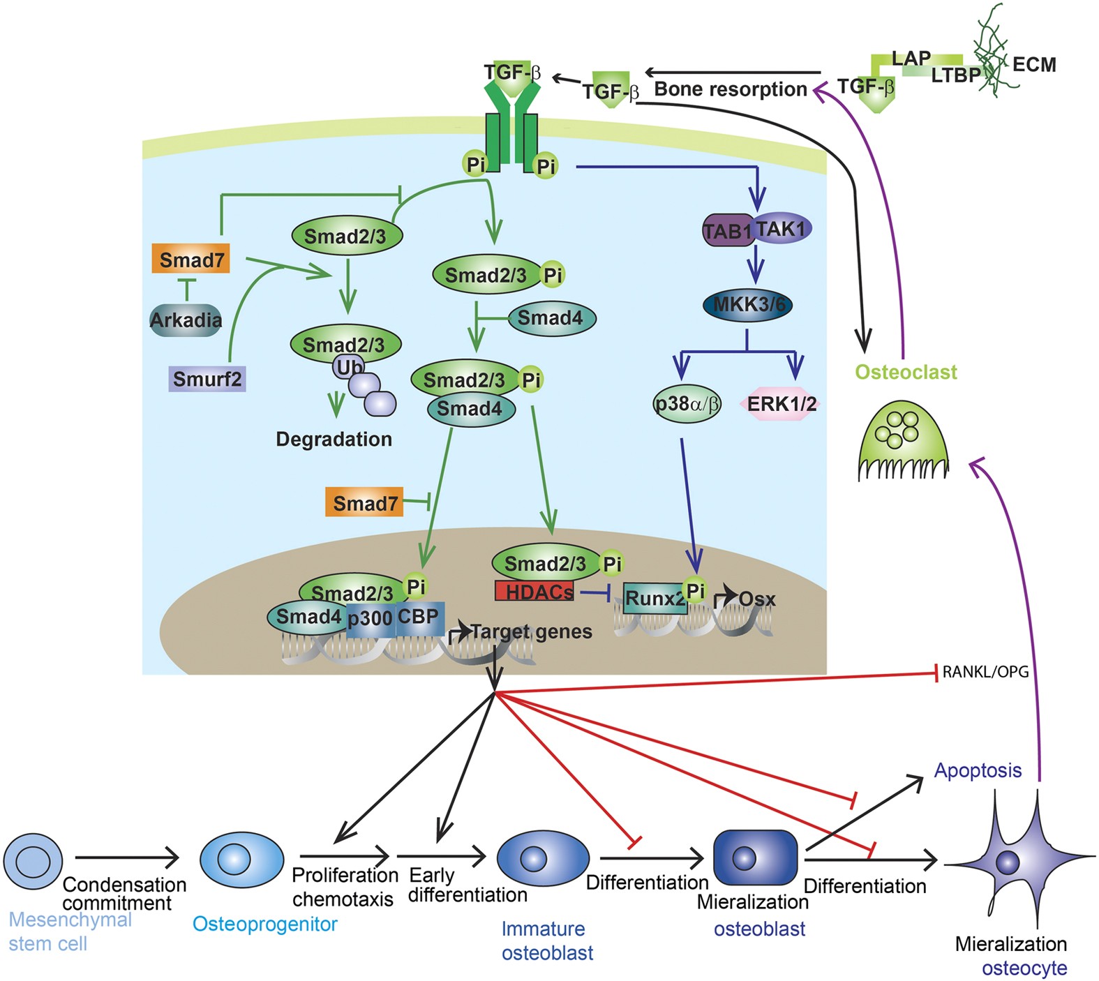
TGF-β and BMP signaling in osteoblast, skeletal development, and bone formation, homeostasis and disease
Transforming growth factor-beta (TGF-β) and bone morphogenic protein (BMP) signaling has fundamental roles in both embryonic skeletal development and postnatal bone homeostasis. TGF-βs and BMPs, acting on a tetrameric receptor complex, transduce signals to both the canonical Smad-dependent signaling pathway (that is, TGF-β/BMP ligands, receptors, and Smads) and the non-canonical-Smad-independent signaling pathway (that is, p38 mitogen-activated protein kinase/p38 MAPK) to regulate mesenchymal stem cell differentiation during skeletal development, bone formation and bone homeostasis. Both the Smad and p38 MAPK signaling pathways converge at transcription factors, for example, Runx2 to promote osteoblast differentiation and chondrocyte differentiation from mesenchymal precursor cells. TGF-β and BMP signaling is controlled by multiple factors, including the ubiquitin–proteasome system, epigenetic factors, and microRNA. Dysregulated TGF-β and BMP signaling result in a number of bone disorders in humans. Knockout or mutation of TGF-β and BMP signaling-related genes in mice leads to bone abnormalities of varying severity, which enable a better understanding of TGF-β/BMP signaling in bone and the signaling networks underlying osteoblast differentiation and bone formation. There is also crosstalk between TGF-β/BMP signaling and several critical cytokines’ signaling pathways (for example, Wnt, Hedgehog, Notch, PTHrP, and FGF) to coordinate osteogenesis, skeletal development, and bone homeostasis. This review summarizes the recent advances in our understanding of TGF-β/BMP signaling in osteoblast differentiation, chondrocyte differentiation, skeletal development, cartilage formation, bone formation, bone homeostasis, and related human bone diseases caused by the disruption of TGF-β/BMP signaling.
Rapid generation of maternal mutants via oocyte transgenic expression of CRISPR-Cas9 and sgRNAs in zebrafish
A time-saving and deletion-prone method facilitates functional study of maternal factors in zebrafish.

Highly Efficient CRISPR-Cas9-Based Methods for Generating Deletion Mutations and F0 Embryos that Lack Gene Function in Zebrafish
Cas9 RNP complexes consisting of synthetic crRNA:tracrRNA duplex guide RNAs consistently induce mutations in virtually all copies of a targeted gene in zebrafish embryos. Hoshijima et al. show these tools allow effective screening of individual or combinations of gene function in F0 embryos and the facile induction of deletion mutations.

Osteoblast-Osteoclast Communication and Bone Homeostasis
Bone remodeling is tightly regulated by a cross-talk between bone-forming osteoblasts and bone-resorbing osteoclasts. Osteoblasts and osteoclasts communicate with each other to regulate cellular behavior, survival and differentiation through direct cell-to-cell contact or through secretory proteins. A direct interaction between osteoblasts and osteoclasts allows bidirectional transduction of activation signals through EFNB2-EPHB4, FASL-FAS or SEMA3A-NRP1, regulating differentiation and survival of osteoblasts or osteoclasts. Alternatively, osteoblasts produce a range of different secretory molecules, including M-CSF, RANKL/OPG, WNT5A, and WNT16, that promote or suppress osteoclast differentiation and development. Osteoclasts also influence osteoblast formation and differentiation through secretion of soluble factors, including S1P, SEMA4D, CTHRC1 and C3. Here we review the current knowledge regarding membrane bound- and soluble factors governing cross-talk between osteoblasts and osteoclasts.
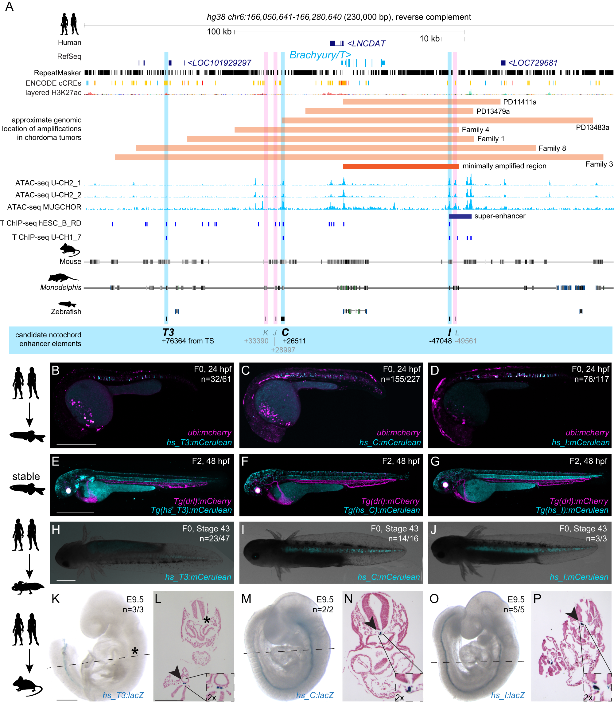
Conserved enhancers control notochord expression of vertebrate Brachyury
The cell type-specific expression of key transcription factors is central to development and disease. Brachyury/T/TBXT is a major transcription factor for gastrulation, tailbud patterning, and notochord formation; however, how its expression is controlled in the mammalian notochord has remained elusive. Here, we identify the complement of notochord-specific enhancers in the mammalian Brachyury/T/TBXT gene. Using transgenic assays in zebrafish, axolotl, and mouse, we discover three conserved Brachyury-controlling notochord enhancers, T3, C, and I, in human, mouse, and marsupial genomes. Acting as Brachyury-responsive, auto-regulatory shadow enhancers, in cis deletion of all three enhancers in mouse abolishes Brachyury/T/Tbxt expression selectively in the notochord, causing specific trunk and neural tube defects without gastrulation or tailbud defects. The three Brachyury-driving notochord enhancers are conserved beyond mammals in the brachyury/tbxtb loci of fishes, dating their origin to the last common ancestor of jawed vertebrates. Our data define the vertebrate enhancers for Brachyury/T/TBXTB notochord expression through an auto-regulatory mechanism that conveys robustness and adaptability as ancient basis for axis development.
Regulation of osteoblastogenesis and bone mass by Wnt10b
Wnts comprise a family of secreted signaling proteins that regulate diverse developmental processes. Activation of Wnt signaling by Wnt10b inhibits differentiation of preadipocytes and blocks adipose tissue development; however, the effect of Wnt10b on other mesenchymal lineages has not been defined. To explore the physiological role of Wnt signaling in bone development, we analyzed FABP4-Wnt10b mice, which express the Wnt10b transgene in marrow. Femurs from FABP4-Wnt10b mice have almost four times as much bone in the distal metaphyses and are mechanically stronger. These mice maintain elevated bone mass at least through 23 months of age. In addition, FABP4-Wnt10b mice are protected from the bone loss characteristic of estrogen deficiency. We used pharmacological and genetic approaches to demonstrate that canonical Wnt signaling stimulates osteoblastogenesis and inhibits adipogenesis of bipotential mesenchymal precursors. Wnt10b shifts cell fate toward the osteoblast lineage by induction of the osteoblastogenic transcription factors Runx2, Dlx5, and osterix and suppression of the adipogenic transcription factors C/EBPα and PPARγ. One mechanism whereby Wnt10b promotes osteoblastogenesis is suppression of PPARγ expression. Finally, Wnt10b-/- mice have decreased trabecular bone and serum osteocalcin, confirming that Wnt10b is an endogenous regulator of bone formation.

Highly efficient genome editing using oocyte-specific zcas9 transgenic zebrafish. Liu Y, Zhang C, Zhang Y, Lin S, Shi DL, Shao M
Highly efficient genome editing using oocyte-specific zcas9 transgenic zebrafish Since its first application to induce mutations in mammalian cells (Cong et al., 2013; Mali et al., 2013), CRISPR/Cas9 rapidly becomes a routine technique to perform genome editing in a variety of biological systems due to its facile, robust, and multiplexable features (Hwang et al., 2013; Guo et al., 2014; Wang et al., 2013). In every system, Cas9 and sgRNA should co-express to induce mutations. This is typically achieved by injection of the sgRNA mixed with Cas9 mRNA or protein into the fertilized eggs in zebrafish. As the in vivo translation of capped RNAs delay the presence of sufficient Cas9 protein, the mRNA injection produces more mosaic animals and shows relatively lower efficiency when compared to Cas9 protein injection (Burger et al., 2016). However, it is much more cost effective than using commercially available Cas9 protein, especially in the case of large-scale mutation screens. To further simplify this procedure, lower the cost and maintain an acceptable high genome editing efficiency in zebrafish, we set out to generate a transgenic line with stable and specific Cas9 expression in the egg. zpc (zp3b) promoter (zpc0.5) was reported to drive robust and specific GFP expression in the oocyte (Onichtchouk et al., 2003), we thus fused it with a zebrafish codon optimized cas9 (zcas9) followed by an SV40 3' UTR (Fig. 1A and File S1; Liu et al., 2014), and introduced this construct to zebrafish genome by Tol2 mediated transgenesis (Urasaki et al., 2006). To screen female F0 fish harboring this transgene, the F0 female founders were outcrossed with the wild-type male, the resulting eggs were injected with an sgRNA targeting slc45a2 (Moreno-Mateos et al., 2015), a gene essential for melanin synthesis, and assayed for their pigmentation phenotype. We found that the offspring of one founder showed a high proportion of pigmentation defect. Some of them exhibited a homozygous mutant like albino phenotype, suggesting a robust Cas9 expression in the egg and a very high efficient gene disruption. To examine the specificity of zcas9 expression in the transgenic line, the offspring (F1) of this F0 founder were raised to adulthood, and the ovaries of female F1 transgenic fish were dissected and subjected to in situ hybridization (ISH) employing the probe of zcas9. We found that the transcripts of zcas9 were specifically distributed in oocytes with a diameter of 40-200 µm, at around IB-II stages, while in WT embryos, no hybridization signals were detected (Fig. 1B, C). We did not detect zcas9 expression in stage IA oocytes or in larger ones after stage II. Accordingly, by RT-PCR, we also failed
Zebrafish hox paralogue group 2 genes function redundantly as selector genes to pattern the second pharyngeal arch
The pharyngeal arches are one of the defining features of the vertebrates, with the first arch forming the mandibles of the jaw and the second forming jaw support structures. The cartilaginous elements of each arch are formed from separate migratory neural crest cell streams, which derive from the dorsal aspect of the neural tube. The second and more posterior crest streams are characterized by specific Hox gene expression. The zebrafish has a larger overall number of Hox genes than the tetrapod vertebrates, as the result of a duplication event in its lineage. However, in both zebrafish and mouse, there are just two members of Hox paralogue group 2 (PG2): Hoxa2 and Hoxb2. Here, we show that morpholino-mediated "knock-down" of both zebrafish Hox PG2 genes results in major defects in second pharyngeal arch cartilages, involving replacement of ventral elements with a mirror-image duplication of first arch structures, and accompanying changes to pharyngeal musculature. In the mouse, null mutants of Hoxa2 have revealed that this single Hox gene is required for normal second arch patterning. By contrast, loss-of-function of either zebrafish Hox PG2 gene individually has no phenotypic consequence, showing that these two genes function redundantly to confer proper pattern to the second pharyngeal arch. We have also used hoxb1a mis-expression to induce localized ectopic expression of zebrafish Hox PG2 genes in the first arch; using this strategy, we find that ectopic expression of either Hox PG2 gene can confer second arch identity onto first arch structures, suggesting that the zebrafish Hox PG2 genes act as "selector genes."
Zebrafish Hox Paralogue Group 2 Genes Function Redundantly as Selector Genes to Pattern the Second Pharyngeal Arch
The pharyngeal arches are one of the defining features of the vertebrates, with the first arch forming the mandibles of the jaw and the second forming jaw support structures. The cartilaginous elements of each arch are formed from separate migratory neural crest cell streams, which derive from the dorsal aspect of the neural tube. The second and more posterior crest streams are characterized by specific Hox gene expression. The zebrafish has a larger overall number of Hox genes than the tetrapod vertebrates, as the result of a duplication event in its lineage. However, in both zebrafish and mouse, there are just two members of Hox paralogue group 2 (PG2): Hoxa2 and Hoxb2. Here, we show that morpholino-mediated “knock-down” of both zebrafish Hox PG2 genes results in major defects in second pharyngeal arch cartilages, involving replacement of ventral elements with a mirror-image duplication of first arch structures, and accompanying changes to pharyngeal musculature. In the mouse, null mutants of Hoxa2 have revealed that this single Hox gene is required for normal second arch patterning. By contrast, loss-of-function of either zebrafish Hox PG2 gene individually has no phenotypic consequence, showing that these two genes function redundantly to confer proper pattern to the second pharyngeal arch. We have also used hoxb1a mis-expression to induce localized ectopic expression of zebrafish Hox PG2 genes in the first arch; using this strategy, we find that ectopic expression of either Hox PG2 gene can confer second arch identity onto first arch structures, suggesting that the zebrafish Hox PG2 genes act as “selector genes.”
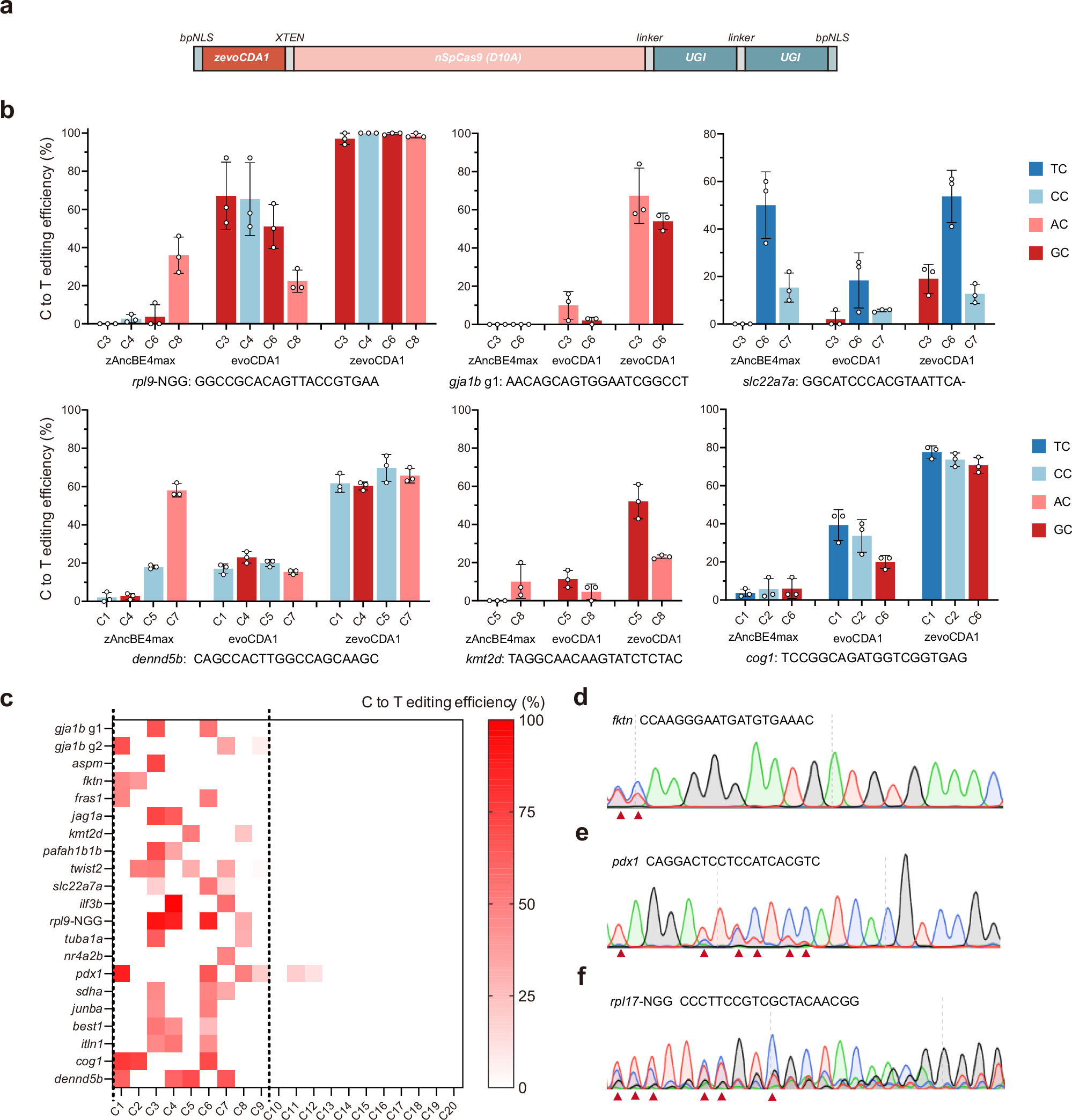
Cytosine base editors with increased PAM and deaminase motif flexibility for gene editing in zebrafish
Cytosine base editing is a powerful tool for making precise single nucleotide changes in cells and model organisms like zebrafish, which are valuable for studying human diseases. However, current base editors struggle to edit cytosines in certain DNA contexts, particularly those with GC and CC pairs, limiting their use in modelling disease-related mutations. Here we show the development of zevoCDA1, an optimized cytosine base editor for zebrafish that improves editing efficiency across various DNA contexts and reduces restrictions imposed by the protospacer adjacent motif. We also create zevoCDA1-198, a more precise editor with a narrower editing window of five nucleotides, minimizing off-target effects. Using these advanced tools, we successfully generate zebrafish models of diseases that were previously challenging to create due to sequence limitations. This work enhances the ability to introduce human pathogenic mutations in zebrafish, broadening the scope for genomic research with improved precision and efficiency.
Molecular Signaling Pathways and Essential Metabolic Elements in Bone Remodeling: An Implication of Therapeutic Targets for Bone Diseases
Bone is one of the dynamic tissues in the human body that undergoes continuous remodelling through subsequent actions of bone cells, osteoclasts, and osteoblasts. Several signal transduction pathways are involved in the transition of mesenchymal stem cells into osteoblasts. These primarily include Runx2, ATF4, Wnt signaling and sympathetic signalling. The differentiation of osteoclasts is controlled by M-CSF, RANKL, and costimulatory signalling. It is well known that bone remodelling is regulated through receptor activator of nuclear factor-kappa B ligand followed by the binding to RANK, which eventually induces the differentiation of osteoclasts. The resorbing osteoclasts secrete TRAP, cathepsin K, MMP-9 and gelatinase to digest the proteinaceous matrix of type I collagen and form a saucer-shaped lacuna along with resorption tunnels in the trabecular bone. Osteoblasts secrete a soluble decoy receptor, osteoprotegerin that prevents the binding of RANK/RANKL and thus moderating osteoclastogenesis. Moreover, bone homeostasis is also regulated by several growth factors, cytokines, calciotropic hormones, parathyroid hormone and sex steroids. The current review presents a correlation of the probable molecular targets underlying the regulation of bone mass and the role of essential metabolic elements in bone remodelling. Targeting these signaling pathways may help design newer therapies for treating bone diseases.

Stage- and tissue-specific gene editing using 4-OHT–inducible Cas9 in whole organism
Li et al. report a novel Cas9-based spatiotemporal gene-editing approach in zebrafish. Given the limited germline transmission efficiency of knock-in allel

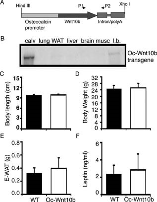
Wnt10b Increases Postnatal Bone Formation by Enhancing Osteoblast Differentiation
Abstract Overexpression of Wnt10b from the osteocalcin promoter in transgenic mice increases postnatal bone mass. Increases in osteoblast perimeter, mineralizing surface, and bone formation rate without detectable changes in pre-osteoblast proliferation, osteoblast apoptosis, or osteoclast number and activity suggest that, in this animal model, Wnt10b primarily increases bone mass by stimulating osteoblastogenesis. Introduction: Wnt signaling regulates many aspects of development including postnatal accrual of bone. Potential mechanisms for how Wnt signaling increases bone mass include regulation of osteoblast and/or osteoclast number and activity. To help differentiate between these possibilities, we studied mice in which Wnt10b is expressed specifically in osteoblast lineage cells or in mice devoid of Wnt10b. Materials and Methods: Transgenic mice, in which mouse Wnt10b is expressed from the human osteocalcin promoter (Oc-Wnt10b), were generated in C57BL/6 mice. Transgene expression was evaluated by RNase protection assay. Quantitative assessment of bone variables was done by radiography, μCT, and static and dynamic histomorphometry. Mechanisms of bone homeostasis were evaluated with assays for BrdU, TUNEL, and TRACP5b activity, as well as serum levels of C-terminal telopeptide of type I collagen (CTX). The endogenous role of Wnt10b in bone was assessed by dynamic histomorphometry in Wnt10b−/− mice. Results: Oc-Wnt10b mice have increased mandibular bone and impaired eruption of incisors during postnatal development. Analyses of femoral distal metaphyses show significantly higher BMD, bone volume fraction, and trabecular number. Increased bone formation is caused by increases in number of osteoblasts per bone surface, rate of mineral apposition, and percent mineralizing surface. Although number of osteoclasts per bone surface is not altered, Oc-Wnt10b mice have increased total osteoclast activity because of higher bone mass. In Wnt10b−/− mice, changes in mineralizing variables and osteoblast perimeter in femoral distal metaphyses were not observed; however, bone formation rate is reduced because of decreased total bone volume and trabecular number. Conclusions: High bone mass in Oc-Wnt10b mice is primarily caused by increased osteoblastogenesis, with a minor contribution from elevated mineralizing activity of osteoblasts.
Osteoclast differentiation and activation
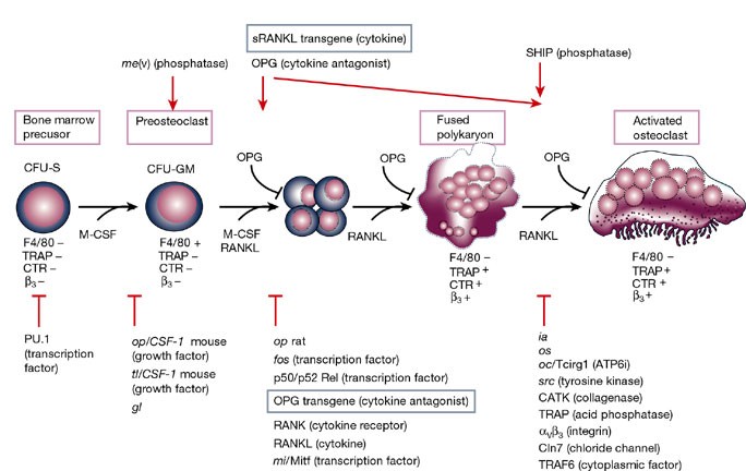
Osteoclasts are specialized cells derived from the monocyte/macrophage haematopoietic lineage that develop and adhere to bone matrix, then secrete acid and lytic enzymes that degrade it in a specialized, extracellular compartment. Discovery of the RANK signalling pathway in the osteoclast has provided insight into the mechanisms of osteoclastogenesis and activation of bone resorption, and how hormonal signals impact bone structure and mass. Further study of this pathway is providing the molecular basis for developing therapeutics to treat osteoporosis and other diseases of bone loss.
Transgene driving GFP expression from the promoter of the zona pellucida gene zpc is expressed in oocytes and provides an early marker for gonad differentiation in zebrafish
Although mechanisms of sex differentiation have been studied intensely in mammals, insects, and worms, little is known about this process in lower vertebrates. To establish a marker for female gonad ...
