High-grade serous carcinoma tumours present poor survival rates, often associated with immunologically excluded environments driven by hypoxia and extensive extracellular matrix remodelling that disrupt tumour-stromal-immune interactions. Current experimental models fail to fully capture these microenvironmental features, limiting understanding of tumour-immune dynamics and drug development. Here, we present bioengineered patient-derived tumour-immune models to mimic physiologically relevant oxygen levels and extracellular matrix remodelling. Cancer cells are co-cultured with cancer-associated fibroblasts within human plasma-3D matrices or grown on decellularized human ovaries. Immune cells are either included within the 3D constructs to study multi-cellular interactions or challenged to infiltrate the matrices. We demonstrate that intratumoural hypoxia acts as a friend and a foe enhancing the activation and cytotoxicity of CD8 + T cells while inducing stromal/matrix dysregulation associated with impaired immune infiltration. Targeting TGF-β signalling attenuates the hypoxia-driven stromal-mediated immune exclusion. These relevant models may aid the development of targeted therapies to transform immunologically cold tumours into immunogenic to benefit female patients.
Temporal tissue dynamics from a spatial snapshot
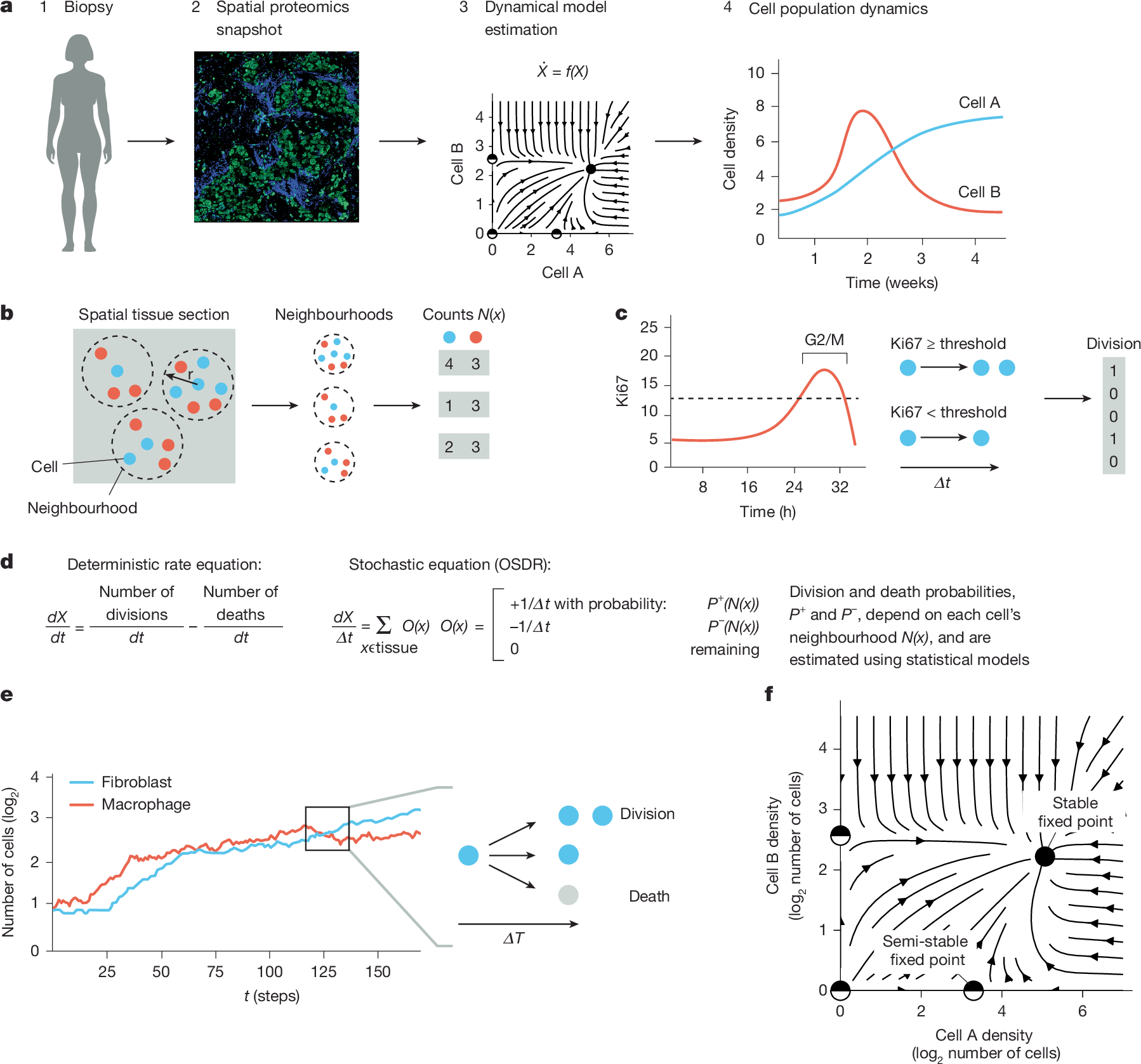
Physiological and pathological processes such as inflammation and cancer emerge from interactions between cells over time1. However, methods to follow cell populations over time within the native context of a human tissue are lacking because a biopsy offers only a single snapshot. Here we present one-shot tissue dynamics reconstruction (OSDR), an approach to estimate a dynamical model of cell populations based on a single tissue sample. OSDR uses spatial proteomics to learn how the composition of cellular neighbourhoods influences division rate, providing a dynamical model of cell population change over time. We apply OSDR to human breast cancer data2–4, and reconstruct two fixed points of fibroblasts and macrophage interactions5,6. These fixed points correspond to hot and cold fibrosis7, in agreement with co-culture experiments that measured these dynamics directly8. We then use OSDR to discover a pulse-generating excitable circuit of T and B cells in the tumour microenvironment, suggesting temporal flares of anticancer immune responses. Finally, we study longitudinal biopsies from a triple-negative breast cancer clinical trial3, in which OSDR predicts the collapse of the tumour cell population in responders but not in non-responders, based on early-treatment biopsies. OSDR can be applied to a wide range of spatial proteomics assays to enable analysis of tissue dynamics based on patient biopsies.
Updates on Osteoimmunology: What's New on the Cross-Talk Between Bone and Immune System
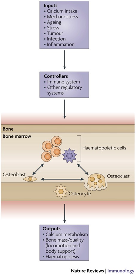
The term osteoimmunology was coined many years ago to describe the research field that deals with the cross-regulation between bone cells and the immune system. As a matter of fact, many factors that are classically considered immune-related, such as interleukins (i.e. IL-6, -11,-17 and -23), tumor necrosis factor (TNF)-α, receptor-activator of nuclear factor kappa B (RANK) and its ligand (RANKL), nuclear factor of activated t-cell, cytoplasmatic-1 (NFATc1) and others have all been found to be crucial in osteoclast and osteoblast biology. Conversely, bone cells, which we used to think would only regulate each other and take care of remodeling bone, actually regulate immune cells, by creating the so-called “endosteal niche”. Both osteoblasts and osteoclasts participate to this niche, either by favoring engraftment, or mobilization of hematopoietic stem cells (HSCs). In this review, we will describe the main milestones at the base of the osteoimmunology and present the key cellular players of the bone-immune system cross-talk, including HSCs, osteoblasts, osteoclasts, bone marrow macrophages, osteomacs, T- and B-lymphocytes, dendritic cells and neutrophils. We will also briefly describe some pathological conditions in which the bone-immune system cross-talk plays a crucial role, with the final aim to portray the state of the art in the mechanisms regulating the bone-immune system interplay, and some of the latest molecular players in the field. This is important to encourage investigation in this field, to identify new targets in the treatment of bone and immune diseases.

Mathematical Discovery of Potential Therapeutic Targets: Application to Rare Melanomas
Patients with rare types of melanoma such as acral, mucosal, or uveal melanoma, have lower survival rates than patients with cutaneous melanoma; these lower survival rates reflect the lower objective response rates to immunotherapy compared to cutaneous melanoma. Understanding tumor-immune dynamics in rare melanomas is critical for the development of new therapies and for improving response rates to current cancer therapies. Progress has been hindered by the lack of clinical data and the need for better preclinical models of rare melanomas. Canine melanoma provides a valuable comparative oncology model for rare types of human melanomas. We analyzed RNA sequencing data from canine melanoma patients and combined this with literature information to create a novel mechanistic mathematical model of melanoma-immune dynamics. Sensitivity analysis of the mathematical model indicated influential pathways in the dynamics, providing support for potential new therapeutic targets and future combinations of therapies. We share our learnings from this work, to help enable the application of this proof-of-concept workflow to other rare disease settings with sparse available data.

OSTEOIMMUNOLOGY: Interplay Between the Immune System and Bone Metabolism
Studies of bone and the immune system have converged in recent years under the banner of osteoimmunology. The immune system is spawned in the bone marrow reservoir, and investigators now recognize that important niches also exist there for memory lymphocytes. At the same time, various factors produced during immune responses are capable of profoundly affecting regulation of bone. Mechanisms have evolved to prevent excessive interference by the immune system with bone homeostasis, yet pathologic bone loss is a common sequela associated with autoimmunity and cancer. There are also developmental links, or parallels, between bone and the immune system. Cells that regulate bone turnover share a common precursor with inflammatory immune cells and may restrict themselves anatomically, in part by utilizing a signaling network analogous to lymphocyte costimulation. Efforts are currently under way to further characterize how these two organ systems overlap and to develop therapeutic strategies that benefit from this understanding.

Immunoporosis: Role of Innate Immune Cells in Osteoporosis
Osteoporosis or porous bone disorder is the result of an imbalance in an otherwise highly balanced physiological process known as ‘bone remodeling’. The immune system is intricately involved in bone physiology as well as pathologies. Inflammatory diseases are often correlated with osteoporosis. Inflammatory mediators such as reactive oxygen species (ROS), and pro-inflammatory cytokines and chemokines directly or indirectly act on the bone cells and play a role in the pathogenesis of osteoporosis. Recently, Srivastava et al. (Srivastava RK, Dar HY, Mishra PK. Immunoporosis: Immunology of Osteoporosis-Role of T Cells. Frontiers in immunology. 2018;9:657) have coined the term “immunoporosis” to emphasize the role of immune cells in the pathology of osteoporosis. Accumulated pieces of evidence suggest both innate and adaptive immune cells contribute to osteoporosis. However, innate cells are the major effectors of inflammation. They sense various triggers to inflammation such as pathogen-associated molecular patterns (PAMPS), damage-associated molecular patterns (DAMPS), cellular stress, etc., thus producing pro-inflammatory mediators that play a critical role in the pathogenesis of osteoporosis. In this review, we have discussed the role of the innate immune cells in great detail and divided these cells into different sections in a systemic manner. In the beginning, we talked about cells of the myeloid lineage, including macrophages, monocytes, and dendritic cells. This group of cells explicitly influences the skeletal system by the action of production of pro-inflammatory cytokines and can transdifferentiate into osteoclast. Other cells of the myeloid lineage, such as neutrophils, eosinophils, and mast cells, largely impact osteoporosis via the production of pro-inflammatory cytokines. Further, we talked about the cells of the lymphoid lineage, including natural killer cells and innate lymphoid cells, which share innate-like properties and play a role in osteoporosis. In addition to various innate immune cells, we also discussed the impact of classical pro-inflammatory cytokines on osteoporosis. We also highlighted the studies regarding the impact of physiological and metabolic changes in the body, which results in chronic inflammatory conditions such as ageing, ultimately triggering osteoporosis.

Mathematical Modeling of the Gut–Bone Axis and Implications of Butyrate Treatment on Osteoimmunology
Butyrate, a short-chain fatty acid produced by the gut microbiota, has pivotal roles in the regulation of the immune system. Recent studies have revealed that butyrate increases the differentiation of peripheral regulatory T cells in the gut–bone axis and promotes osteoblasts’ bone forming activity. However, the mechanism of the therapeutic benefit of butyrate in bone remodeling remains incompletely understood. Here, we develop a multicompartment mathematical model to quantitatively predict the contribution of butyrate on the expansion of regulatory T cells in the gut, blood, and bone compartments. We investigate the interplay between regulatory T cell-derived TGF-β and CD8+ T cell-derived Wnt-10b with changes in gut butyrate concentration. In addition, we connect our model to a detailed model of bone metabolism to study the impacts of butyrate and Wnt-10b on trabecular bone volume. Our results indicate both direct and indirect immune-mediated impacts of butyrate on bone metabolism.

Hierarchical classification of immune cell transcriptomes at population-scale
Accurate immune cell classification is essential for interpreting single-cell RNA sequencing (scRNA-seq) data. However, progress is constrained by the lack of independent, high-resolution benchmarks, as the routine integration of datasets introduces statistical dependencies that artificially inflate model generalizability. Here, we present the single-cell universal classification omnibus (Suco), a resource of independent, uniform expert annotations, and Compocyte, a modular hierarchical classifier. Together, they establish a framework designed for the scale of human population immunology. This approach substantially outperforms existing classifiers while facilitating expert review of ambiguous annotations. Applying Compocyte across 50 studies, including three newly generated datasets, we classified 15.6 million leukocytes from 3,965 patients. Within this expansive cohort, we identified a new tumor-associated resorptive macrophage phenotype, a non-canonical monocyte subtype in subclinical cytokine release syndrome, and the programmatic erosion of T cell memory stemness across metastatic sites. Suco and Compocyte thus provide a generalizable architecture and benchmark capable of sustaining high-resolution annotation across massive clinical cohorts. ### Competing Interest Statement CMR has consulted regarding oncology drug development with Amgen, AstraZeneca, Daiichi Sankyo, Genentech, Merck, and Novartis, and has received licensing and royalty payments for DLL3-directed therapeutics. T.W. reports stock ownership for Roche, Astra Zeneca, Bayer, Innate Pharma, Kyntra, Illumina, 10x Genomics, and Merck KGaA as well as research funding from Atrandi Biosciences, Vilnius, Lithuania; CanVirex AG, Basel Switzerland; and Institut fuer Klinische Krebsforschung GmbH, Frankfurt, Germany, and travel funding from Roche, Basel, Switzerland. S.Z. reports advisory board membership and honoraria from Amgen, Astellas, AstraZeneca, Bayer, Bristol-Myers Squibb, Daiichi Sankyo, Eisai, EUSA, Gilead, Ipsen, Johnson&Johnson, Lilly, MedSir, Medtoday, Merck, MSD, Novartis, Pfizer, Roche, Sanofi Aventis, StreamedUp, Urotrials, Urotube, Zentiva and resarch funding from Eisai. S.Z. reports clinical trial support from Amgen, AstraZeneca, AVEO, Bayer, Biontech, Bristol-Myers Squibb, Calithera, Exelixis, Gilead, Lilly, MSD, Novartis, Pfizer, Roche, Seagen/Astellas, Urotrials and travels & conference support from Amgen, Astellas, AstraZeneca, Bayer, EISAI, Ipsen, Johnson&Johnson, Merck, MSD, Pfizer. All remaining authors declare no relevant competing interests. Spanish Association Against Cancer, PI049999 Federal Ministry of Research, Technology and Space, 001001KT2322 National Cancer Institute, R35 CA263816 National Cancer Institute, U24 CA213274 National Cancer Institute, P30 CA008748 Research Council of Lithuania, P-MIP-24-93

Immunoporosis: Immunology of Osteoporosis—Role of T Cells
The role of immune system in various bone pathologies such as osteoporosis, osteoarthritis and rheumatoid arthritis is now well established. This had led to the emergence of a modern field of systems biology called as osteoimmunology, an integrated research between fields of immunology and bone biology under one umbrella. Osteoporosis is one of the most common inflammatory bone-loss conditions with more than 200 million individuals affected worldwide. T helper cells along with various other immune cells are major players involved in bone homeostasis. In the present review, we specifically discuss the role of various defined T lymphocyte subsets (Th cells comprising Th1, Th2, Th9, Th17, Th22, regulatory T cells, follicular helper T cells, natural killer T cells, γδ T cells and CD8+ T cells) in the pathophysiology of osteoporosis. The study of the specific role of immune system in osteoporosis has now been proposed by our group as “Immunoporosis: The immunology of osteoporosis” with special emphasis on the role of various subsets of T lymphocytes. The establishment of this new field had been the need of the hour due to the emergence of novel roles of various T cell lymphocytes in accelerated bone loss observed during osteoporosis. Activated T cells either directly or indirectly through the secretion of various cytokines and factors modulate bone health and thereby regulate bone remodelling. Various studies have summarized the role of inflammation in pathogenesis of osteoporosis, but very few reports had delineated the precise role of various T cell subsets in the pathobiology of osteoporosis. The present review thus for the first time clearly highlights and summarizes the role of various T lymphocytes in the development and pathophysiology of osteoporosis, giving birth to a new field of biology termed as “Immunoporosis”. This novel field will thus provide an overview of the nexus between the cellular components of both bone and immune systems, responsible for the observed bone loss in osteoporosis. A molecular insight into the upcoming and novel field of immunoporosis would thus lead to development of innovative approaches for the prevention and treatment of osteoporosis.

Computational Modeling of Interactions between Multiple Myeloma and the Bone Microenvironment
Multiple Myeloma (MM) is a B-cell malignancy that is characterized by osteolytic bone lesions. It has been postulated that positive feedback loops in the interactions between MM cells and the bone microenvironment form reinforcing ‘vicious cycles’, resulting in more bone resorption and MM cell population growth in the bone microenvironment. Despite many identified MM-bone interactions, the combined effect of these interactions and their relative importance are unknown. In this paper, we develop a computational model of MM-bone interactions and clarify whether the intercellular signaling mechanisms implemented in this model appropriately drive MM disease progression. This new computational model is based on the previous bone remodeling model of Pivonka et al. [1], and explicitly considers IL-6 and MM-BMSC (bone marrow stromal cell) adhesion related pathways, leading to formation of two positive feedback cycles in this model. The progression of MM disease is simulated numerically, from normal bone physiology to a well established MM disease state. Our simulations are consistent with known behaviors and data reported for both normal bone physiology and for MM disease. The model results suggest that the two positive feedback cycles identified for this model are sufficient to jointly drive the MM disease progression. Furthermore, quantitative analysis performed on the two positive feedback cycles clarifies the relative importance of the two positive feedback cycles, and identifies the dominant processes that govern the behavior of the two positive feedback cycles. Using our proposed quantitative criteria, we identify which of the positive feedback cycles in this model may be considered to be ‘vicious cycles’. Finally, key points at which to block the positive feedback cycles in MM-bone interactions are identified, suggesting potential drug targets.
Osteoprotegerin in Bone Metastases: Mathematical Solution to the Puzzle
Bone is a common site for cancer metastasis. To create space for their growth, cancer cells stimulate bone resorbing osteoclasts. Cytokine RANKL is a key osteoclast activator, while osteoprotegerin (OPG) is a RANKL decoy receptor and an inhibitor of osteoclastogenesis. Consistently, systemic application of OPG decreases metastatic tumor burden in bone. However, OPG produced locally by cancer cells was shown to enhance osteolysis and tumor growth. We propose that OPG produced by cancer cells causes a local reduction in RANKL levels, inducing a steeper RANKL gradient away from the tumor and towards the bone tissue, resulting in faster resorption and tumor expansion. We tested this hypothesis using a mathematical model of nonlinear partial differential equations describing the spatial dynamics of OPG, RANKL, PTHrP, osteoclasts, tumor and bone mass. We demonstrate that at lower expression rates, tumor-derived OPG enhances the chemotactic RANKL gradient and osteolysis, whereas at higher expression rates OPG broadly inhibits RANKL and decreases osteolysis and tumor burden. Moreover, tumor expression of a soluble mediator inducing RANKL in the host tissue, such as PTHrP, is important for correct orientation of the RANKL gradient. A meta-analysis of OPG, RANKL and PTHrP expression in normal prostate, carcinoma and metastatic tissues demonstrated an increase in expression of OPG, but not RANKL, in metastatic prostate cancer, and positive correlation between OPG and PTHrP in metastatic prostate cancer. The proposed mechanism highlights the importance of the spatial distribution of receptors, decoys and ligands, and can be applied to other systems involving regulation of spatially anisotropic processes.
A unified network systems approach uncovers a core program underlying T follicular helper cell differentiation
Characterizing multi-scale processes underlying immune-state progression is critical for defining their function. This becomes pertinent for functionally diverse and plastic immune cells, such as T follicular helper (Tfh) cells. Here, we adopt a multi-scale network-systems approach that incorporates both regulatory and protein-protein interactions. This approach integrates diverse data types, captures regulation across levels of immune system organization, and recapitulates known Tfh differentiation drivers. Further, we present CoreNet, a core Tfh gene set that is conserved between humans and mice, across tissue types and disease contexts, and is consistent across data modalities. Using CoreNet, we implicate NR3C1 and interleukin (IL)-12 in the regulation of Tfh differentiation. Notably, IL-12 is permissive for differentiation of Tfh precursors but blocks differentiation into germinal center Tfh cells. Overall, this work elucidates networks with unexplored roles governing Tfh differentiation across species and tissues, while providing a generalizable framework. CoreNet is accessible through an interactive web server: https://pitt-csi.shinyapps.io/tfhcorenet/.
Mathematical modelling of the pathogenesis of multiple myeloma‐induced bone disease
SUMMARY Multiple myeloma (MM) is the second most common haematological malignancy and results in destructive bone lesions. The interaction between MM cells and the bone microenvironment plays an important role in the development of the tumour cells and MM‐induced bone disease and forms a ‘vicious cycle’ of tumour development and bone destruction, intensified by suppression of osteoblast activity and promotion of osteoclast activity. In this paper, a mathematical model is proposed to simulate how the interaction between MM cells and the bone microenvironment facilitates the development of the tumour cells and the resultant bone destruction. It includes both the roles of inhibited osteoblast activity and stimulated osteoclast activity. The model is able to mimic the temporal variation of bone cell concentrations and resultant bone volume after the invasion and then removal of the tumour cells and explains why MM‐induced bone lesions rarely heal even after the complete removal of MM cells. The behaviour of the model compares well with published experimental data. The model serves as a first step to understand the development of MM‐induced bone disease and could be applied further to evaluate the current therapies against MM‐induced bone disease and even suggests new potential therapeutic targets. © 2014 The Authors. International Journal for Numerical Methods in Biomedical Engineering published by John Wiley & Sons Ltd

A T Cell View of the Bone Marrow
The majority of T cells present in the bone marrow (BM) represent an activated/memory phenotype and most of these, if not all, are circulating T cells. Their lodging in the bone marrow keeps them activated, turning the bone marrow microenvironment into a “memory reservoir”. This article will focus on how T cell activation in the marrow results in both direct and indirect effect on the hematopoiesis. The hematopoietic stem cell niche will be presented, with its main components and organization, along with the role-played by T-lymphocytes in basal and pathologic conditions and their effect on the bone remodeling process. Also discussed herein will be how “normal” bone mass peak is achieved only in the presence of an intact adaptive immune system, with T and B cells playing critical roles in this process. Our main hypothesis is that the partnership between T cells and cells of the bone marrow microenvironment orchestrates numerous processes regulating immunity, hematopoiesis and bone remodeling.

Osteoimmunology: shared mechanisms and crosstalk between the immune and bone systems
Osteoimmunology is an interdisciplinary field, that covers the shared mechanisms and interactions between bone cells and immune cellsReceptor activator of nuclear factor-κB (NF-κB) ligand (RANKL) is an osteoclast-differentiation factor that links the activated immune system and bone loss. In addition, abnormal bone homeostasis has been observed in various mice deficient in immunomodulatory molecules.Osteoclast differentiation is dependent on the transcription factor nuclear factor of activated T cells, cytoplasmic 1 (NFATc1), which is induced and activated by RANKL and its co-stimulatory (immunoglobulin-like) receptors.Interleukin-17 (IL-17)-producing T helper cells (TH17 cells) are the key T-cell subset that links T-cell activation and bone destruction in autoimmune arthritis.Bone cells are involved in the maintenance and mobilization of haematopoietic stem cells.Osteoimmunology is becoming increasingly important for understanding the pathogenesis of, and developing new therapeutic strategies for, diseases that affect both systems.
Comparing phenotypic manifolds with Kompot: Detecting differential abundance and gene expression at single-cell resolution
Single-cell studies are frequently designed to compare across conditions such as health and disease. However, existing computational approaches typically rely on grouping cells into discrete populations before making comparisons, which can limit resolution for detecting state-dependent changes. Here, we introduce Kompot, a statistical framework for comparative analysis of multi-condition single-cell data. Kompot quantifies both differential abundance, capturing how cells redistribute across the phenotypic space, and differential expression, identifying condition-specific transcriptional changes that may be localized, heterogeneous, or oppositely regulated across states. By modeling cell density and gene expression as continuous functions over a shared cell-state representation, Kompot enables single-cell–resolution inference with principled uncertainty estimates, without requiring predefined clusters or cell types. Applying Kompot to aging murine bone marrow, we identified a continuum of shifts in hematopoietic stem cell and mature cell states, transcriptional remodeling of monocytes independent of compositional changes, and divergent regulation of oxidative stress response genes across cell types. We demonstrate the utility of Kompot in disease settings by identifying cell-state and gene expression changes associated with improved efficacy of combinatorial immunotherapy in melanoma. Additionally, Kompot enables multi-sample comparative analysis by accounting for sample-to-sample heterogeneity. By capturing both global and cell-state–specific effects of perturbation, the Kompot framework is broadly applicable to dissecting condition-specific effects in complex single-cell landscapes. ### Competing Interest Statement The authors have declared no competing interest. National Institutes of Health, R35GM147125, R01CA292932, T32GM136534, S10OD028685 The Mark Foundation for Cancer Research, https://ror.org/00v7th354, Endeavor Award Edward P. Evans Foundation, https://ror.org/03h22gm35, Discovery Research Grant Brotman Baty Institute, https://ror.org/03jxvbk42, Pilot Award
Modeling the Human Bone–Tumor Niche: Reducing and Replacing the Need for Animal Data
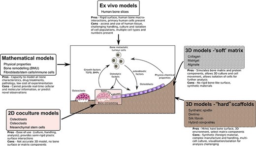
ABSTRACT Bone is the most common site for cancer metastasis. Understanding the interactions within the complex, heterogeneous bone–tumor microenvironment is essential for the development of new therapeutics. Various animal models of tumor‐induced bone disease are routinely used to provide valuable information on the relationship between cancer cells and the skeleton. However, new model systems exist that offer an alternative approach to the use of animals and might more accurately reveal the cellular interactions occurring within the human bone–tumor niche. This review highlights replacement models that mimic the bone microenvironment and where cancer metastases and tumor growth might be assessed alongside bone turnover. Such culture models include the use of calcified regions of animal tissue and scaffolds made from bone mineral hydroxyapatite, synthetic polymers that can be manipulated during manufacture to create structures resembling trabecular bone surfaces, gel composites that can be modified for stiffness and porosity to resemble conditions in the tumor–bone microenvironment. Possibly the most accurate model system involves the use of fresh human bone samples, which can be cultured ex vivo in the presence of human tumor cells and demonstrate similar cancer cell–bone cell interactions as described in vivo. In addition, the use of mathematical modeling and computational biology approaches provide an alternative to preliminary animal testing. The use of such models offers the capacity to mimic significant elements of the human bone–tumor environment, and complement, refine, or replace the use of preclinical models. © 2020 The Authors. JBMR Plus published by Wiley Periodicals, Inc. on behalf of American Society for Bone and Mineral Research.