


Connections between the gut and brain monitor the intestinal tissue and its microbial and dietary content1, regulating both physiological intestinal functions such as nutrient absorption and motility2,3, and brain-wired feeding behaviour2. It is therefore plausible that circuits exist to detect gut microorganisms and relay this information to areas of the central nervous system that, in turn, regulate gut physiology4. Here we characterize the influence of the microbiota on enteric-associated neurons by combining gnotobiotic mouse models with transcriptomics, circuit-tracing methods and functional manipulations. We find that the gut microbiome modulates gut-extrinsic sympathetic neurons: microbiota depletion leads to increased expression of the neuronal transcription factor cFos, and colonization of germ-free mice with bacteria that produce short-chain fatty acids suppresses cFos expression in the gut sympathetic ganglia. Chemogenetic manipulations, translational profiling and anterograde tracing identify a subset of distal intestine-projecting vagal neurons that are positioned to have an afferent role in microbiota-mediated modulation of gut sympathetic neurons. Retrograde polysynaptic neuronal tracing from the intestinal wall identifies brainstem sensory nuclei that are activated during microbial depletion, as well as efferent sympathetic premotor glutamatergic neurons that regulate gastrointestinal transit. These results reveal microbiota-dependent control of gut-extrinsic sympathetic activation through a gut–brain circuit.
Brain-Gut Circuit Lets Microbiota Directly Affect the Sympathetic Nervous System
Gut bacteria play a direct role in neuronal communications between the gut and the brain, according to new research.

A microbiome-dependent gut–brain pathway regulates motivation for exercise
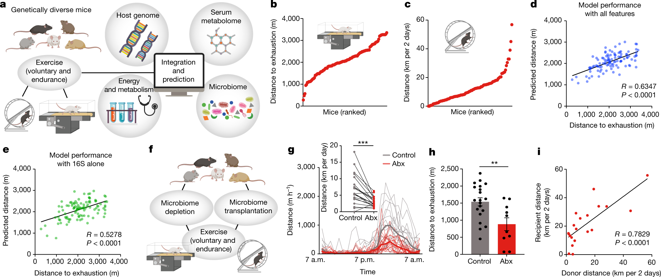
Exercise exerts a wide range of beneficial effects for healthy physiology1. However, the mechanisms regulating an individual’s motivation to engage in physical activity remain incompletely understood. An important factor stimulating the engagement in both competitive and recreational exercise is the motivating pleasure derived from prolonged physical activity, which is triggered by exercise-induced neurochemical changes in the brain. Here, we report on the discovery of a gut–brain connection in mice that enhances exercise performance by augmenting dopamine signalling during physical activity. We find that microbiome-dependent production of endocannabinoid metabolites in the gut stimulates the activity of TRPV1-expressing sensory neurons and thereby elevates dopamine levels in the ventral striatum during exercise. Stimulation of this pathway improves running performance, whereas microbiome depletion, peripheral endocannabinoid receptor inhibition, ablation of spinal afferent neurons or dopamine blockade abrogate exercise capacity. These findings indicate that the rewarding properties of exercise are influenced by gut-derived interoceptive circuits and provide a microbiome-dependent explanation for interindividual variability in exercise performance. Our study also suggests that interoceptomimetic molecules that stimulate the transmission of gut-derived signals to the brain may enhance the motivation for exercise.
Gut microbial taxa elevated by dietary sugar disrupt memory function
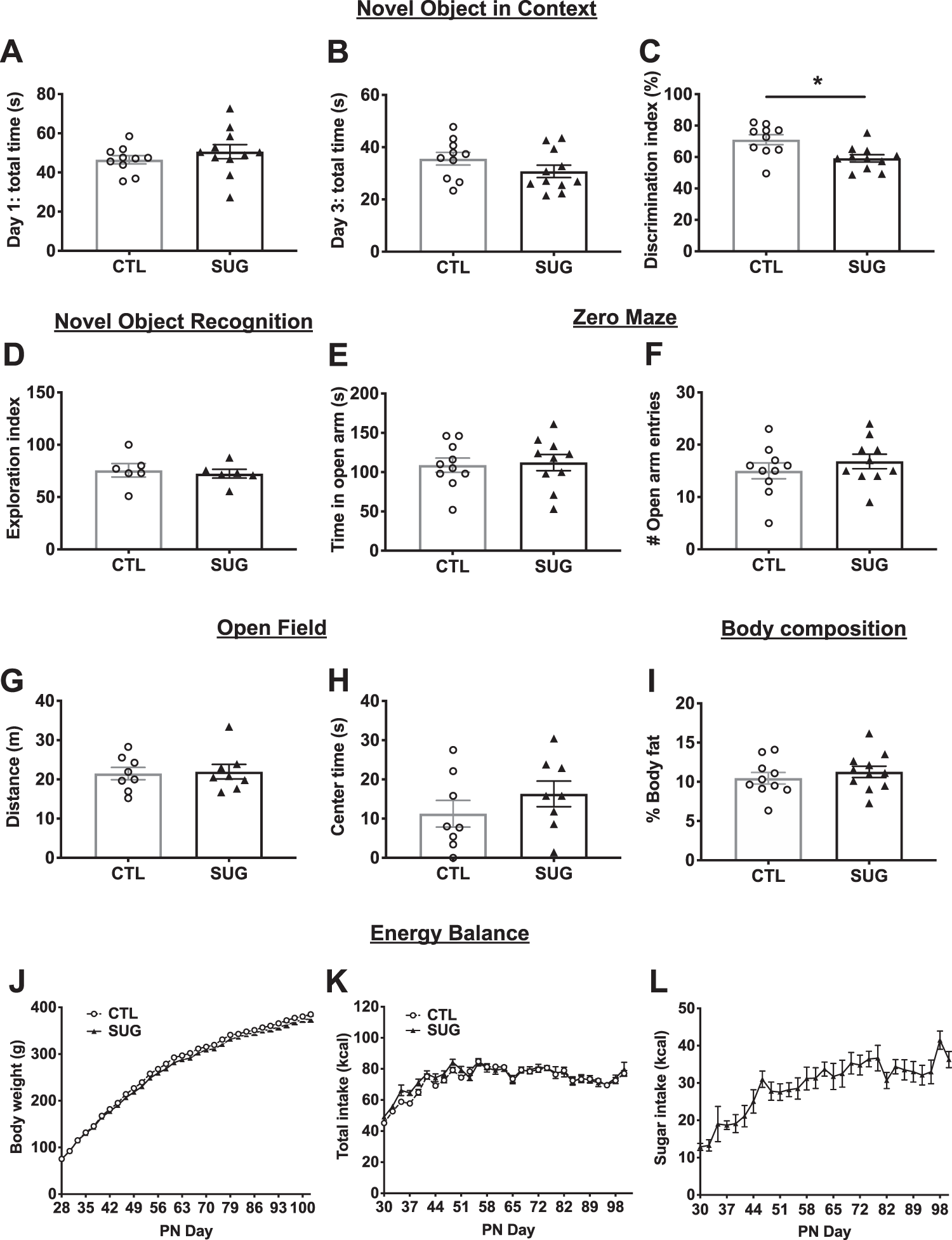
Emerging evidence highlights a critical relationship between gut microbiota and neurocognitive development. Excessive consumption of sugar and other unhealthy dietary factors during early life developmental periods yields changes in the gut microbiome as well as neurocognitive impairments. However, it is unclear whether these two outcomes are functionally connected. Here we explore whether excessive early life consumption of added sugars negatively impacts memory function via the gut microbiome. Rats were given free access to a sugar-sweetened beverage (SSB) during the adolescent stage of development. Memory function and anxiety-like behavior were assessed during adulthood and gut bacterial and brain transcriptome analyses were conducted. Taxa-specific microbial enrichment experiments examined the functional relationship between sugar-induced microbiome changes and neurocognitive and brain transcriptome outcomes. Chronic early life sugar consumption impaired adult hippocampal-dependent memory function without affecting body weight or anxiety-like behavior. Adolescent SSB consumption during adolescence also altered the gut microbiome, including elevated abundance of two species in the genus Parabacteroides (P. distasonis and P. johnsonii) that were negatively correlated with hippocampal function. Transferred enrichment of these specific bacterial taxa in adolescent rats impaired hippocampal-dependent memory during adulthood. Hippocampus transcriptome analyses revealed that early life sugar consumption altered gene expression in intracellular kinase and synaptic neurotransmitter signaling pathways, whereas Parabacteroides microbial enrichment altered gene expression in pathways associated with metabolic function, neurodegenerative disease, and dopaminergic signaling. Collectively these results identify a role for microbiota “dysbiosis” in mediating the detrimental effects of early life unhealthy dietary factors on hippocampal-dependent memory function.
Bacteria in the gut have a direct line to the brain
With its 100 million neurons, the gut has earned a reputation as the body's "second brain"—corresponding with the real brain to manage things like intestinal muscle activity and enzyme secretions. A growing ...

Role of diet and its effects on the gut microbiome in the pathophysiology of mental disorders
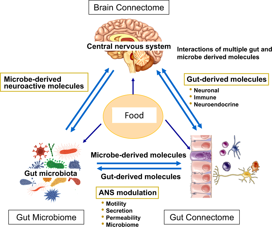
There is emerging evidence that diet has a major modulatory influence on brain-gut-microbiome (BGM) interactions with important implications for brain health, and for several brain disorders. The BGM system is made up of neuroendocrine, neural, and immune communication channels which establish a network of bidirectional interactions between the brain, the gut and its microbiome. Diet not only plays a crucial role in shaping the gut microbiome, but it can modulate structure and function of the brain through these communication channels. In this review, we summarize the evidence available from preclinical and clinical studies on the influence of dietary habits and interventions on a selected group of psychiatric and neurologic disorders including depression, cognitive decline, Parkinson’s disease, autism spectrum disorder and epilepsy. We will particularly address the role of diet-induced microbiome changes which have been implicated in these effects, and some of which are shared between different brain disorders. While the majority of these findings have been demonstrated in preclinical and in cross-sectional, epidemiological studies, to date there is insufficient evidence from mechanistic human studies to make conclusions about causality between a specific diet and microbially mediated brain function. Many of the dietary benefits on microbiome and brain health have been attributed to anti-inflammatory effects mediated by the microbial metabolites of dietary fiber and polyphenols. The new attention given to dietary factors in brain disorders has the potential to improve treatment outcomes with currently available pharmacological and non-pharmacological therapies.
The Microbial Metabolites, Short-Chain Fatty Acids, Regulate Colonic T<sub>reg</sub> Cell Homeostasis
Protecting the Guts Regulatory T cells (T regs ) in the gut are important sentinels in maintaining the peace between our gut and its trillions of resident bacteria and have been shown to be regulated by specific strains of bacteria in mouse models. Smith et al. (p. 569 , published online 4 July; see the Perspective by Bollrath and Powrie ) asked whether metabolite(s) generated by resident bacterial species may regulate T regs in the gut. Indeed, short-chain fatty acids (SCFAs), bacterial fermentation products of dietary fibers produced by a range of bacteria, restored colonic T reg numbers in mice devoid of a gut microbiota and increased T reg numbers in colonized mice. The effects of SCFAs on T regs were mediated through GPCR43, a receptor for SCFAs, which is expressed on colonic T regs . Mice fed SCFAs were protected against experimentally induced colitis in a manner that was dependent on GPR43-expressing T regs . , Bacterial fermentation products regulate the number and function of regulatory T cells in the mouse colon. [Also see Perspective by Bollrath and Powrie ] , Regulatory T cells (T regs ) that express the transcription factor Foxp3 are critical for regulating intestinal inflammation. Candidate microbe approaches have identified bacterial species and strain-specific molecules that can affect intestinal immune responses, including species that modulate T reg responses. Because neither all humans nor mice harbor the same bacterial strains, we posited that more prevalent factors exist that regulate the number and function of colonic T regs . We determined that short-chain fatty acids, gut microbiota–derived bacterial fermentation products, regulate the size and function of the colonic T reg pool and protect against colitis in a Ffar2 -dependent manner in mice. Our study reveals that a class of abundant microbial metabolites underlies adaptive immune microbiota coadaptation and promotes colonic homeostasis and health.

Regulation of Intestinal Barrier Function by Microbial Metabolites
The human gastrointestinal tract (GI) harbors a diverse population of microbial life that continually shapes host pathophysiological responses. Despite readily available abundant metagenomic data, the functional dynamics of gut microbiota remain to be explored in various health and disease conditions. Microbiota generate a variety of metabolites from dietary products that influence host health and pathophysiological functions. Since gut microbial metabolites are produced in close proximity to gut epithelium, presumably they have significant impact on gut barrier function and immune responses.

The gut microbiome may influence brain aging, mouse study suggests
A communication pathway between the brain and the gut may be integral to how well the brain holds on to memories
Germ-free mice exhibit profound gut microbiota-dependent alterations of intestinal endocannabinoidome signaling
The gut microbiota is a unique ecosystem of microorganisms interacting with the host through several biochemical mechanisms. The endocannabinoidome (eCBome), a complex signaling system including the endocannabinoid system, approximately 50 receptors and metabolic enzymes, and more than 20 lipid mediators with important physiopathologic functions, modulates gastrointestinal tract function and may mediate host cell-microbe communications there. Germ-free (GF) mice, which lack an intestinal microbiome and so differ drastically from conventionally raised (CR) mice, offer a unique opportunity to explore the eCBome in a microbe-free model and in the presence of a reintroduced functional gut microbiome through fecal microbiota transplant (FMT). We aimed to gain direct evidence for a link between the microbiome and eCBome systems by investigating eCBome alterations in the gut in GF mice before and after FMT. Basal eCBome gene expression and lipid profiles were measured in various segments of the intestine of GF and CR mice at juvenile and adult ages using targeted quantitative PCR transcriptomics and LC-MS/MS lipidomics. GF mice exhibited age-dependent modifications in intestinal eCBome gene expression and lipid mediator levels. FMT from CR donor mice to age-matched GF male mice reversed several of these alterations, particularly in the ileum and jejunum, after only 1 week, demonstrating that the gut microbiome directly impacts the host eCBome and providing a cause-effect relationship between the presence or absence of intestinal microbes and eCBome signaling. These results open the way to new studies investigating the mechanisms through which intestinal microorganisms exploit eCBome signaling to exert some of their physiopathologic functions.

Gut–brain link grabs neuroscientists
Idea that intestinal bacteria affect mental health gains ground.
Got food cravings? What’s living in your gut may be responsible
A new Pitt study shows that the gut microbiome of mice influences their preferred diet. The results, researchers say, could apply to humans, too.

Species-specific CD4+ T cells enable prediction of mucosal immune phenotypes from microbiota composition
Determining the mechanisms by which the gut microbiome modulates the host immune system has translational potential for treating or preventing immune-mediated diseases. A key challenge is identifying the immunogenic bacterial strains in the setting of a complex microbiota. We use a combination of anaerobic culturing, in vitro T cell assays, gnotobiotic mouse models, and ex vivo T cell restimulations to explore the influence of species and strain diversity on the specificity of mucosal T cells. Our approach efficiently predicts the relative impact of individual bacterial strains within a complex community and can be used to parse microbiota-dependent phenotypes into component fractions., How bacterial strains within a complex human microbiota collectively shape intestinal T cell homeostasis is not well understood. Methods that quickly identify effector strains or species that drive specific mucosal T cell phenotypes are needed to define general principles for how the microbiota modulates host immunity. We colonize germ-free mice with defined communities of cultured strains and profile antigen-specific responses directed toward individual strains ex vivo. We find that lamina propria T cells are specific to bacterial strains at the species level and can discriminate between strains of the same species. Ex vivo restimulations consistently identify the strains within complex communities that induce Th17 responses in vivo, providing the potential to shape baseline immune tone via community composition. Using an adoptive transfer model of colitis, we find that lamina propria T cells respond to different bacterial strains in conditions of inflammation versus homeostasis. Collectively, our approach represents a unique method for efficiently predicting the relative impact of individual bacterial strains within a complex community and for parsing microbiota-dependent phenotypes into component fractions.
Host Immunity Influences the Composition of Murine Gut Microbiota
The influence of gut microbiota on host immunity is widely studied, and its disturbance has been linked to several immune-mediated disorders. Conversely, whether and how inherently disturbed canonical Th1 (pro-inflammatory) and/or Th2 (anti-inflammatory) immune pathways modify the host microbiome is not sufficiently investigated. Here, we characterized the humoral, cellular, and cytokine immunity, and associated alterations in gut microbiota of naïve wild-type mice (C57BL/6 and BALB/c), and mice with deficiencies in Th2 responses (IL-4Rα and IL-33 knockout mice) or in both Th1 and Th2 responses (NOD scid gamma, NSG mice). A global analysis by de novo clustering of 16S rRNA profiles of the gut microbiota independently grouped wild-type immunocompetent (C57BL/6 and BALB/c), Th2-deficient (IL-4Rα-/- and IL-33-/-), and severely immunodeficient (NSG) mice; where wild-type mice, but not Th2 or severely immunodeficient mice, were enriched in gut bacteria that produce short-chain fatty acids. These include members of phyla Firmicutes, Verrucomicrobia, and Bacteroidetes such as Lactobacillus spp., Akkermansia muciniphila, and Odoribacter spp. Further comparison of the two naïve wild-type mouse strains showed higher microbial diversity (Shannon), primarily linked to higher richness (Chao1), as well as a distinct difference in microbial composition (weighted UniFrac) in BALB/c mice compared to C57BL/6. T-cell and blood cytokine analyses demonstrated a Th1-polarization in naïve adaptive immunity in C57BL/6 animals compared to BALB/c mice, and an expected Th2 deficient cellular response in IL-4Rα-/- and IL-33-/- mice compared to its genetic background BALB/c strain. Together, these data suggest that alterations in the Th1/Th2 balance or a complete ablation of Th1/Th2 responses can lead to major alterations in gut microbiota composition and function. Given the similarities between the human and mouse immune systems and gut microbiota, our finding that immune status is a strong driver of gut microbiota composition has important consequences for human immunodeficiency studies.
Think Twice: How the Gut's "Second Brain" Influences Mood and Well-Being
The emerging and surprising view of how the enteric nervous system in our bellies goes far beyond just processing the food we eat
Meta-omics analysis of elite athletes identifies a performance-enhancing microbe that functions via lactate metabolism
The human gut microbiome is linked to many states of human health and disease1. The metabolic repertoire of the gut microbiome is vast, but the health implications of these bacterial pathways are poorly understood. In this study, we identify a link between members of the genus Veillonella and exercise performance. We observed an increase in Veillonella relative abundance in marathon runners postmarathon and isolated a strain of Veillonella atypica from stool samples. Inoculation of this strain into mice significantly increased exhaustive treadmill run time. Veillonella utilize lactate as their sole carbon source, which prompted us to perform a shotgun metagenomic analysis in a cohort of elite athletes, finding that every gene in a major pathway metabolizing lactate to propionate is at higher relative abundance postexercise. Using 13C3-labeled lactate in mice, we demonstrate that serum lactate crosses the epithelial barrier into the lumen of the gut. We also show that intrarectal instillation of propionate is sufficient to reproduce the increased treadmill run time performance observed with V. atypica gavage. Taken together, these studies reveal that V. atypica improves run time via its metabolic conversion of exercise-induced lactate into propionate, thereby identifying a natural, microbiome-encoded enzymatic process that enhances athletic performance.

Activation of Gpr109a, Receptor for Niacin and the Commensal Metabolite Butyrate, Suppresses Colonic Inflammation and Carcinogenesis
Commensal gut microflora and dietary fiber protect against colonic inflammation and colon cancer through unknown targets. Butyrate, a bacterial product from fermentation of dietary fiber in the colon, has been implicated in this process. GPR109A (encoded by Niacr1) is a receptor for butyrate in the colon. GPR109A is also a receptor for niacin, which is also produced by gut microbiota and suppresses intestinal inflammation. Here we showed that Gpr109a signaling promoted anti-inflammatory properties in colonic macrophages and dendritic cells and enabled them to induce differentiation of Treg cells and IL-10-producing T cells.
