


What determines the functional organization of cortex? One hypothesis is that innate connectivity patterns, either structural or functional connectivity, set up a scaffold upon which functional specialization can later take place. We tested this hypothesis by asking whether the visual word form area (VWFA), an experience-driven region, was already functionally connected to proto language networks in neonates scanned within one week of birth. Using the data from the Human Connectone Project (HCP) and the Developing Human Connectome Project (dHCP), we calculated intrinsic functional connectivity during resting-state functional magnetic resonance imaging (fMRI), and found that neonates showed similar functional connectivity patterns to adults. We observed that (1) language regions connected more strongly with the putative VWFA than other adjacent ventral visual regions that also show foveal bias, and (2) the VWFA connected more strongly with frontotemporal language regions than with regions adjacent to these language regions. These data suggest that the location of the VWFA is earmarked at birth due to its connectivity with the language network, providing evidence that innate connectivity instructs the later refinement of cortex.
An investigation across 45 languages and 12 language families reveals a universal language network
To understand the architecture of human language, it is critical to examine diverse languages; yet most cognitive neuroscience research has focused on a handful of primarily Indo-European languages. Here, we report an investigation of the fronto-temporo-parietal language network across 45 languages and establish the robustness to cross-linguistic variation of its topography and key functional properties, including left-lateralization, strong functional integration among its brain regions, and functional selectivity for language processing.
Brain Hyperconnectivity in Children with Autism and its Links to Social Deficits
In this study, Supekar, Menon, and colleagues shed light on a longstanding question about the nature of brain connectivity aberrations in autism. Using functional neuroimaging data, they demonstrate that the brains of children with autism are hyperconnected in ways that are related to the severity of social impairment exhibited by these children. They replicate these findings in two additional cohorts of children, challenging current theories of brain connectivity in autism and providing unique insights into the brain mechanisms underlying the disorder.

The neuroimaging correlates of depression established across six large-scale population datasets
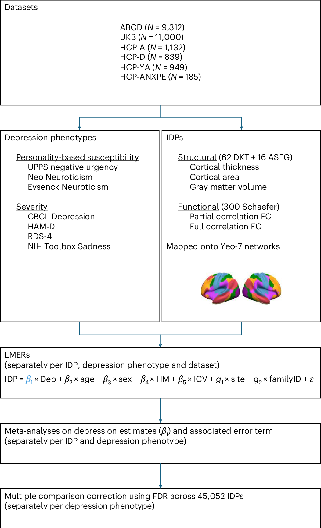
Depression has been linked to reduced size of subcortical regions and abnormal functional connectivity in frontal and default mode networks. However, recent meta-analyses have failed to identify significant converging correlates of depression across the literature such that a conclusive mapping of the neuroimaging correlates of depression remains elusive. We leveraged 23,417 participants across 6 population datasets to comprehensively establish the neuroimaging correlates of depression. We found reductions in gray matter volume/cortical surface area associated with depression in the frontal cortex, anterior cingulate and insula, confirming previous studies showing the importance of prefrontal and default mode regions in depression. Our findings demonstrate multiple surprising results, including a lack of depression correlates in subcortical brain regions and significant depression correlates in somatomotor and visual regions. Overall, these results shed new light on key brain regions involved in the pathophysiology of depression, updating our understanding of the neuroimaging correlates of depression symptoms.
The neuroimaging correlates of depression established across six large-scale population datasets
Depression has been linked to reduced size of subcortical regions and abnormal functional connectivity in frontal and default mode networks. However, recent meta-analyses have failed to identify significant converging correlates of depression across the literature such that a conclusive mapping of the neuroimaging correlates of depression remains elusive. We leveraged 23,417 participants across 6 population datasets to comprehensively establish the neuroimaging correlates of depression. We found reductions in gray matter volume/cortical surface area associated with depression in the frontal cortex, anterior cingulate and insula, confirming previous studies showing the importance of prefrontal and default mode regions in depression. Our findings demonstrate multiple surprising results, including a lack of depression correlates in subcortical brain regions and significant depression correlates in somatomotor and visual regions. Overall, these results shed new light on key brain regions involved in the pathophysiology of depression, updating our understanding of the neuroimaging correlates of depression symptoms.
The brain functional connectome is robustly altered by lack of sleep
Sleep is a universal phenomenon necessary for maintaining homeostasis and function across a range of organs. Lack of sleep has severe health-related consequences affecting whole-body functioning, yet no other organ is as severely affected as the brain. The neurophysiological mechanisms underlying these deficits are poorly understood. Here, we characterize the dynamic changes in brain connectivity profiles inflicted by sleep deprivation and how they deviate from regular daily variability. To this end, we obtained functional magnetic resonance imaging data from 60 young, adult male participants, scanned in the morning and evening of the same day and again the following morning. 41 participants underwent total sleep deprivation before the third scan, whereas the remainder had another night of regular sleep. Sleep deprivation strongly altered the connectivity of several resting-state networks, including dorsal attention, default mode, and hippocampal networks. Multivariate classification based on connectivity profiles predicted deprivation state with high accuracy, corroborating the robustness of the findings on an individual level. Finally, correlation analysis suggested that morning-to-evening connectivity changes were reverted by sleep (control group)-a pattern which did not occur after deprivation. We conclude that both, a day of waking and a night of sleep deprivation dynamically alter the brain functional connectome.
Embracing the suboptimal organization of the human brain
Human brain architecture is guiding brain-inspired artificial intelligence (AI) and has been treated as an optimal template, whose deviations could mark different psychiatric and neurological conditions. We argue this premise is wrong: under any single goal (e.g., minimal wiring cost or maximal communication efficiency), the human connectome is suboptimal. Instead, its organization reflects multi-objective trade-offs navigated over evolution and development under biological and environmental constraints. For psychopathology, atypical trajectories are not distances from an ideal brain but reweighted compromises in the same trade-off space. For neuro-AI, directly duplicating the brain’s connectivity risks copying its irrelevant compromises. Treating brains and models as products of multi-objective optimization and co-tuning relevant objectives offers a more powerful framework for interpreting clinical phenotypes and designing next-generation AI.

Handwriting but not typewriting leads to widespread brain connectivity: a high-density EEG study with implications for the classroom
As traditional handwriting is progressively being replaced by digital devices, it is essential to investigate the implications for the human brain. Brain electrical activity was recorded in 36 university students as they were handwriting visually presented words using a digital pen and typewriting the words on a keyboard. Connectivity analyses were performed on EEG data recorded with a 256-channel sensor array. When writing by hand, brain connectivity patterns were far more elaborate than when typewriting on a keyboard, as shown by widespread theta/alpha connectivity coherence patterns between network hubs and nodes in parietal and central brain regions. Existing literature indicates that connectivity patterns in these brain areas and at such frequencies are crucial for memory formation and for encoding new information and, therefore, are beneficial for learning. Our findings suggest that the spatiotemporal pattern from visual and proprioceptive information obtained through the precisely controlled hand movements when using a pen, contribute extensively to the brain's connectivity patterns that promote learning. We urge that children, from an early age, must be exposed to handwriting activities in school to establish the neuronal connectivity patterns that provide the brain with optimal conditions for learning. Although it is vital to maintain handwriting practice at school, it is also important to keep up with continuously developing technological advances. Therefore, both teachers and students should be aware of which practice has the best learning effect in what context, for example when taking lecture notes or when writing an essay.

🚨 We're very happy to introduce TRIBE v2: a foundation model of the human brain's responses to sight, sound, and language. Leveraging 1,000+ hours of fMRI across 720 subjects, it generalizes… | Stéphane d'Ascoli | 16 comments
🚨 We're very happy to introduce TRIBE v2: a foundation model of the human brain's responses to sight, sound, and language. Leveraging 1,000+ hours of fMRI across 720 subjects, it generalizes zero-shot to new stimuli, tasks and people, finetunes efficiently, and enables in-silico experiments. ❓How does it work? Stemming from our v1, which won the Algonauts 2025 challenge, TRIBE v2 combines video, audio, and language embeddings to predict brain activity for any brain, then adapts to each individual. Key results: 📊 High-quality predictions — TRIBE v2 predicts brain activity across cortical and subcortical regions, significantly better than standard linear models, with a log-linear scaling law and no plateau in sight. 🎯 Zero-shot generalization — Without retraining, the predictions of TRIBE v2 are more correlated with group-averaged brain responses than almost any individual fMRI scan! A short finetuning step vastly improves over linear models trained, from scratch, on each individual. 🧪 In-silico experiments — Can we do useful experiments with TRIBE v2? Yes: classic vision and language paradigms replicate in-silico. It zero-shot recovers the FFA, PPA, EBA, VWFA, Broca's lateralization, and syntactic responses in STG — all without training on these artificial tasks. 🔍 Interpretability & multimodality — ICA on the weights rediscovers known functional networks (auditory, language, motion, default mode, visual) from naturalistic data alone. Ablating modalities further maps how vision, audition, and language integrate, with the largest gains at the temporo-parietal-occipital junction. 🧠 This effort is a step toward a foundation model of the human brain. Much remains to be understood, but we hope this opens a path for neuroscience, AI, and medical research alike. All code, weights, and a live demo are open — find it useful or mistaken in some conditions? Let us know, new test cases can only help improving this effort. 📄 Paper: https://lnkd.in/e7cbunJp 💻 Code: https://lnkd.in/ebwBVuJp ▶️ Demo: https://lnkd.in/eEUVxP4S 🤗 Model: https://lnkd.in/e2T8nPJP Joint work with Jérémy RAPIN, Yohann Benchetrit, Teon Brooks, Katie Begany, Joséphine Raugel, Hubert Banville and Jean-Rémi King. 🙏 Special thanks to Elisa Cascardi, Diego Marcos, Dominic Giardini, AI at Meta, and the open-source and neuroscience communities (in particular Lune Bellec and Bertrand Thirion for the amazing Courtois NeuroMod and IBC datasets) | 16 comments on LinkedIn
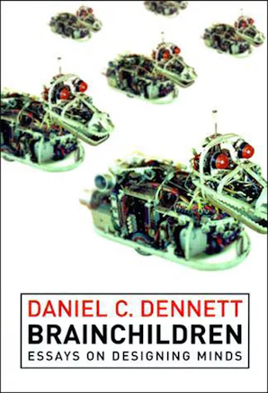
Brainchildren
Minds are complex artifacts, partly biological and partly social; only a unified, multidisciplinary approach will yield a realistic theory of how they came i...
Understanding the Limits of Vision Language Models Through the Lens of the Binding Problem
Recent work has documented striking heterogeneity in the performance of state-of-the-art vision language models (VLMs), including both multimodal language models and text-to-image models. These models are able to describe and generate a diverse array of complex, naturalistic images, yet they exhibit surprising failures on basic multi-object reasoning tasks -- such as counting, localization, and simple forms of visual analogy -- that humans perform with near perfect accuracy. To better understand this puzzling pattern of successes and failures, we turn to theoretical accounts of the binding problem in cognitive science and neuroscience, a fundamental problem that arises when a shared set of representational resources must be used to represent distinct entities (e.g., to represent multiple objects in an image), necessitating the use of serial processing to avoid interference. We find that many of the puzzling failures of state-of-the-art VLMs can be explained as arising due to the binding problem, and that these failure modes are strikingly similar to the limitations exhibited by rapid, feedforward processing in the human brain.
NeuroAI’s Next Steps: Balancing Computation and Embodiment
Deep neural networks trained at scale now predict neural activity across sensory cortices, raising a question that divides neuroscience: does the convergence between artificial and biological representations constitute understanding, or does understanding require models of biological processes grounded in the embodied, evolved character of natural intelligence? In this Dual Perspectives article, Bing Brunton and Patrick Mineault articulate two research programs—Process and Convergence—to engage this question about the future of NeuroAI research. The conversation is organized around four questions that probe what scaled model–brain alignment reveals, what the brain is coupled to and evolved to do, what success beyond the lab looks like, and where the field should invest over the coming decade. Their exchanges reveal the distance between the two programs to be primarily about the level of analysis and priorities for what to build next. The conversation converges on shared ground: measure broadly, then distill to causal principles.

re:vision | Replication Initiative for Visual Neuroscience
Replicate and generalize visual-neuroscience findings using the LAION-fMRI (LfMRI) 7T dataset.

Verbalizable Representations Form a Global Workspace in Language Models
If the mind is an ocean, we spend our lives floating at the surface. Beneath us, an enormous amount of processing takes place without our knowledge: our visual systems parsing the contours of a face, our motor circuits maintaining our posture. At any given moment, only a small fraction of this neural activity is accessible to us. Yet it is this privileged sliver of activity that we rely on to reason deliberately: to plan what ingredients to buy for a recipe, or to puzzle out why an engine won’t start. Such thoughts can be articulated out loud, deliberately held in mind, and brought to bear on whatever task the moment demands. This distinction, between our accessible thoughts and our unconscious processing, is perhaps the most striking feature of human cognition.
Verbalizable Representations Form a Global Workspace in Language Models
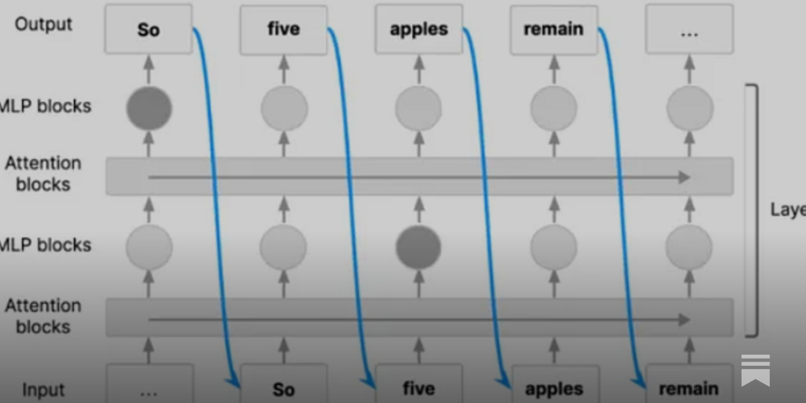
If the mind is an ocean, we spend our lives floating at the surface. Beneath us, an enormous amount of processing takes place without our knowledge: our visual systems parsing the contours of a face, our motor circuits maintaining our posture. At any given moment, only a small fraction of this neural activity is accessible to us. Yet it is this privileged sliver of activity that we rely on to reason deliberately: to plan what ingredients to buy for a recipe, or to puzzle out why an engine won’t start. Such thoughts can be articulated out loud, deliberately held in mind, and brought to bear on whatever task the moment demands. This distinction, between our accessible thoughts and our unconscious processing, is perhaps the most striking feature of human cognition.
Introducing TRIBE v2: A Predictive Foundation Model Trained to Understand How the Human Brain Processes Complex Stimuli | Keith Doelling
This is some very cool work by some awesome colleagues Jean-Rémi King, and Teon Brooks! Seriously not enough good things can be said about how cool it is. You should enjoy it and play with it. And kudos to Meta for open sourcing it. At the same time, I'm already seeing posts about how the model will replace fMRI experiments as researchers will simulate how the brain "really works" instead of running costly experiments. I think this goes WELL beyond what its creators intend. We are already seeing that use of AI in science allows you to explore charted ideas more thoroughly and much more rapidly but slows us down in finding novel ideas (https://lnkd.in/eMR2akqt). At the same time, there is growing concern that LLM performance will collapse as they are increasingly trained on their own output (https://lnkd.in/eavgfyuY). Leaving neuroscience to AI simulations risks following the same fate, where we generate seemingly new findings without gaining new meaning. A mechanistic understanding of how the brain works (if that is still your goal) will be found at the margins, in errors and idiosyncrasies of neural function. What TRIBE provides is a super useful and cool instantiation of our current understanding on how and where neural activity is instantiated in the brain. But it won't help us make groundbreaking new findings of how neural circuits lead to cognition and behavior. Experiments on real human brains, may be costly, but they will always be necessary!