

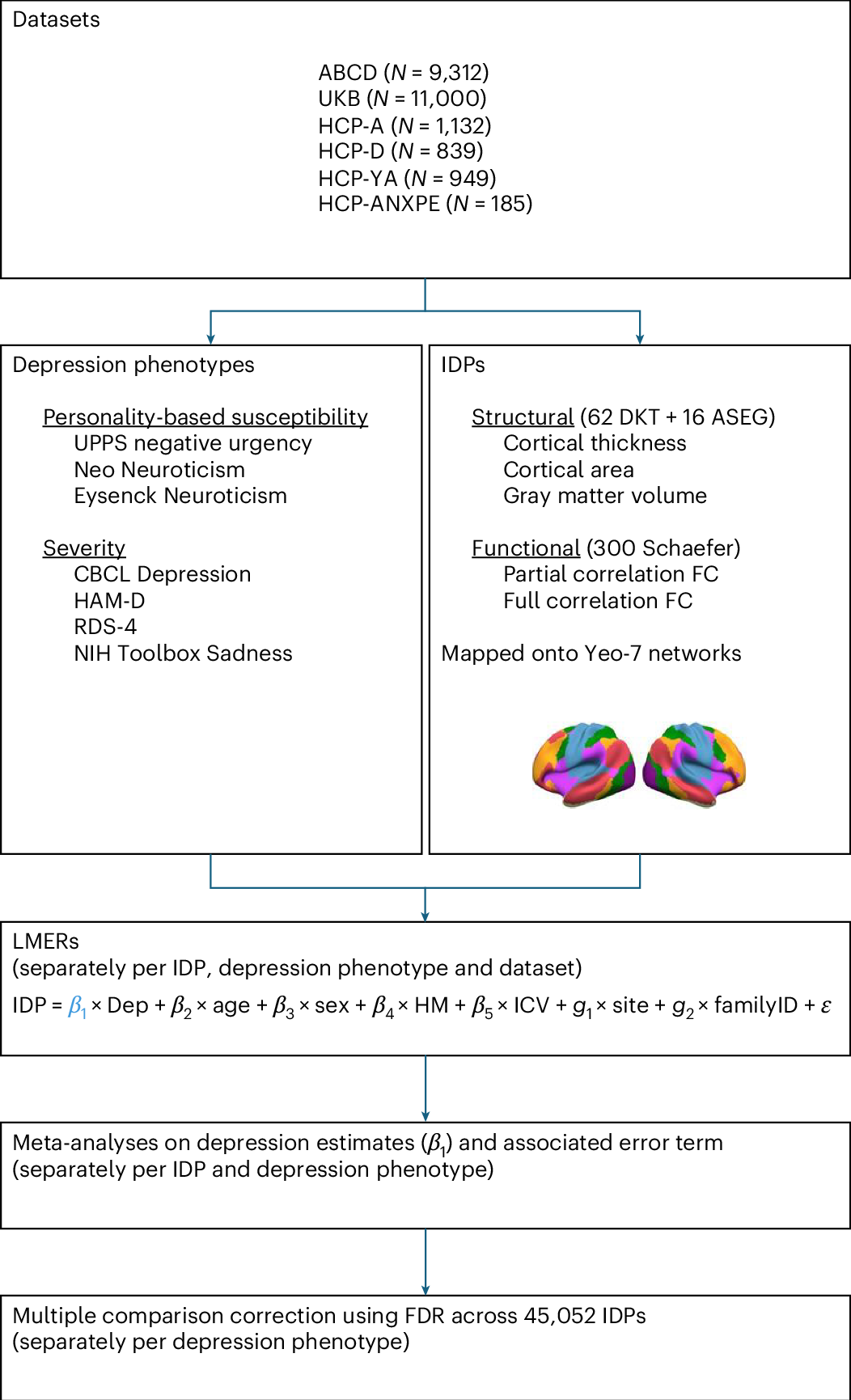
Depression has been linked to reduced size of subcortical regions and abnormal functional connectivity in frontal and default mode networks. However, recent meta-analyses have failed to identify significant converging correlates of depression across the literature such that a conclusive mapping of the neuroimaging correlates of depression remains elusive. We leveraged 23,417 participants across 6 population datasets to comprehensively establish the neuroimaging correlates of depression. We found reductions in gray matter volume/cortical surface area associated with depression in the frontal cortex, anterior cingulate and insula, confirming previous studies showing the importance of prefrontal and default mode regions in depression. Our findings demonstrate multiple surprising results, including a lack of depression correlates in subcortical brain regions and significant depression correlates in somatomotor and visual regions. Overall, these results shed new light on key brain regions involved in the pathophysiology of depression, updating our understanding of the neuroimaging correlates of depression symptoms.
The neuroimaging correlates of depression established across six large-scale population datasets
Depression has been linked to reduced size of subcortical regions and abnormal functional connectivity in frontal and default mode networks. However, recent meta-analyses have failed to identify significant converging correlates of depression across the literature such that a conclusive mapping of the neuroimaging correlates of depression remains elusive. We leveraged 23,417 participants across 6 population datasets to comprehensively establish the neuroimaging correlates of depression. We found reductions in gray matter volume/cortical surface area associated with depression in the frontal cortex, anterior cingulate and insula, confirming previous studies showing the importance of prefrontal and default mode regions in depression. Our findings demonstrate multiple surprising results, including a lack of depression correlates in subcortical brain regions and significant depression correlates in somatomotor and visual regions. Overall, these results shed new light on key brain regions involved in the pathophysiology of depression, updating our understanding of the neuroimaging correlates of depression symptoms.
Depression doesn't shrink the brain like we thought it did | New Scientist
For the past decade, depression has been linked to a shrunken hippocampus, which has a vital role in memory. But a large study using up-to-date scanning techniques suggests this may not be the case

What stress does to the brain - Neuroscience News
A new study sheds light on how the brain changes as a result of chronic stress. Selective noradrenaline release rewires connectivity patterns between different brain regions. Activity in the amygdala and networks that process sensory stimuli increased.

Dysregulated adult hippocampal neurogenesis in major depressive disorder
Major depressive disorder (MDD) is associated with reduced hippocampal volume, altered connectivity and negative memory bias, suggesting disrupted hippocampal plasticity. Dysregulated adult hippocampal neurogenesis is a potential contributor, but its relevance in humans and role in MDD remain unclear. Here we investigated the molecular basis of hippocampal dysfunction in nonmedicated individuals with MDD by integrating analyses of neurogenic trajectories, cell-type- and subfield-specific gene expression, chromatin accessibility and protein expression. We identify a neurogenic lineage in the adult human hippocampal subgranular zone and provide evidence for a stalled neurogenic process in MDD, associated with transcriptional regulation, stress-related reprogramming and interferon signaling across developmental stages. Excitatory and inhibitory neurons show dysregulation of transcription factor networks affecting cell states. Cellular stress, excitatory–inhibitory imbalance, impaired synaptic plasticity, reduced metabolic capacity and immune activation, underlie impaired neurogenesis and reduced hippocampus circuit plasticity. Findings indicate genetic and epigenetic regulation of gene expression in MDD, and overlapping pathogenetic mechanisms with autoimmune, neurodevelopmental and neurodegenerative diseases. This work provides a new understanding of the pathogenesis of hippocampus-dependent cognitive symptoms in MDD and suggests potential therapeutic targets.
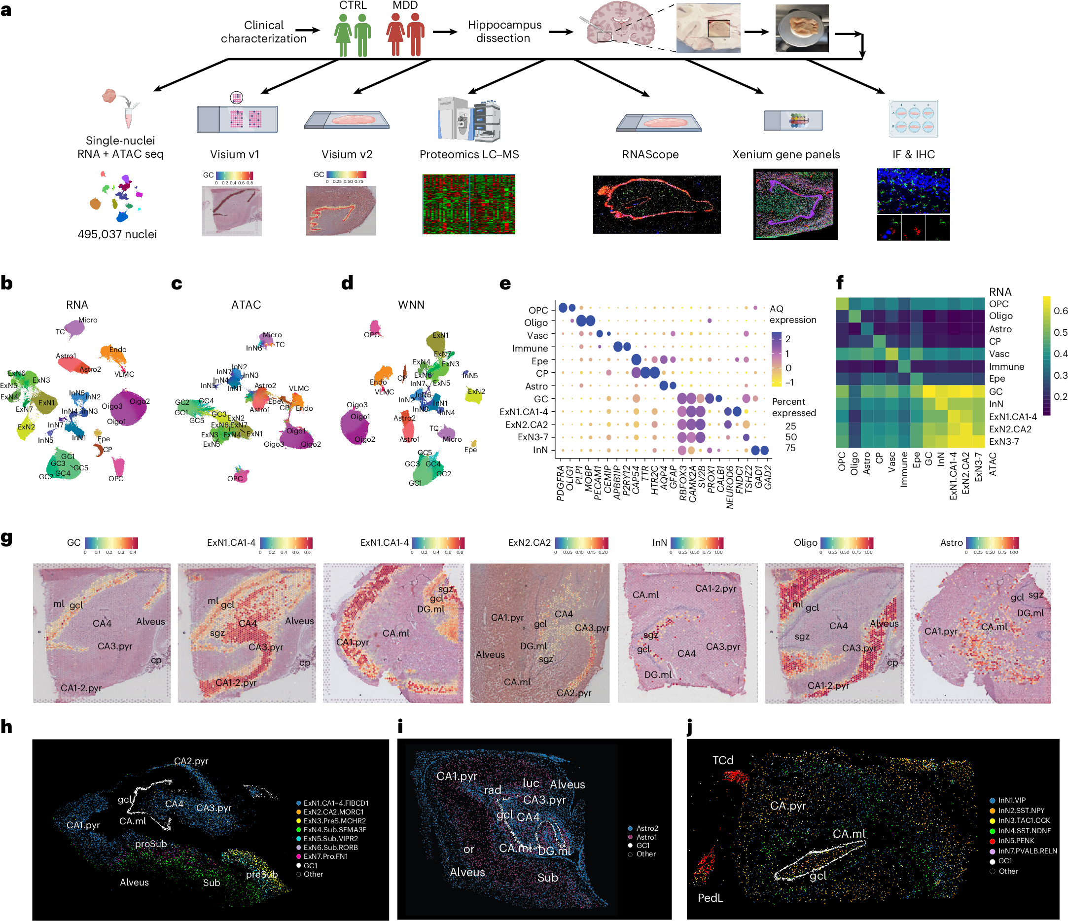
Dysregulated adult hippocampal neurogenesis in major depressive disorder
Major depressive disorder (MDD) is associated with reduced hippocampal volume, altered connectivity and negative memory bias, suggesting disrupted hippocampal plasticity. Dysregulated adult hippocampal neurogenesis is a potential contributor, but its relevance in humans and role in MDD remain unclear. Here we investigated the molecular basis of hippocampal dysfunction in nonmedicated individuals with MDD by integrating analyses of neurogenic trajectories, cell-type- and subfield-specific gene expression, chromatin accessibility and protein expression. We identify a neurogenic lineage in the adult human hippocampal subgranular zone and provide evidence for a stalled neurogenic process in MDD, associated with transcriptional regulation, stress-related reprogramming and interferon signaling across developmental stages. Excitatory and inhibitory neurons show dysregulation of transcription factor networks affecting cell states. Cellular stress, excitatory–inhibitory imbalance, impaired synaptic plasticity, reduced metabolic capacity and immune activation, underlie impaired neurogenesis and reduced hippocampus circuit plasticity. Findings indicate genetic and epigenetic regulation of gene expression in MDD, and overlapping pathogenetic mechanisms with autoimmune, neurodevelopmental and neurodegenerative diseases. This work provides a new understanding of the pathogenesis of hippocampus-dependent cognitive symptoms in MDD and suggests potential therapeutic targets.
Generative AI Use and Depressive Symptoms Among US Adults
This survey study examines the associations of generative artificial intelligence (AI) use with negative affective symptoms in a large nationally representative sample of US adults.

Brain Hyperconnectivity in Children with Autism and its Links to Social Deficits
In this study, Supekar, Menon, and colleagues shed light on a longstanding question about the nature of brain connectivity aberrations in autism. Using functional neuroimaging data, they demonstrate that the brains of children with autism are hyperconnected in ways that are related to the severity of social impairment exhibited by these children. They replicate these findings in two additional cohorts of children, challenging current theories of brain connectivity in autism and providing unique insights into the brain mechanisms underlying the disorder.

The Entangled Brain: How Perception, Cognition, and Emotion Are Woven Together
A new vision of the brain as a fully integrated, networked organ.Popular neuroscience accounts often focus on specific mind-brain aspects like addiction, c
Embracing the suboptimal organization of the human brain
Human brain architecture is guiding brain-inspired artificial intelligence (AI) and has been treated as an optimal template, whose deviations could mark different psychiatric and neurological conditions. We argue this premise is wrong: under any single goal (e.g., minimal wiring cost or maximal communication efficiency), the human connectome is suboptimal. Instead, its organization reflects multi-objective trade-offs navigated over evolution and development under biological and environmental constraints. For psychopathology, atypical trajectories are not distances from an ideal brain but reweighted compromises in the same trade-off space. For neuro-AI, directly duplicating the brain’s connectivity risks copying its irrelevant compromises. Treating brains and models as products of multi-objective optimization and co-tuning relevant objectives offers a more powerful framework for interpreting clinical phenotypes and designing next-generation AI.

#precision #scaling #generalization #impact | Jean-Rémi King
Well, we did not expect that: in 10 days: 1.6K ⭐ on Github, 70K downloads on HuggingFace 🤗, >6M views on socials. 🧠 TRIBE v2, our new foundation model of the human brain responses to sight, sound, and language, has led to a surge of community demos, dozens of PRs and issues, and a level of engagement that rivals major flagships model at Meta (e.g. Llama4: 3.6M views, and DINOv3: 900K): https://lnkd.in/eQgqyDvf As this raised several questions, let me emphasize some elements of clarification: 1. #Precision: This model exclusively uses fMRI recordings. While powerful, these data are slow-paced proxy measurements of brain activity, and aggregate responses ~100k-3M neurons per data point. This means we are very far from a neuronal-level modeling of the brain. 2. #Scaling: We leveraged ~1,000 hours of naturalistic fMRI data. This is very substantial for a functional imaging study with naturalistic data; but still this is "small data" compared to medical or biology foundation models in general (e.g. structural MRI models are typically tens of thousands of individual brains). 3. #Generalization: The model shows surprising out-of-domain generalization, but it is **not** a replacement for new data collection. The model will almost surely fail in areas it hasn't seen: task-specific behaviors, memory protocols, touch sensation etc. And these are the "known unknowns" - I expect we'll discover many more unknown unknowns along the way. 4. #Impact: The goal of this model, and of our team in general, is fundamental research in neuroscience. While I am optimistic about how such foundation models will help clinical diagnosis, prognosis, and patient care, the physics of MRI is such that there is no clear path for making this kind of technology directly usable to consumer/wearable products. We're in for the science, hence the open sourcing: 📄 Paper: https://lnkd.in/e7cbunJp 💻 Code: https://lnkd.in/ebwBVuJp ▶️ Demo: https://lnkd.in/eEUVxP4S 🤗 Model: https://lnkd.in/e2T8nPJP

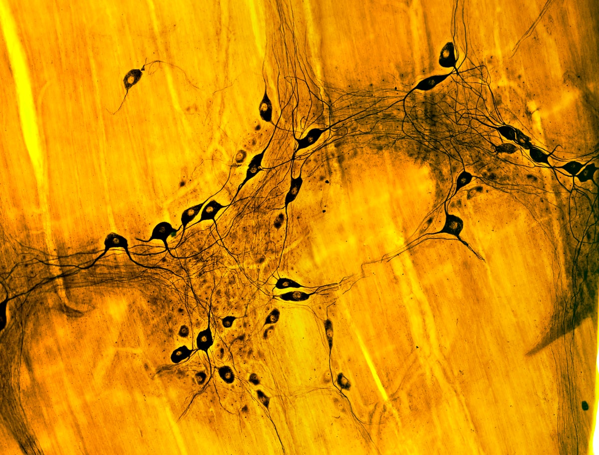
Think Twice: How the Gut's "Second Brain" Influences Mood and Well-Being
The emerging and surprising view of how the enteric nervous system in our bellies goes far beyond just processing the food we eat
Social Media and Depression Symptoms: a Meta-Analysis
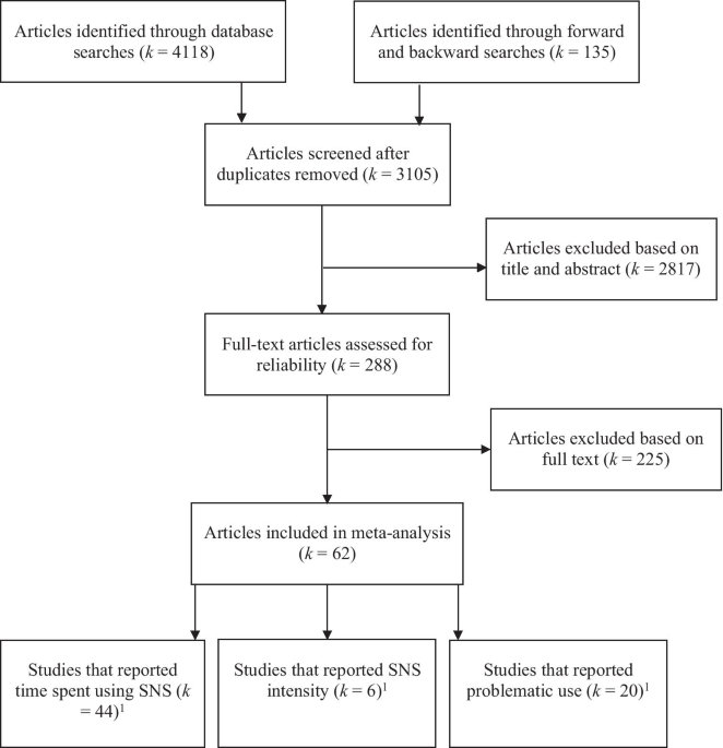
Social Networking Sites (SNS) have close to 3 billion users worldwide. Recently, however, SNS have come under media scrutiny for their potential association with depression. Two previous meta-analyses failed to find evidence for a robust concurrent association between SNS use and depression symptoms. However, these analyses focused primarily on the time spent using SNS. The current meta-analysis is the first to consider the multi-dimensional nature of SNS use, and examines separately the quantitative associations of depression symptoms to SNS use in three types of SNS studies examining three distinct constructs of SNS use: time spent using SNS, intensity of SNS use, and problematic SNS use. Sixty-two studies (N = 451, 229) met inclusion criteria. Depression symptoms were significantly, but weakly, associated with time spent using SNS (r = 0.11) and intensity of SNS use (r = 0.09). However, the association of depression symptoms to problematic SNS use was moderate (r = 0.29), was significantly higher than for time spent using SNS (Qbetween = 35.85, p < 0.001) or intensity of SNS use (Qbetween = 13.95, p < 0.001), and was not significantly moderated by age, gender, year of study publication, or mode of recruitment. These results suggest that future research examining causal models of the relation of SNS use and depression, as well as research on intervention and prevention, should focus in more detail on individuals who are engaging in a pattern of problematic SNS use.
Dance training is superior to repetitive physical exercise in inducing brain plasticity in the elderly
Animal research indicates that a combination of physical activity and sensory enrichment has the largest and the only sustaining effect on adult neuroplasticity. Dancing has been suggested as a human homologue to this combined intervention as it poses demands on both physical and cognitive functions. For the present exploratory study, we designed an especially challenging dance program in which our elderly participants constantly had to learn novel and increasingly difficult choreographies. This six-month-long program was compared to conventional fitness training matched for intensity. An extensive pre/post-assessment was performed on the 38 participants (63–80 y), covering general cognition, attention, memory, postural and cardio-respiratory performance, neurotrophic factors and–most crucially–structural MRI using an exploratory analysis. For analysis of MRI data, a new method of voxel-based morphometry (VBM) designed specifically for pairwise longitudinal group comparisons was employed. Both interventions increased physical fitness to the same extent. Pronounced differences were seen in the effects on brain volumes: Dancing compared to conventional fitness activity led to larger volume increases in more brain areas, including the cingulate cortex, insula, corpus callosum and sensorimotor cortex. Only dancing was associated with an increase in plasma BDNF levels. Regarding cognition, both groups improved in attention and spatial memory, but no significant group differences emerged. The latter finding may indicate that cognitive benefits may develop later and after structural brain changes have taken place. The present results recommend our challenging dance program as an effective measure to counteract detrimental effects of aging on the brain.
nubrain - A foundation model for neural decoding
Building the world's largest dataset of human brain activity.

Has anyone looked at this paper more closely? I've seen many of my bachelor students refer to it when it comes to depressive disorders. Authors find worldwide depression rates doubled from 1990 to 2020, which seems like a lot. pubmed.ncbi.nlm.nih.gov/40313474/

Trends in Prevalent Cases and Disability-Adjusted Life-Years of Depressive Disorders Worldwide: Findings From the Global Burden of Disease Study From 1990 to 2021 - PubMed
pubmed.ncbi.nlm.nih.gov