During hindbrain development, segmental regulation of the paralogous Hoxa2 and Hoxb2 genes in rhombomeres (r) 3 and 5 involves Krox20-dependent enhancers that have been conserved during the duplication of the vertebrate Hox clusters from a common ancestor. Examining these evolutionarily related control regions could provide important insight into the degree to which the basic Krox20-dependent mechanisms, cis-regulatory components, and their organization have been conserved. Toward this goal we have performed a detailed functional analysis of a mouse Hoxa2 enhancer capable of directing reporter expression in r3 and r5. The combined activities of five separate cis-regions, in addition to the conserved Krox20 binding sites, are involved in mediating enhancer function. A CTTT (BoxA) motif adjacent to the Krox20 binding sites is important for r3/r5 activity. The BoxA motif is similar to one (Box1) found in the Hoxb2 enhancer and indicates that the close proximity of these Box motifs to Krox20 sites is a common feature of Krox20 targets in vivo. Two other rhombomeric elements (RE1 and RE3) are essential for r3/r5 activity and share common TCT motifs, indicating that they interact with a similar cofactor(s). TCT motifs are also found in the Hoxb2 enhancer, suggesting that they may be another common feature of Krox20-dependent control regions. The two remaining Hoxa2 cis-elements, RE2 and RE4, are not conserved in the Hoxb2 enhancer and define differences in some of components that can contribute to the Krox20-dependent activities of these enhancers. Furthermore, analysis of regulatory activities of these enhancers in a Krox20 mutant background has uncovered differences in their degree of dependence upon Krox20 for segmental expression. Together, this work has revealed a surprising degree of complexity in the number of cis-elements and regulatory components that contribute to segmental expression mediated by Krox20 and sheds light on the diversity and evolution of Krox20 target sites and Hox regulatory elements in vertebrates.
The zinc finger gene Krox20 regulates HoxB2 (Hox2.8) during hindbrain segmentation
The zinc finger gene Krox20 and many Hox homeobox genes are expressed in segment-restricted domains in the hindbrain. The restricted expression patterns appear before morphological segmentation, suggesting that these transcription factors may play an early role in the establishment and identity of rhombomeric segments. In this paper, we show that the HoxB2 (Hox2.8) gene is normally upregulated in rhombomeres (r) 3, 4, and 5, and we identify an enhancer region upstream of the gene that imposes r3/r5 expression in transgenic mice.

Segmental expression of Hoxb-1 is controlled by a highly conserved autoregulatory loop dependent upon exd/pbx
Comparison of Hoxb-1 regulatory regions from different vertebrates identified three related sequence motifs critical for rhombomere 4 (r4) expression in the hindbrain. Functional analysis in transgenic mice and Drosophila embryos demonstrated that the conserved elements are involved in a positive autoregulatory loop dependent on labial (lab) family members. Binding of Hoxb-1 to these elements in vitro requires cofactors, and the motifs closely resemble the consensus binding site for pbxi, a homolog of the Drosophila extradenticle (exd) homeodomain protein.
Zebrafish hox paralogue group 2 genes function redundantly as selector genes to pattern the second pharyngeal arch
The pharyngeal arches are one of the defining features of the vertebrates, with the first arch forming the mandibles of the jaw and the second forming jaw support structures. The cartilaginous elements of each arch are formed from separate migratory neural crest cell streams, which derive from the dorsal aspect of the neural tube. The second and more posterior crest streams are characterized by specific Hox gene expression. The zebrafish has a larger overall number of Hox genes than the tetrapod vertebrates, as the result of a duplication event in its lineage. However, in both zebrafish and mouse, there are just two members of Hox paralogue group 2 (PG2): Hoxa2 and Hoxb2. Here, we show that morpholino-mediated "knock-down" of both zebrafish Hox PG2 genes results in major defects in second pharyngeal arch cartilages, involving replacement of ventral elements with a mirror-image duplication of first arch structures, and accompanying changes to pharyngeal musculature. In the mouse, null mutants of Hoxa2 have revealed that this single Hox gene is required for normal second arch patterning. By contrast, loss-of-function of either zebrafish Hox PG2 gene individually has no phenotypic consequence, showing that these two genes function redundantly to confer proper pattern to the second pharyngeal arch. We have also used hoxb1a mis-expression to induce localized ectopic expression of zebrafish Hox PG2 genes in the first arch; using this strategy, we find that ectopic expression of either Hox PG2 gene can confer second arch identity onto first arch structures, suggesting that the zebrafish Hox PG2 genes act as "selector genes."
Zebrafish Hox Paralogue Group 2 Genes Function Redundantly as Selector Genes to Pattern the Second Pharyngeal Arch
The pharyngeal arches are one of the defining features of the vertebrates, with the first arch forming the mandibles of the jaw and the second forming jaw support structures. The cartilaginous elements of each arch are formed from separate migratory neural crest cell streams, which derive from the dorsal aspect of the neural tube. The second and more posterior crest streams are characterized by specific Hox gene expression. The zebrafish has a larger overall number of Hox genes than the tetrapod vertebrates, as the result of a duplication event in its lineage. However, in both zebrafish and mouse, there are just two members of Hox paralogue group 2 (PG2): Hoxa2 and Hoxb2. Here, we show that morpholino-mediated “knock-down” of both zebrafish Hox PG2 genes results in major defects in second pharyngeal arch cartilages, involving replacement of ventral elements with a mirror-image duplication of first arch structures, and accompanying changes to pharyngeal musculature. In the mouse, null mutants of Hoxa2 have revealed that this single Hox gene is required for normal second arch patterning. By contrast, loss-of-function of either zebrafish Hox PG2 gene individually has no phenotypic consequence, showing that these two genes function redundantly to confer proper pattern to the second pharyngeal arch. We have also used hoxb1a mis-expression to induce localized ectopic expression of zebrafish Hox PG2 genes in the first arch; using this strategy, we find that ectopic expression of either Hox PG2 gene can confer second arch identity onto first arch structures, suggesting that the zebrafish Hox PG2 genes act as “selector genes.”
A Hox regulatory network of hindbrain segmentation is conserved to the base of vertebrates
Nested Hox expression domains are found in jawed vertebrates and in non-vertebrate chordates, but it is unclear whether there is a link between brain regionalization and Hox expression in jawless vertebrates; here, Hox expression is shown to be integrated with hindbrain segmentation in lampreys.

Enhancer redundancy provides phenotypic robustness in mammalian development
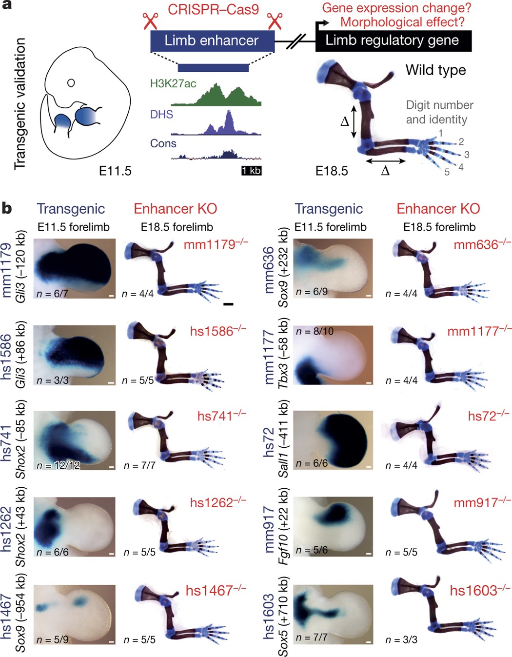
Gene enhancer knockout phenotypes and analysis of enhancer activity patterns show that developmental genes are regulated by multiple redundant enhancers in mouse embryos.
Conserved enhancers control notochord expression of vertebrate Brachyury
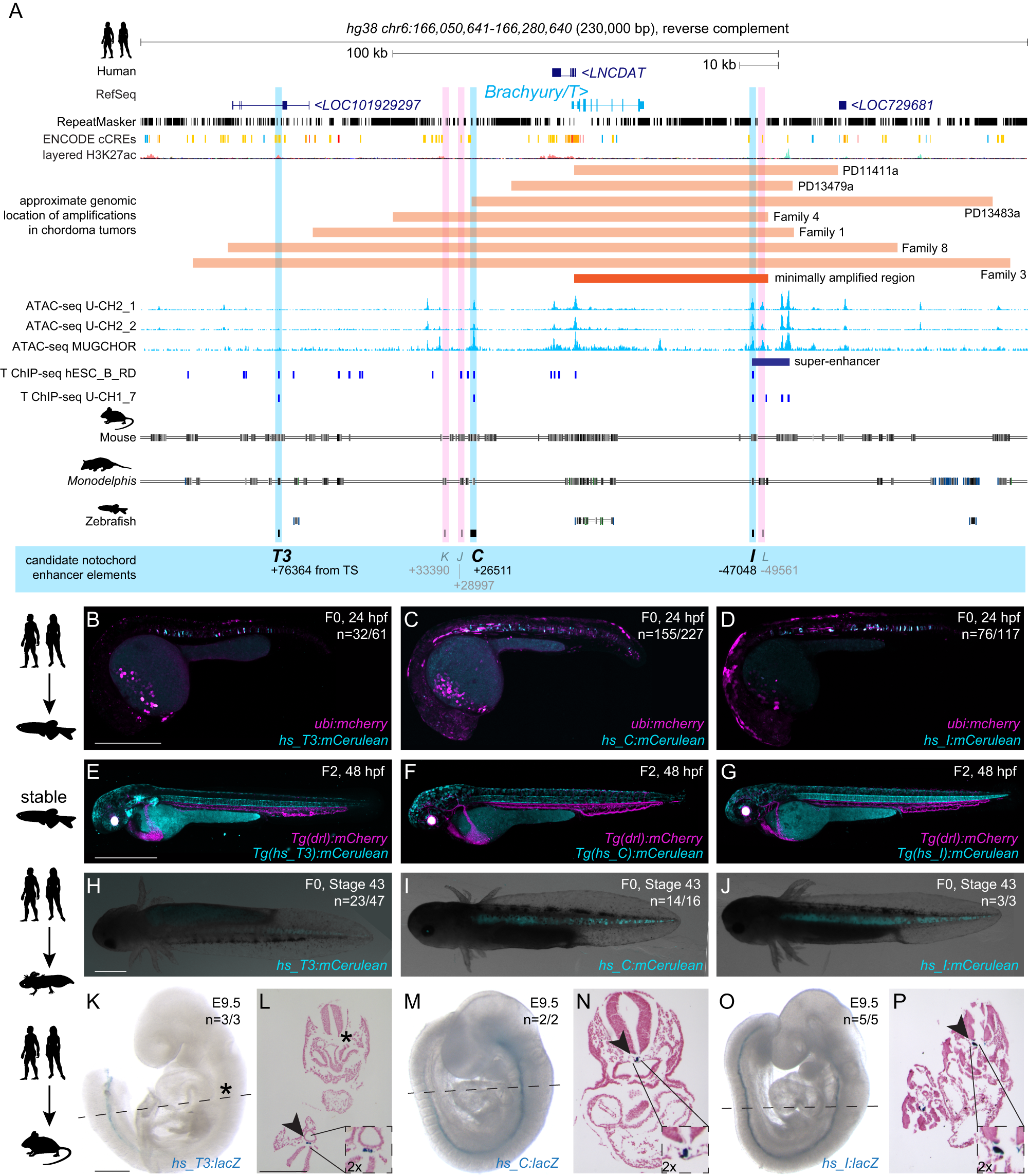
The cell type-specific expression of key transcription factors is central to development and disease. Brachyury/T/TBXT is a major transcription factor for gastrulation, tailbud patterning, and notochord formation; however, how its expression is controlled in the mammalian notochord has remained elusive. Here, we identify the complement of notochord-specific enhancers in the mammalian Brachyury/T/TBXT gene. Using transgenic assays in zebrafish, axolotl, and mouse, we discover three conserved Brachyury-controlling notochord enhancers, T3, C, and I, in human, mouse, and marsupial genomes. Acting as Brachyury-responsive, auto-regulatory shadow enhancers, in cis deletion of all three enhancers in mouse abolishes Brachyury/T/Tbxt expression selectively in the notochord, causing specific trunk and neural tube defects without gastrulation or tailbud defects. The three Brachyury-driving notochord enhancers are conserved beyond mammals in the brachyury/tbxtb loci of fishes, dating their origin to the last common ancestor of jawed vertebrates. Our data define the vertebrate enhancers for Brachyury/T/TBXTB notochord expression through an auto-regulatory mechanism that conveys robustness and adaptability as ancient basis for axis development.
Osteoblast-Osteoclast Communication and Bone Homeostasis
Bone remodeling is tightly regulated by a cross-talk between bone-forming osteoblasts and bone-resorbing osteoclasts. Osteoblasts and osteoclasts communicate with each other to regulate cellular behavior, survival and differentiation through direct cell-to-cell contact or through secretory proteins. A direct interaction between osteoblasts and osteoclasts allows bidirectional transduction of activation signals through EFNB2-EPHB4, FASL-FAS or SEMA3A-NRP1, regulating differentiation and survival of osteoblasts or osteoclasts. Alternatively, osteoblasts produce a range of different secretory molecules, including M-CSF, RANKL/OPG, WNT5A, and WNT16, that promote or suppress osteoclast differentiation and development. Osteoclasts also influence osteoblast formation and differentiation through secretion of soluble factors, including S1P, SEMA4D, CTHRC1 and C3. Here we review the current knowledge regarding membrane bound- and soluble factors governing cross-talk between osteoblasts and osteoclasts.

BMP–Smad1/9 signaling plays a critical role in regulating zebrafish PGC proliferation
The germ cell fate in zebrafish is determined by germ plasm, whereas mammalian germ cell fate is induced by bone morphogenetic protein (BMP) signaling. It remains elusive whether BMP signaling is implicated in zebrafish germ cell development. Here, we demonstrate that BMP–Smad1/9 signaling plays a critical role in zebrafish primordial germ cell (PGC) maintenance rather than fate determination. BMP inhibition or smad1/9 knockdown reduces PGC numbers. Furthermore, we generated PGC-specific smad1/9 knockouts using a transgenic approach with PGC-specifically expressed Cas9 and ubiquitously expressed guide RNAs. Smad1/9 deficiency in PGCs leads to impaired PGC proliferation and increased apoptosis, consequently reducing PGC numbers. Transcriptome analysis revealed unchanged PGC-specific gene expression, but a marked upregulation of DNA damage response-related genes, which is validated by ectopic ATR–pChk1 activation in PGCs and PGC restoring by ATR inhibition. Collectively, these findings underscore conserved but functionally distinct roles of BMP signaling in vertebrate PGC development.

Targeted mutagenesis of specific genomic DNA sequences in animals for the in vivo generation of variant libraries
Understanding how the number, placement and affinity of transcription factor binding sites dictates gene regulatory programs remains a major unsolved challenge in biology, particularly in the context of multicellular organisms. To uncover these rules, it is first necessary to find the binding sites within a regulatory region with high precision, and then to systematically modulate this binding site arrangement while simultaneously measuring the effect of this modulation on output gene expression. Massively parallel reporter assays (MPRAs), where the gene expression stemming from 10,000s of in vitro-generated regulatory sequences is measured, have made this feat possible in high-throughput in single cells in culture. However, because of lack of technologies to incorporate DNA libraries, MPRAs are limited in whole organisms. To enable MPRAs in multicellular organisms, we generated tools to create a high degree of mutagenesis in specific genomic loci in vivo using base editing. Targeting GFP integrated in the genome of Drosophila cell culture and whole animals as a case study, we show that the base editor AIDevoCDA1 stemming from sea lamprey fused to nCas9 is highly mutagenic. Surprisingly, longer gRNAs increase mutation efficiency and expand the mutating window, which can allow the introduction of mutations in previously untargetable sequences. Finally, we demonstrate arrays of >20 gRNAs that can efficiently introduce mutations along a 200bp sequence, making it a promising tool to test enhancer function in vivo in a high throughput manner.

Comparing phenotypic manifolds with Kompot: Detecting differential abundance and gene expression at single-cell resolution
Single-cell studies are frequently designed to compare across conditions such as health and disease. However, existing computational approaches typically rely on grouping cells into discrete populations before making comparisons, which can limit resolution for detecting state-dependent changes. Here, we introduce Kompot, a statistical framework for comparative analysis of multi-condition single-cell data. Kompot quantifies both differential abundance, capturing how cells redistribute across the phenotypic space, and differential expression, identifying condition-specific transcriptional changes that may be localized, heterogeneous, or oppositely regulated across states. By modeling cell density and gene expression as continuous functions over a shared cell-state representation, Kompot enables single-cell–resolution inference with principled uncertainty estimates, without requiring predefined clusters or cell types. Applying Kompot to aging murine bone marrow, we identified a continuum of shifts in hematopoietic stem cell and mature cell states, transcriptional remodeling of monocytes independent of compositional changes, and divergent regulation of oxidative stress response genes across cell types. We demonstrate the utility of Kompot in disease settings by identifying cell-state and gene expression changes associated with improved efficacy of combinatorial immunotherapy in melanoma. Additionally, Kompot enables multi-sample comparative analysis by accounting for sample-to-sample heterogeneity. By capturing both global and cell-state–specific effects of perturbation, the Kompot framework is broadly applicable to dissecting condition-specific effects in complex single-cell landscapes. ### Competing Interest Statement The authors have declared no competing interest. National Institutes of Health, R35GM147125, R01CA292932, T32GM136534, S10OD028685 The Mark Foundation for Cancer Research, https://ror.org/00v7th354, Endeavor Award Edward P. Evans Foundation, https://ror.org/03h22gm35, Discovery Research Grant Brotman Baty Institute, https://ror.org/03jxvbk42, Pilot Award

Osteoclasts Provide Coupling Signals to Osteoblast Lineage Cells Through Multiple Mechanisms
Bone remodeling is essential for the repair and replacement of damaged and old bone. The major principle underlying this process is that osteoclast-mediated resorption of a quantum of bone is followed by osteoblast precursor recruitment; these cells differentiate to matrix-producing osteoblasts, which form new bone to replace what was resorbed. Evidence from osteopetrotic syndromes indicate that osteoclasts not only resorb bone, but also provide signals to promote bone formation. Osteoclasts act upon osteoblast lineage cells throughout their differentiation by facilitating growth factor release from resorbed matrix, producing secreted proteins and microvesicles, and expressing membrane-bound factors. These multiple mechanisms mediate the coupling of bone formation to resorption in remodeling. Additional interactions of osteoclasts with osteoblast lineage cells, including interactions with canopy and reversal cells, are required to achieve coordination between bone formation and resorption during bone remodeling.

Regulation of osteoblastogenesis and bone mass by Wnt10b
Wnts comprise a family of secreted signaling proteins that regulate diverse developmental processes. Activation of Wnt signaling by Wnt10b inhibits differentiation of preadipocytes and blocks adipose tissue development; however, the effect of Wnt10b on other mesenchymal lineages has not been defined. To explore the physiological role of Wnt signaling in bone development, we analyzed FABP4-Wnt10b mice, which express the Wnt10b transgene in marrow. Femurs from FABP4-Wnt10b mice have almost four times as much bone in the distal metaphyses and are mechanically stronger. These mice maintain elevated bone mass at least through 23 months of age. In addition, FABP4-Wnt10b mice are protected from the bone loss characteristic of estrogen deficiency. We used pharmacological and genetic approaches to demonstrate that canonical Wnt signaling stimulates osteoblastogenesis and inhibits adipogenesis of bipotential mesenchymal precursors. Wnt10b shifts cell fate toward the osteoblast lineage by induction of the osteoblastogenic transcription factors Runx2, Dlx5, and osterix and suppression of the adipogenic transcription factors C/EBPα and PPARγ. One mechanism whereby Wnt10b promotes osteoblastogenesis is suppression of PPARγ expression. Finally, Wnt10b-/- mice have decreased trabecular bone and serum osteocalcin, confirming that Wnt10b is an endogenous regulator of bone formation.

Shadow Enhancers Are Pervasive Features of Developmental Regulatory Networks
Cannavò et al. examine redundant (shadow) enhancers genome wide, finding that the majority of loci have more than two elements with similar activity. Evolutionary analyses show evidence of pervasive stabilizing selection and an ability to buffer mutations, suggesting that shadow enhancers have complex and fundamental roles in developmental networks

The Regulation of Bone Metabolism and Disorders by Wnt Signaling
Wnt, a secreted glycoprotein, has an approximate molecular weight of 40 kDa, and it is a cytokine involved in various biological phenomena including ontogeny, morphogenesis, carcinogenesis, and maintenance of stem cells. The Wnt signaling pathway can be classified into two main pathways: canonical and non-canonical. Of these, the canonical Wnt signaling pathway promotes osteogenesis. Sclerostin produced by osteocytes is an inhibitor of this pathway, thereby inhibiting osteogenesis. Recently, osteoporosis treatment using an anti-sclerostin therapy has been introduced. In this review, the basics of Wnt signaling, its role in bone metabolism and its involvement in skeletal disorders have been covered. Furthermore, the clinical significance and future scopes of Wnt signaling in osteoporosis, osteoarthritis, rheumatoid arthritis and neoplasia are discussed.

TGF-β and BMP signaling in osteoblast, skeletal development, and bone formation, homeostasis and disease
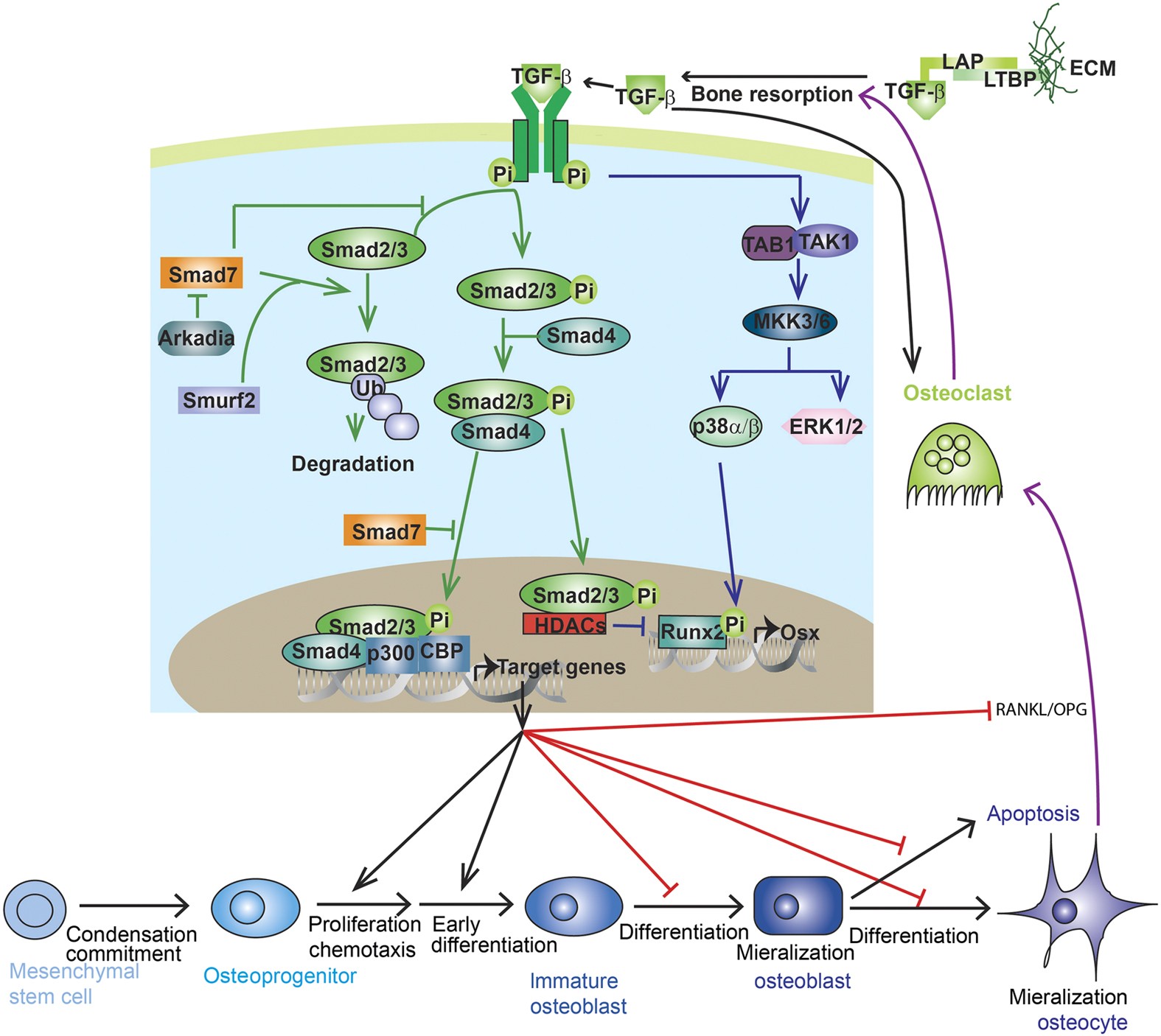
Transforming growth factor-beta (TGF-β) and bone morphogenic protein (BMP) signaling has fundamental roles in both embryonic skeletal development and postnatal bone homeostasis. TGF-βs and BMPs, acting on a tetrameric receptor complex, transduce signals to both the canonical Smad-dependent signaling pathway (that is, TGF-β/BMP ligands, receptors, and Smads) and the non-canonical-Smad-independent signaling pathway (that is, p38 mitogen-activated protein kinase/p38 MAPK) to regulate mesenchymal stem cell differentiation during skeletal development, bone formation and bone homeostasis. Both the Smad and p38 MAPK signaling pathways converge at transcription factors, for example, Runx2 to promote osteoblast differentiation and chondrocyte differentiation from mesenchymal precursor cells. TGF-β and BMP signaling is controlled by multiple factors, including the ubiquitin–proteasome system, epigenetic factors, and microRNA. Dysregulated TGF-β and BMP signaling result in a number of bone disorders in humans. Knockout or mutation of TGF-β and BMP signaling-related genes in mice leads to bone abnormalities of varying severity, which enable a better understanding of TGF-β/BMP signaling in bone and the signaling networks underlying osteoblast differentiation and bone formation. There is also crosstalk between TGF-β/BMP signaling and several critical cytokines’ signaling pathways (for example, Wnt, Hedgehog, Notch, PTHrP, and FGF) to coordinate osteogenesis, skeletal development, and bone homeostasis. This review summarizes the recent advances in our understanding of TGF-β/BMP signaling in osteoblast differentiation, chondrocyte differentiation, skeletal development, cartilage formation, bone formation, bone homeostasis, and related human bone diseases caused by the disruption of TGF-β/BMP signaling.