

Estradiol is the “prototypic” sex hormone of women. Yet, women have another sex hormone, which is often disregarded: Progesterone. The goal of this article is to provide a comprehensive review on progesterone, and its metabolite allopregnanolone, emphasizing three key areas: biological properties, main functions, and effects on mood in women. Recent years of intensive research on progesterone and allopregnanolone have paved the way for new treatment of postpartum depression. However, treatment for premenstrual syndrome and premenstrual dysphoric disorder as well as contraception that women can use without risking mental health problems are still needed. As far as progesterone is concerned, we might be dealing with a two-edged sword: while its metabolite allopregnanolone has been proven useful for treatment of PPD, it may trigger negative symptoms in women with PMS and PMDD. Overall, our current knowledge on the beneficial and harmful effects of progesterone is limited and further research is imperative.
Progesterone and plasma metabolites in women with and in those without premenstrual dysphoric disorder
BACKGROUND: The molecular mechanisms underpinning the progesterone-triggering mood symptoms in women with premenstrual dysphoric disorder (PMDD) are unknown. Cell metabolism is a potential source of variability. Very little is known about the effect of progesterone sensitivity on the metabolome. In this study, we aimed to characterize the effects of progesterone on the global metabolic profile and explore the differences between women with PMDD and controls. METHODS: Plasma was obtained from 12 women with prospectively confirmed PMDD and 25 controls under two hormone conditions: (1) gonadal suppression induced by leuprolide acetate (3.75 mg IM monthly) and (2) add-back phase with leuprolide and progesterone (200 mg twice daily by vaginal suppository). The global metabolic profile was obtained using liquid and gas chromatography followed by mass spectrometry. Differences between groups and time points were tested using repeated measures analysis of variance. The false discovery rate was calculated to account for multiple testing. RESULTS: Amino acids and their derivatives represented 78% (28/36) of the known compounds that were found in significantly lower plasma concentrations after progesterone administration than during gonadal suppression. The concentration of tyrosine was nominally significantly decreased after progesterone add-back in controls, but not in cases (P = 0.02). CONCLUSION: Plasma levels of some amino acids are decreased in response to progesterone. Albeit preliminary, evidence further suggests that progesterone has a different effect on the metabolic profiles of women with PMDD compared to controls. Further research is needed to replicate our findings in a larger sample and to identify the unknown compounds, especially those differentially expressed.

The steroid metabolome in women with premenstrual dysphoric disorder during GnRH agonist-induced ovarian suppression: effects of estradiol and progesterone addback
Clinical evidence suggests that symptoms in premenstrual dysphoric disorder (PMDD) reflect abnormal responsivity to ovarian steroids. This differential steroid sensitivity could be underpinned by abnormal processing of the steroid signal. We used a pharmacometabolomics approach in women with prospectively confirmed PMDD (n=15) and controls without menstrual cycle-related affective symptoms (n=15). All were medication-free with normal menstrual cycle lengths. Notably, women with PMDD were required to show hormone sensitivity in an ovarian suppression protocol. Ovarian suppression was induced for 6 months with gonadotropin-releasing hormone (GnRH)-agonist (Lupron); after 3 months all were randomized to 4 weeks of estradiol (E2) or progesterone (P4). After a 2-week washout, a crossover was performed. Liquid chromatography/tandem mass spectrometry measured 49 steroid metabolites in serum. Values were excluded if >40% were below the limit of detectability (n=21). Analyses were performed with Wilcoxon rank-sum tests using false-discovery rate (q

5α-Reductase Inhibition Prevents the Luteal Phase Increase in Plasma Allopregnanolone Levels and Mitigates Symptoms in Women with Premenstrual Dysphoric Disorder
Changes in neurosteroid levels during the luteal phase of the menstrual cycle may precipitate affective symptoms. To test this hypothesis, we stabilized neurosteroid levels by administering the 5α-reductase inhibitor dutasteride to block conversion of progesterone to its neurosteroid metabolite allopregnanolone in women with premenstrual dysphoric disorder (PMDD) and in asymptomatic control women. Sixteen women with prospectively confirmed PMDD and 16 control women participated in one of two separate randomized, double-blind, placebo-controlled, cross-over trials, each lasting three menstrual cycles. After one menstrual cycle of single-blind placebo, participants were randomized to receive, for the next two menstrual cycles, either double-blind placebo or dutasteride (low-dose 0.5 mg/day in the first eight PMDD and eight control women or high-dose 2.5 mg/day in the second group of women). All women completed the daily rating form (DRF) and were evaluated in clinic during the follicular and luteal phases of each menstrual cycle. Main outcome measures were the DRF symptoms of irritability, sadness, and anxiety. Analyses were performed with SAS PROC MIXED. In the low-dose group, no significant effect of dutasteride on PMDD symptoms was observed compared with placebo (ie, symptom cyclicity maintained), and plasma allopregnanolone levels increased in women with PMDD from follicular to the luteal phases, suggesting the absence of effect of the low-dose dutasteride on 5α-reductase. In contrast, the high-dose group experienced a statistically significant reduction in several core PMDD symptoms (ie, irritability, sadness, anxiety, food cravings, and bloating) on dutasteride compared with placebo. Dutasteride had no effect on mood in controls. Stabilization of allopregnanolone levels from the follicular to the luteal phase of the menstrual cycle by blocking the conversion of progesterone to its 5α-reduced neurosteroid metabolite mitigates symptoms in PMDD. These data provide preliminary support for the pathophysiologic relevance of neurosteroids in this condition.
Premenstrual Dysphoric Disorder Symptoms Following Ovarian Suppression: Triggered by Change in Ovarian Steroid Levels But Not Continuous Stable Levels | American Journal of Psychiatry
Objective:Premenstrual dysphoric disorder (PMDD) symptoms are eliminated by ovarian suppression and stimulated by administration of ovarian steroids, yet they appear with ovarian steroid levels indistinguishable from those in women without PMDD. Thus, ...

Progesterone Is Important for Transgender Women’s Therapy—Applying Evidence for the Benefits of Progesterone in Ciswomen
Current transgender women’s therapy is estradiol/antiandrogen. I suggest adding progesterone, given its antiandrogen, breast-feminizing, bone-formation, sl
When Women Initiate Estrogen Therapy Matters | The Menopause Society
New large-scale analysis shows potential long-term benefits of initiating hormones during perimenopause

The 2022 hormone therapy position statement of The North American Menopause Society
Abstract “The 2022 Hormone Therapy Position Statement of The North American Menopause Society” (NAMS) updates “The 2017 Hormone Therapy Position Statement of The North American Menopause Society” and identifies future research needs. An Advisory Panel of clinicians and researchers expert in the field of women’s health and menopause was recruited by NAMS to review the 2017 Position Statement, evaluate new literature, assess the evidence, and reach consensus on recommendations, using the level of evidence to identify the strength of recommendations and the quality of the evidence. The Advisory Panel’s recommendations were reviewed and approved by the NAMS Board of Trustees. Hormone therapy remains the most effective treatment for vasomotor symptoms (VMS) and the genitourinary syndrome of menopause and has been shown to prevent bone loss and fracture. The risks of hormone therapy differ depending on type, dose, duration of use, route of administration, timing of initiation, and whether a progestogen is used. Treatment should be individualized using the best available evidence to maximize benefits and minimize risks, with periodic reevaluation of the benefits and risks of continuing therapy. For women aged younger than 60 years or who are within 10 years of menopause onset and have no contraindications, the benefit-risk ratio is favorable for treatment of bothersome VMS and prevention of bone loss. For women who initiate hormone therapy more than 10 years from menopause onset or who are aged older than 60 years, the benefit-risk ratio appears less favorable because of the greater absolute risks of coronary heart disease, stroke, venous thromboembolism, and dementia. Longer durations of therapy should be for documented indications such as persistent VMS, with shared decision-making and periodic reevaluation. For bothersome genitourinary syndrome of menopause symptoms not relieved with over-the-counter therapies in women without indications for use of systemic hormone therapy, low-dose vaginal estrogen therapy or other therapies (eg, vaginal dehydroepiandrosterone or oral ospemifene) are recommended.
Estrogen therapy for osteoporosis in the modern era
Menopause predisposes women to osteoporosis due to declining estrogen levels. This results in a decrease in bone mineral density (BMD) and an increase in fractures. Osteoporotic fractures lead to substantial morbidity and mortality, and are considered one of the largest public health priorities by the World Health Organization (WHO). It is therefore essential for menopausal women to receive appropriate guidance for the prevention and management of osteoporosis. The Women’s Health Initiative (WHI) randomized controlled trial first proved hormonal therapy (HT) reduces the incidence of all osteoporosis-related fractures in postmenopausal women. However, the study concluded that the adverse effects outweighed the potential benefits on bone, leading to a significant decrease in HT use for menopausal symptoms. Additionally, HT was not used as first-line therapy for osteoporosis and fractures. Subsequent studies have challenged these initial conclusions and have shown significant efficacy of HT in various doses, durations, regimens, and routes of administration. These studies support that HT improves BMD and reduces fracture risk in women with and without osteoporosis. Furthermore, the studies suggest that low-dose and transdermal HT are less likely associated with the adverse effects of breast cancer, endometrial hyperplasia, coronary artery disease (CAD), and venous thromboembolism (VTE) previously observed in standard-dose oral HT regimens. Given the need for estrogen in menopausal women and evidence supporting the cost effectiveness, safety, and efficacy of HT, we propose that HT should be considered for the primary prevention and treatment of osteoporosis in appropriate candidates. HT should be individualized and the once “lowest dose for shortest period of time” concept should no longer be used. This review will focus on the prior and current studies for various HT formulations used for the prevention and treatment of osteoporosis, exploring the safety profile of low-dose and transdermal HT that have been shown to be safer than oral standard-dose HT.
Menopausal Hormone Therapy and Health Outcomes During the Intervention and Extended Poststopping Phases of the Women’s Health Initiative Randomized Trials
Manson and coauthors provide an update and overview of health outcomes during the intervention and poststopping phases of the Women’s Health Initiative (WHI) hormone therapy trials. In an accompanying Editorial, Nabel discusses how the WHI is a victory for women and their health.

Critical Role of Estrogens on Bone Homeostasis in Both Male and Female: From Physiology to Medical Implications
Bone is a multi-skilled tissue, protecting major organs, regulating calcium phosphate balance and producing hormones. Its development during childhood determines height and stature as well as resistance against fracture in advanced age. Estrogens are key regulators of bone turnover in both females and males. These hormones play a major role in longitudinal and width growth throughout puberty as well as in the regulation of bone turnover. In women, estrogen deficiency is one of the major causes of postmenopausal osteoporosis. In this review, we will summarize the main clinical and experimental studies reporting the effects of estrogens not only in females but also in males, during different life stages. Effects of estrogens on bone involve either Estrogen Receptor (ER)α or ERβ depending on the type of bone (femur, vertebrae, tibia, mandible), the compartment (trabecular or cortical), cell types involved (osteoclasts, osteoblasts and osteocytes) and sex. Finally, we will discuss new ongoing strategies to increase the benefit/risk ratio of the hormonal treatment of menopause.
Selective estrogen receptor modulators in clinical practice: a safety overview
Introduction: Selective estrogen receptor (ER) modulators (SERMs) are a class of nonsteroidal compounds that interact with ERs, each with a distinct tissue-specific profile. Depending upon the degr...

Oestradiol decreases colonic permeability through oestrogen receptor β‐mediated up‐regulation of occludin and junctional adhesion molecule‐A in epithelial cells
Oestradiol modulates paracellular permeability and tight junction (TJ) function in endothelia and reproductive tissues, but whether the ovarian hormones and cycle affect the paracellular pathway in the intestinal epithelium remains unclear. Oestrogen receptors (ERs) are expressed in intestinal epithelial cells, and oestradiol regulates epithelium formation. We examined the effects of oestrous cycle stage, oestradiol benzoate (EB), and progesterone (P) on colonic paracellular permeability (CPP) in the female rat, and whether EB affects expression of the TJ proteins in the rat colon and the human colon cell line Caco‐2. In cyclic rats, CPP was determined through lumen‐to‐blood 51 Cr‐labelled EDTA clearance, and in Ussing chambers for dextran permeability. CPP was also examined in ovariectomized (OVX) rats treated with P or EB, with and without the ER antagonist ICI 182,780, or with the selective agonists for ERα (propyl pyrazole triol; PPT) or ERβ (diarylpropionitrile; DPN). In oestrus rats, CPP was reduced ( P < 0.01) relative to dioestrus. In OVX rats, EB dose‐dependently decreased CPP, an effect mimicked by DPN and blocked by ICI 182,780, whereas P had no effect. Oestradiol increased occludin mRNA and protein in the colon ( P < 0.05), but not zona occludens (ZO)‐1. Further, EB and DPN enhanced occludin and junctional adhesion molecule (JAM)‐A expression in Caco‐2 cells without change in ZO‐1, an effect blocked by ICI 182,780. These data show that oestrogen reinforces intestinal epithelial barrier through ERβ‐mediated up‐regulation of the transmembrane proteins occludin and JAM‐A determining paracellular spaces. These findings highlight the importance of the ERβ pathway in the control of colonic paracellular transport and mucosal homeostasis.

The Role of Estrogen in the Control of Rat Osteocyte Apoptosis
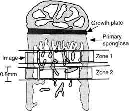
Abstract We have previously shown that estrogen withdrawal by gonadotrophin-releasing hormone analogs (GnRHa) induces osteocyte death via apoptosis in human bone. Although it is likely that the increase in osteocyte death via apoptosis was related to the loss of estrogen, these experiments could not rule out a direct role for the GnRHa. Therefore, in this study, we have used a rat model of ovariectomy (OVX) to determine whether the effect of estrogen withdrawal extends to other species and to clarify the role of estrogen in the maintenance of osteocyte viability. Twelve 9-week-old rats were divided into three treatment groups: sham operated (SHAM) (n = 4), OVX (n = 4), and OVX + estrogen (E2) (25 μg/day) (n = 4). At 3 weeks following the start of treatment, tibial bones were removed. The percentage of osteocytes displaying DNA breaks, using an in situ nick-translation method, was significantly higher in the OVX group compared with the SHAM control in both cortical bone (10.04% vs. 2.31%, respectively; p < 0.0001) and trabecular bone (6.44% vs. 1.58%, respectively; p = 0.003). Addition of estrogen in the OVX animals completely abrogated the increase in osteocyte apoptosis in cortical bone (0.78%) and trabecular bone (1.17%). The percentage of apoptotic osteocytes decreased with increasing distance from the primary/secondary spongiosa interface below the growth plate in the OVX model and the OVX + E2 model. Nuclear morphology and electrophoresis of DNA confirmed the presence of apoptotic cells in the samples. In conclusion, OVX in the rat results in an increase in osteocyte apoptosis as a direct or indirect result of E2 loss. Addition of estrogen in the OVX animals prevents this increase in osteocyte apoptosis. These data confirm an important role for estrogen in the control of osteocyte apoptosis and the maintenance of osteocyte viability. Estrogen deficiency might, through compromising the viability of osteocyte networks, reduce the ability of bone to respond appropriately to loading.
Risks and Benefits of HRT Versus ERT in Order to Separate HRT from ERT
Aim: While the benefits of Hormone Replace Therapy (HRT) and Estrogen Replace Therapy (ERT) might overlap, their risks have to be separated particularly with regard to breast cancer. The risks of HRT are mainly Coronary Heart Disease (CHD) and Breast cancer. The main risk of ERT is the incidence of strokes which can be avoided by not using an oral estrogen but an Intra-uterine contraceptive but reducing the risk of endometrial cancer in women with a womb. Methods: Studies that randomized peri-menopausal and menopausal women to either HRT or ERT versus placebo and one study that included similar women followed up prospectively for the incidence of the development of Alzheimer’s Dementia (AD). Results: The significant risks of HRT included CHD, stroke and pulmonary embolism. The risk of breast cancer only really existed after long term follow-up [(annualized incidence, 0.45% vs 0.36%; Hazard Ratio (HR) 1.28)]. The significant risks of ERT only included strokes. There was a lower risk of breast cancer in the long term [HR 0.77, 95% CI 0.62–0.95; p=0.02]. Conclusions: In women with a uterus, where HRT instead of ERT is mandated, the oral progestogen of HRT can be replaced by intrauterine device loaded with a Progestogen. Similarly, the oral Estrogen of HRT and ERT can be replaced by Estrogen sources like the patch, gel or implants that bypass the liver and bypass potential problems like strokes and pulmonary embolism.
Estrogens and Androgens in Skeletal Physiology and Pathophysiology
Estrogens and androgens influence the growth and maintenance of the mammalian skeleton and are responsible for its sexual dimorphism. Estrogen deficiency at menopause or loss of both estrogens and androgens in elderly men contribute to the development of osteoporosis, one of the most common and impactful metabolic diseases of old age. In the last 20 years, basic and clinical research advances, genetic insights from humans and rodents, and newer imaging technologies have changed considerably the landscape of our understanding of bone biology as well as the relationship between sex steroids and the physiology and pathophysiology of bone metabolism. Together with the appreciation of the side effects of estrogen-related therapies on breast cancer and cardiovascular diseases, these advances have also drastically altered the treatment of osteoporosis. In this article, we provide a comprehensive review of the molecular and cellular mechanisms of action of estrogens and androgens on bone, their influences on skeletal homeostasis during growth and adulthood, the pathogenetic mechanisms of the adverse effects of their deficiency on the female and male skeleton, as well as the role of natural and synthetic estrogenic or androgenic compounds in the pharmacotherapy of osteoporosis. We highlight latest advances on the crosstalk between hormonal and mechanical signals, the relevance of the antioxidant properties of estrogens and androgens, the difference of their cellular targets in different bone envelopes, the role of estrogen deficiency in male osteoporosis, and the contribution of estrogen or androgen deficiency to the monomorphic effects of aging on skeletal involution.

Menopausal hormone therapy and breast cancer: what is the evidence from randomized trials?
The relationship between menopausal hormone therapy (HT) and breast cancer is complex and further complicated by misinformation, perception, and overgeneralization of data. These issues are address...
