Osteoimmunology: The Nexus between bone and immune system
Osteoimmunology is an interdisciplinary research field which combines the existing fields of osteology (bone biology) and immunology under one umbrella. The observation that contributed enormously to the emergence of osteoimmunology as an independent field of investigation was the enhanced bone loss in various inflammatory bone diseases such as rheumatoid arthritis, osteoporosis and periodontitis. T helper cells (Th1, Th2, Treg and Th17) along with various other immune cells (B cells, DC, macrophages etc.) are actively involved in bone homeostasis. The present review thus provides an overview of the nexus between these two prominent systems (Bone and Immune system) of an organism, which reside in a common niche (bone marrow) and thus cross-communicate to modulate their respective development. Investigations in the field of osteoimmunology thus promise the advent of new era in the field with novel therapeutics for bone loss in various inflammatory conditions. A molecular insight into the field of osteoimmunology can lead to novel approaches for the prevention and treatment of diverse inflammatory conditions such as osteoporosis, rheumatoid arthritis and osteoarthritis.
Loss of Bone and Wnt10b Expression in Male Type 1 Diabetic Mice Is Blocked by the Probiotic Lactobacillus reuteri
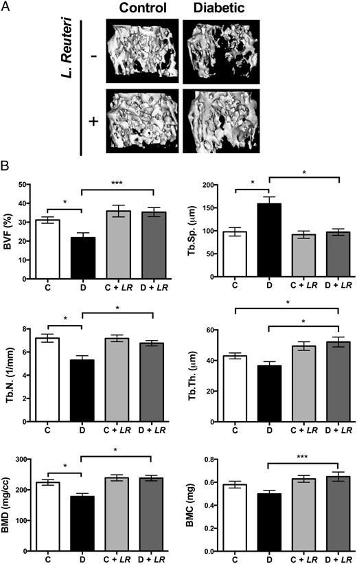
Type 1 diabetes (T1D)–induced osteoporosis is characterized by a predominant suppression of osteoblast number and activity, as well as increased bone marrow adiposity but no change in osteoclast activity. The fundamental mechanisms and alternative anabolic treatments (with few side effects) for T1D bone loss remain undetermined. Recent studies by our laboratory and others indicate that probiotics can benefit bone health. Here, we demonstrate that Lactobacillus reuteri, a probiotic with anti-inflammatory and bone health properties, prevents T1D-induced bone loss and marrow adiposity in mice. We further found that L. reuteri treatment prevented the suppression of Wnt10b in T1D bone. Consistent with a role for attenuated bone Wnt10b expression in T1D osteoporosis, we observed that bone-specific Wnt10b transgenic mice are protected from T1D bone loss. To examine the mechanisms of this protection, we focused on TNF-α, a cytokine up-regulated in T1D that causes suppression of osteoblast Wnt10b expression in vitro. Addition of L. reuteri prevented TNF-α–mediated suppression of Wnt10b and osteoblast maturation markers. Taken together, our findings reveal a mechanism by which T1D causes bone loss and open new avenues for use of probiotics to benefit the bone.
Systems Modeling of Bortezomib and Dexamethasone Combinatorial Effects on Bone Homeostasis in Multiple Myeloma Patients
Osteolytic bone disease is one of the most debilitating manifestations of multiple myeloma (MM). Bortezomib is a proteasome inhibitor that shows both anticancer and bone anabolic properties and is being evaluated for its positive effects in MM patients with skeletal complications. Dexamethasone is a potent corticosteroid that is often given in combination with bortezomib for its antineoplastic effects; however, bone loss and osteoporosis are major adverse effects of long-term steroid-based therapies.

TGF-β and BMP signaling in osteoblast, skeletal development, and bone formation, homeostasis and disease
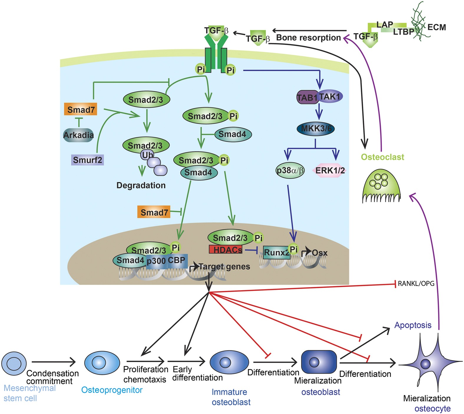
Transforming growth factor-beta (TGF-β) and bone morphogenic protein (BMP) signaling has fundamental roles in both embryonic skeletal development and postnatal bone homeostasis. TGF-βs and BMPs, acting on a tetrameric receptor complex, transduce signals to both the canonical Smad-dependent signaling pathway (that is, TGF-β/BMP ligands, receptors, and Smads) and the non-canonical-Smad-independent signaling pathway (that is, p38 mitogen-activated protein kinase/p38 MAPK) to regulate mesenchymal stem cell differentiation during skeletal development, bone formation and bone homeostasis. Both the Smad and p38 MAPK signaling pathways converge at transcription factors, for example, Runx2 to promote osteoblast differentiation and chondrocyte differentiation from mesenchymal precursor cells. TGF-β and BMP signaling is controlled by multiple factors, including the ubiquitin–proteasome system, epigenetic factors, and microRNA. Dysregulated TGF-β and BMP signaling result in a number of bone disorders in humans. Knockout or mutation of TGF-β and BMP signaling-related genes in mice leads to bone abnormalities of varying severity, which enable a better understanding of TGF-β/BMP signaling in bone and the signaling networks underlying osteoblast differentiation and bone formation. There is also crosstalk between TGF-β/BMP signaling and several critical cytokines’ signaling pathways (for example, Wnt, Hedgehog, Notch, PTHrP, and FGF) to coordinate osteogenesis, skeletal development, and bone homeostasis. This review summarizes the recent advances in our understanding of TGF-β/BMP signaling in osteoblast differentiation, chondrocyte differentiation, skeletal development, cartilage formation, bone formation, bone homeostasis, and related human bone diseases caused by the disruption of TGF-β/BMP signaling.

The gut-bone axis: how bacterial metabolites bridge the distance
1Department of Internal Medicine 3, Rheumatology and Immunology, Friedrich-Alexander University Erlangen-Nürnberg (FAU) and Universitätsklinikum Erlangen, Erlangen, Germany.
TRANCE Is a Novel Ligand of the Tumor Necrosis Factor Receptor Family That Activates c-Jun N-terminal Kinase in T Cells *
A novel member of the tumor necrosis factor (TNF) cytokine family, designated TRANCE, was cloned during a search for apoptosis-regulatory genes using a somatic cell genetic approach in T cell hybridomas. The TRANCEgene encodes a type II membrane protein of 316 amino acids with a predicted molecular mass of 35 kDa. Its extracellular domain is most closely related to TRAIL, FasL, and TNF. TRANCE is an immediate early gene up-regulated by TCR stimulation and is controlled by calcineurin-regulated transcription factors.

Bone and the Immune System
Osteoporosis increases fracture risk, a cause of crippling morbidity and mortality. The immunoskeletal interface (ISI) is a centralization of cell and cytokine effectors shared between skeletal and immune systems. Consequently, the immune system mediates powerful effects on bone turnover. Physiologically, B cells secrete osteoprotegerin (OPG), a potent anti-osteoclastogenic factor that preserves bone mass. However, activated T cells and B cells secrete pro-osteoclastogenic factors including receptor activator of Nuclear factor-kappaB (NF-kB) ligand (RANKL), Interleukin (IL)-17A, and tumor necrosis factor (TNF)-α promoting bone loss in inflammatory states such as rheumatoid arthritis. Recently, ISI disruption has been linked to osteoporosis in human immunodeficiency virus (HIV) infection/acquired immunodeficiency syndrome (AIDS), where elevated B cell RANKL and diminished OPG drive bone resorption. HIV-antiretroviral therapy paradoxically intensifies bone loss during disease reversal, as immune reconstitution produces osteoclastogenic cytokines. Interestingly, in estrogen deficiency, activated T cells secrete RANKL, TNF, and IL-17A that amplify bone resorption and contribute to postmenopausal osteoporosis. T cell–produced TNF and IL-17A further contribute to bone loss in hyperparathyroidism, while T cell production of the anabolic Wingless integration site (Wnt) ligand, Wnt10b, promotes bone formation in response to anabolic parathyroid hormone and the immunomodulatory costimulation inhibitor cytotoxic T lymphocyte–associated protein-4-IgG (abatacept). These findings provide a window into the workings of the ISI and suggest novel targets for future therapeutic interventions to reduce fracture risk.

Physiological and pathophysiological bone turnover — role of the immune system
Receptor activator of NF-κB ligand (RANKL) and osteoprotegerin (OPG) are key downstream effectors of bone resorption; tumour necrosis factor (TNF) might synergize with RANKL to superinduce osteoclastic bone resorptionB cells, regulated by T cells, are a key source of basal OPG whereas activated T cells and B cells are key sources of TNF and RANKL in inflammatory conditionsShort-term antiresorptive therapies might safely prevent bone loss associated with combination antiretroviral therapyAnergic and parathyroid-hormone-treated T cells secrete the protein Wnt-10b, which promotes bone formationNovel therapeutic strategies targeting the immune system might promote bone formation and decrease bone resorption to manage osteoporotic bone loss and prevent fracture

Regulation of Bone Remodeling by Parathyroid Hormone
Parathyroid hormone (PTH) exerts profound effects on skeletal homeostasis through multiple cellular and molecular mechanisms. Continuous hyperparathyroidism causes net loss of bone mass, despite accelerating bone formation by osteoblasts. Intermittent treatment with PTH analogs represents the only Food and Drug Administration (FDA)-approved bone anabolic osteoporosis treatment strategy. Functional PTH receptors are present on cells of the osteoblast lineage, ranging from early skeletal stem cells to matrix-embedded osteocytes. In addition, bone remodeling by osteoclasts liberates latent growth factors present within bone matrix. Here, we will provide an overview of the multiple cellular and molecular mechanisms through which PTH influences bone homeostasis. Notably, net skeletal effects of continuous versus intermittent can differ significantly. Where possible, we will highlight mechanisms through which continuous hyperparathyroidism leads to bone loss, and through which intermittent hyperparathyroidism boosts bone mass. Given the therapeutic usage of intermittent PTH (iPTH) treatment for osteoporosis, particular attention will be paid toward mechanisms underlying the bone anabolic effects of once daily PTH administration.
<i>In silico</i> models of bone remodeling from macro to nano—from organ to cell
Abstract Computational modeling is a tool through which researchers can achieve a greater understanding of the mechanisms governing biological systems. In the field of bone biology, a plethora of models exist which attempt to replicate and investigate bone's dynamic behavior at different scales. At organ level, models are continuum based and describe the variation of bone's apparent density as a function of both biological and external mechanical stimuli. At tissue level, models include bone microarchitecture and more descriptive parameters such as trabecular thickness, osteoclast resorption depth, and activation frequency. Finally, at cell level, models employ partial differential equations to describe complex cellular interactions in the temporal domain. Although informative, these models exist in isolation. Consequently, their interpretation is limited. In this review, we present an overview of the organ‐, tissue‐, and cell‐level models and assess their ability to reflect bone's metabolic processes reliably. Existing interscale synergies are then presented along with a computational framework which could be exploited to achieve a fully integrated, multiscale modeling approach. WIREs Syst Biol Med 2011 3 241–251 DOI: 10.1002/wsbm.115 This article is categorized under: Analytical and Computational Methods > Computational Methods Models of Systems Properties and Processes > Organ, Tissue, and Physiological Models

Galactooligosaccharides Improve Mineral Absorption and Bone Properties in Growing Rats through Gut Fermentation
Galactooligosaccharides (GOS), prebiotic nondigestible oligosaccharides derived from lactose, have the potential for improving mineral balance and bone properties. This study examined the dose–response effect of GOS supplementation on calcium and magnesium absorption, mineral retention, bone properties, and gut microbiota in growing rats. Seventy-five 4-week-old male Sprague–Dawley rats were randomized into one of five treatment groups (n = 15/group) and fed a diet containing 0, 2, 4, 6, or 8% GOS by weight for 8 weeks. Dietary GOS significantly decreased cecal pH and increased cecal wall weight and content weight in a dose-dependent manner (p < 0.0001). Fingerprint patterns of the 16S rRNA gene PCR-DGGE from fecal DNA indicated the variance of bacterial community structure, which was primarily explained by GOS treatments (p = 0.0001). Quantitative PCR of the samples revealed an increase in the relative proportion of bifidobacteria with GOS (p = 0.0001). Net calcium absorption was increased in a dose–response manner (p < 0.01) with GOS supplementation. Dietary GOS also increased (p < 0.02) net magnesium absorption, femur 45Ca uptake, calcium and magnesium retention, and femur and tibia breaking strength. Distal femur total and trabecular volumetric bone mineral density (vBMD) and area and proximal tibia vBMD increased (p < 0.02) with GOS supplementation. Trabecular-rich bones, that is, those that rapidly turn over, were most benefited. Regression modeling showed that GOS benefited calcium and magnesium utilization and vBMD through decreased cecal pH, increased cecal wall and content weight, and increased proportion of bifidobacteria.
A theoretical study of bone remodelling under PEMF at cellular level
Pulsed electromagnetic field (PEMF) devices have been used clinically to slow down osteoporosis and accelerate the healing of bone fractures for many years. However, the underlying mechanism by wh...

Biology of the RANKL–RANK–OPG System in Immunity, Bone, and Beyond
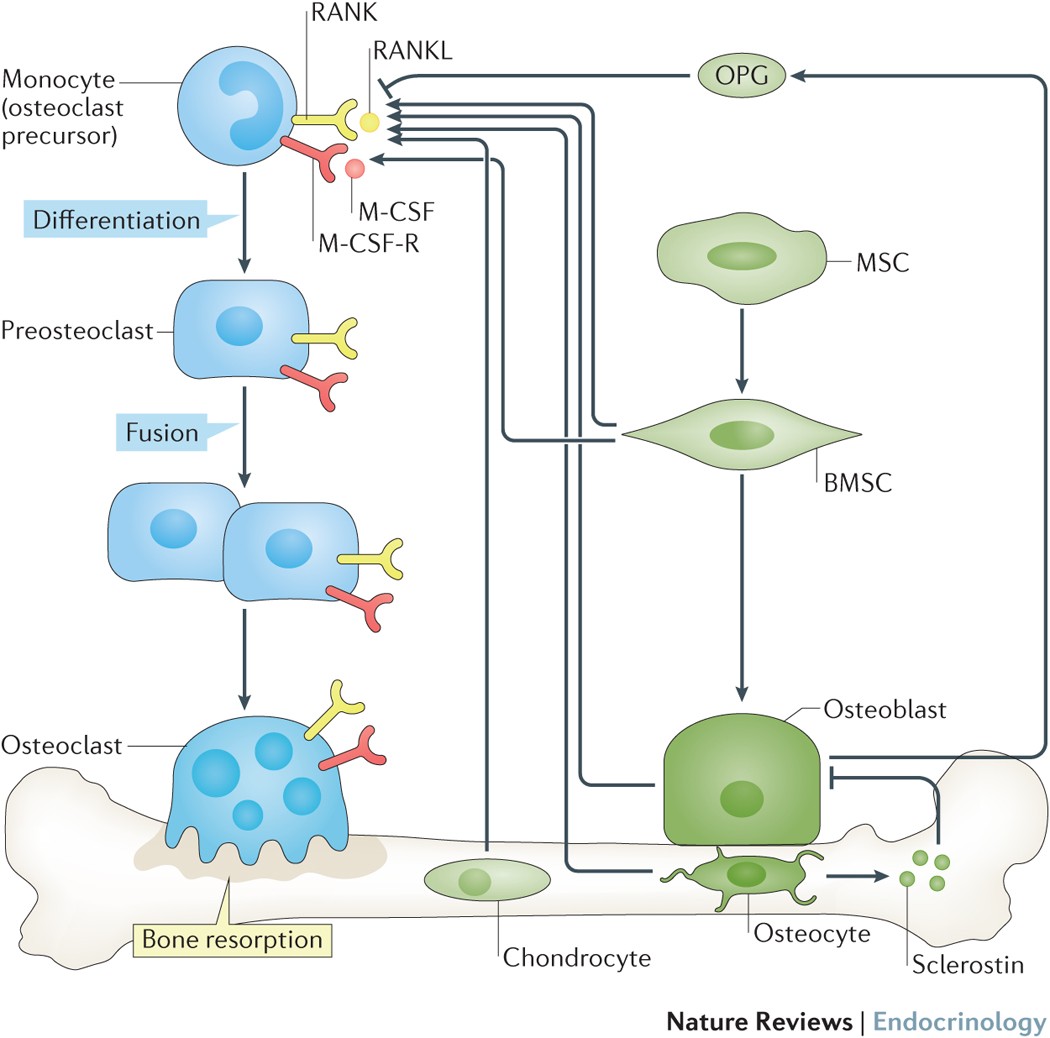
Discovery and characterization of the cytokine receptor-cytokine-decoy receptor triad formed by RANKL-RANK-OPG have led not only to immense advances in understanding the biology of bone homeostasis, but have also crystalized appreciation of the critical regulatory relationship that exists between bone and immunity, resulting in the emergence of the burgeoning field of osteoimmunology. RANKL-RANK-OPG are members of the tumor necrosis factor (TNF) and TNF receptor superfamilies, and share signaling characteristics common to many members of each. Developmentally regulated and cell-type specific expression patterns of each of these factors have revealed key regulatory functions for RANKL-RANK-OPG in bone homeostasis, organogenesis, immune tolerance and cancer. Successful efforts at designing and developing therapeutic agents targeting RANKL-RANK-OPG have been undertaken for osteoporosis, and additional efforts are underway for other conditions. In this review, we will summarize the basic biology of the RANKL-RANK-OPG system, relate its cell-type specific functions to system-wide mechanisms of development and homeostasis, and highlight emerging areas of interest for this cytokine group.

Computational Modeling of Interactions between Multiple Myeloma and the Bone Microenvironment
Multiple Myeloma (MM) is a B-cell malignancy that is characterized by osteolytic bone lesions. It has been postulated that positive feedback loops in the interactions between MM cells and the bone microenvironment form reinforcing ‘vicious cycles’, resulting in more bone resorption and MM cell population growth in the bone microenvironment. Despite many identified MM-bone interactions, the combined effect of these interactions and their relative importance are unknown. In this paper, we develop a computational model of MM-bone interactions and clarify whether the intercellular signaling mechanisms implemented in this model appropriately drive MM disease progression. This new computational model is based on the previous bone remodeling model of Pivonka et al. [1], and explicitly considers IL-6 and MM-BMSC (bone marrow stromal cell) adhesion related pathways, leading to formation of two positive feedback cycles in this model. The progression of MM disease is simulated numerically, from normal bone physiology to a well established MM disease state. Our simulations are consistent with known behaviors and data reported for both normal bone physiology and for MM disease. The model results suggest that the two positive feedback cycles identified for this model are sufficient to jointly drive the MM disease progression. Furthermore, quantitative analysis performed on the two positive feedback cycles clarifies the relative importance of the two positive feedback cycles, and identifies the dominant processes that govern the behavior of the two positive feedback cycles. Using our proposed quantitative criteria, we identify which of the positive feedback cycles in this model may be considered to be ‘vicious cycles’. Finally, key points at which to block the positive feedback cycles in MM-bone interactions are identified, suggesting potential drug targets.
OSTEOIMMUNOLOGY: Interplay Between the Immune System and Bone Metabolism
Studies of bone and the immune system have converged in recent years under the banner of osteoimmunology. The immune system is spawned in the bone marrow reservoir, and investigators now recognize that important niches also exist there for memory lymphocytes. At the same time, various factors produced during immune responses are capable of profoundly affecting regulation of bone. Mechanisms have evolved to prevent excessive interference by the immune system with bone homeostasis, yet pathologic bone loss is a common sequela associated with autoimmunity and cancer. There are also developmental links, or parallels, between bone and the immune system. Cells that regulate bone turnover share a common precursor with inflammatory immune cells and may restrict themselves anatomically, in part by utilizing a signaling network analogous to lymphocyte costimulation. Efforts are currently under way to further characterize how these two organ systems overlap and to develop therapeutic strategies that benefit from this understanding.

150 Years of the Mass Action Law
This year we celebrate the 150th anniversary of the law of mass action. This law is often assumed to have been “there” forever, but it has its own history, background, and a definite starting point. The law has had an impact on chemistry, biochemistry, biomathematics, and systems biology that is difficult to overestimate. It is easily recognized that it is the direct basis for computational enzyme kinetics, ecological systems models, and models for the spread of diseases. The article reviews the explicit and implicit role of the law of mass action in systems biology and reveals how the original, more general formulation of the law emerged one hundred years later ab initio as a very general, canonical representation of biological processes.
Collection
Generated from a Zotero collection.